. 2015 Mar 20;10(3):e0118450.
doi: 10.1371/journal.pone.0118450. eCollection 2015.

Mid-embryo patterning and precision in Drosophila segmentation: Krüppel dual regulation of hunchback

Affiliations

Mid-embryo patterning and precision in Drosophila segmentation: Krüppel dual regulation of hunchback

David M Holloway et al. PLoS One. .

Abstract

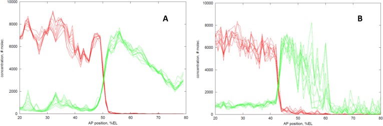
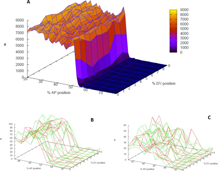
In early development, genes are expressed in spatial patterns which later define cellular identities and tissue locations. The mechanisms of such pattern formation have been studied extensively in early Drosophila (fruit fly) embryos. The gap gene hunchback (hb) is one of the earliest genes to be expressed in anterior-posterior (AP) body segmentation. As a transcriptional regulator for a number of downstream genes, the spatial precision of hb expression can have significant effects in the development of the body plan. To investigate the factors contributing to hb precision, we used fine spatial and temporal resolution data to develop a quantitative model for the regulation of hb expression in the mid-embryo. In particular, modelling hb pattern refinement in mid nuclear cleavage cycle 14 (NC14) reveals some of the regulatory contributions of simultaneously-expressed gap genes. Matching the model to recent data from wild-type (WT) embryos and mutants of the gap gene Krüppel (Kr) indicates that a mid-embryo Hb concentration peak important in thoracic development (at parasegment 4, PS4) is regulated in a dual manner by Kr, with low Kr concentration activating hb and high Kr concentration repressing hb. The processes of gene expression (transcription, translation, transport) are intrinsically random. We used stochastic simulations to characterize the noise generated in hb expression. We find that Kr regulation can limit the positional variability of the Hb mid-embryo border. This has been recently corroborated in experimental comparisons of WT and Kr- mutant embryos. Further, Kr regulation can decrease uncertainty in mid-embryo hb expression (i.e. contribute to a smooth Hb boundary) and decrease between-copy transcriptional variability within nuclei. Since many tissue boundaries are first established by interactions between neighbouring gene expression domains, these properties of Hb-Kr dynamics to diminish the effects of intrinsic expression noise may represent a general mechanism contributing to robustness in early development.


Conflict of interest statement

Competing Interests: The authors have declared that no competing interests exist.

Figures

Fig 1
Fig 1. Maturation of hb expression patterns in NC14.
Data from whole embryos staged, fixed and stained for segmentation gene products, from the BDTNP database (http://bdtnp.lbl.gov/Fly-Net/bioimaging.jsp). Plots show fluorescence intensity (proportional to concentration) on the vertical axis (normalized to maximum intensity for each signal) against the anterior (left)—posterior (right) axis on the horizontal (in relative units of percent egg length, %EL). Nuclear intensities (each dot) are from a 10% dorsoventral (DV) strip along the lateral midline of the embryo. (A) hunchback (hb) mRNA, NC14 onset (embryo 12781-29fe08-22; 0% membrane invagination). (B) Hb (red) and Krüppel (Kr, green) protein, NC14 onset (12120-17se07-17, 0% invagination). (C) hb mRNA, early-mid NC14 (10541-18mr05-32, 20% invagination). (D) Hb (red) and Kr (green) protein, early-mid NC14 (12057-20jn07-12, 20% invagination). (E) hb mRNA, mid NC14, in the MBT (10875-27de05-05, 40% invagination). (F) Hb (red) and Kr (green) protein, mid NC14, in the MBT (Hb: 12824-19mr08-23, 42% invagination; Kr: 12057-20jn07-08, 40% invagination). (A, B) show the early, smooth, Bcd-dependent hb profiles; (C, D) show the transition to the sharper, peaked expression established by mid NC14 (E, F).
Fig 2
Fig 2. Hb-Kr regulatory model.
Schematic of hb (top, red) and Kr (bottom, green) genes (black bars); regulatory reactions represented by arrows, with rate constants k. A complete tabulation of the elementary reactions of the model and the k values are given in Tables 1–3. Protein TFs bind regulatory regions for each gene (in reversible reactions); transcription rates depend on the bound state of the TF BSs (cyan; e.g. h1-kr1 indicates that the hb cis-regulatory region has 1 Hb BS bound and 1 Kr BS bound). hb: overall transcription rate consists of Bcd- and Hb-dependent components (Table 1; and Fig. 2 of [37]), and Kr-dependent components (shown, top; note Hb as a co-factor; see also Table 2). Dual regulation—the Kr contribution is highest when a single Kr is bound (k 16 term), and goes to 0 when a 2nd Kr is bound. Kr: Bcd protein activates Kr transcription (bottom, k 24 term); Hb protein inhibits Kr transcription (0 transcription when Hb bound; see also Table 3). (In the Hb dual and dual-dual variations of the model, Kr is activated not by Bcd, but by the 1st Hb binding.) hb and Kr mRNA are translated, and both mRNA and proteins decay. Spatially, Hb and Kr protein diffuse between nuclei.
Fig 3
Fig 3. Hb PS4 formation by Kr dual regulation (activation/inhibition).
Red curves: Hb protein concentration profiles along the AP axis, at 10, 20, 30, 40 minutes into NC14. Profiles at 10–20 minutes show the early ‘step function’ expression observed experimentally; later profiles show the development of the PS4 stripe, on the correct timescale. Green curves: Kr protein concentration profiles, at the same times. Blue curve: Hb expression (at 40 minutes) in Kr-. See Fig. 1BDF for comparison to experimental data over this period.
Fig 4
Fig 4. Hb-Kr interactions limit noise at their interface.
(A) Stochastic solution of the Kr dual PS4 model. Overlay of results at 1 minute intervals over 10 minutes (30–40 minutes into NC14; cf. t = 30, t = 40 of deterministic solution in Fig. 3). Substantial fluctuations are evident on this timescale in the activated domains, but the mid-embryo interface exhibits much lower noise. (B) The ‘reverse’ Hb dual mechanism exhibits comparable noise reduction (but doesn’t form PS4). Same time intervals as (A). Higher overall noise is tested in (B), but the interface remains precise.
Fig 5
Fig 5. Effect of intrinsic noise on between-embryo variability.
Top curves: 25 stochastic simulations of WT (Kr dual PS4 model), thick black line is the deterministic result for comparison (cf. Fig. 3). Bottom curves: 25 stochastic simulations of Kr-, thick black line—deterministic solution. Arrows indicate the range of positions at which profiles cross half-height. WT has a significantly lower standard deviation in this position than Kr-. t = 40 minutes into NC14.
Fig 6
Fig 6. Kr reduction of within-nucleus noise.
Stochastic simulations of the Kr dual PS4 model with 2 transcription centres per nucleus. (A) WT Hb protein expression surface (note: DV shown stretched relative to AP; actual computational subunits have equal AP, DV dimensions). Vertical axis (and colour scale), number of molecules. (B, C) within-nucleus variability, ±5%EL from the Hb boundary: red and green are hb mRNA levels (number of molecules) produced from each of 2 transcription centres per nucleus. Noise is calculated from the relative differences between the red and green levels at each position (Eq. 1). (B) WT; (C) Kr-. WT has relatively sharper slope than Kr-, and the average within-nucleus noise is significantly lower in WT than in Kr-. The simulations shown have average noise levels: 35% for WT (B); 51% for Kr- (C). t = 40 minutes into NC14.

References

    1. Wolpert L. Positional information and spatial pattern of cellular differentiation. J Theor Biol. 1969;25: 1–47. - PubMed
    1. Ephrussi A, St Johnston D. Seeing is believing: The bicoid morphogen gradient matures. Cell 2004;116: 143–152. - PubMed
    1. Hülskamp M, Pfeifle C, Tautz D. A morphogenetic gradient of hunchback protein organizes the expression of the gap genes Krüppel and knirps in the early Drosophila embryo. Nature 1990;346: 577–580. - PubMed
    1. Yu D, Small S. Precise registration of gene expression boundaries by a repressive morphogen in Drosophila . Curr Biol. 2008; 18: 868–876. 10.1016/j.cub.2008.05.050 - DOI - PMC - PubMed
    1. Bonneton F, Shaw PJ, Fazakerley C, Shi M, Dover GA. Comparison of bicoid-dependent regulation of hunchback between Musca domestica and Drosophila melanogaster . Mech Dev. 1997;66: 143–156. - PubMed

Publication types

MeSH terms

LinkOut - more resources