. 2015 Mar 20;10(3):e0119392.
doi: 10.1371/journal.pone.0119392. eCollection 2015.

Expression of dominant-negative thyroid hormone receptor alpha1 in Leydig and Sertoli cells demonstrates no additional defect compared with expression in Sertoli cells only

Affiliations

Expression of dominant-negative thyroid hormone receptor alpha1 in Leydig and Sertoli cells demonstrates no additional defect compared with expression in Sertoli cells only

Betty Fumel et al. PLoS One. .

Abstract

Background: In the testis, thyroid hormone (T3) regulates the number of gametes produced through its action on Sertoli cell proliferation. However, the role of T3 in the regulation of steroidogenesis is still controversial.

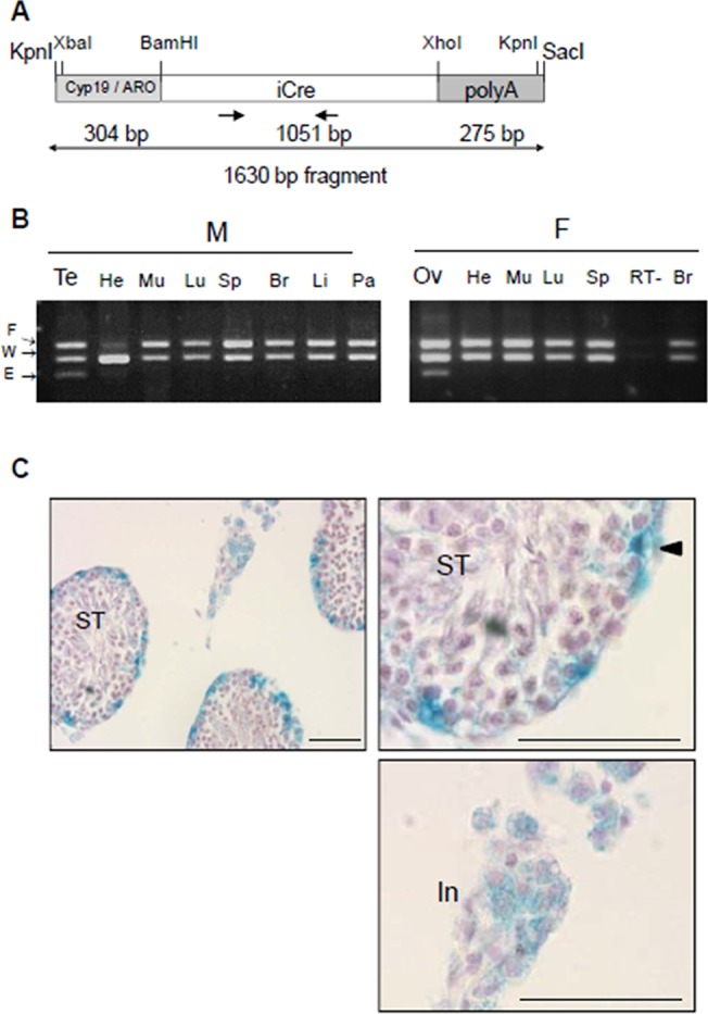
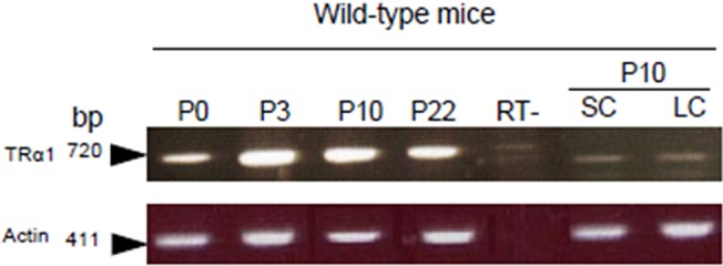
Methods: The TRαAMI knock-in allele allows the generation of transgenic mice expressing a dominant-negative TRα1 (thyroid receptor α1) isoform restricted to specific target cells after Cre-loxP recombination. Here, we introduced this mutant allele in both Sertoli and Leydig cells using a novel aromatase-iCre (ARO-iCre) line that expresses Cre recombinase under control of the human Cyp19(IIa)/aromatase promoter.

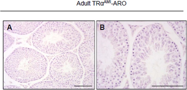
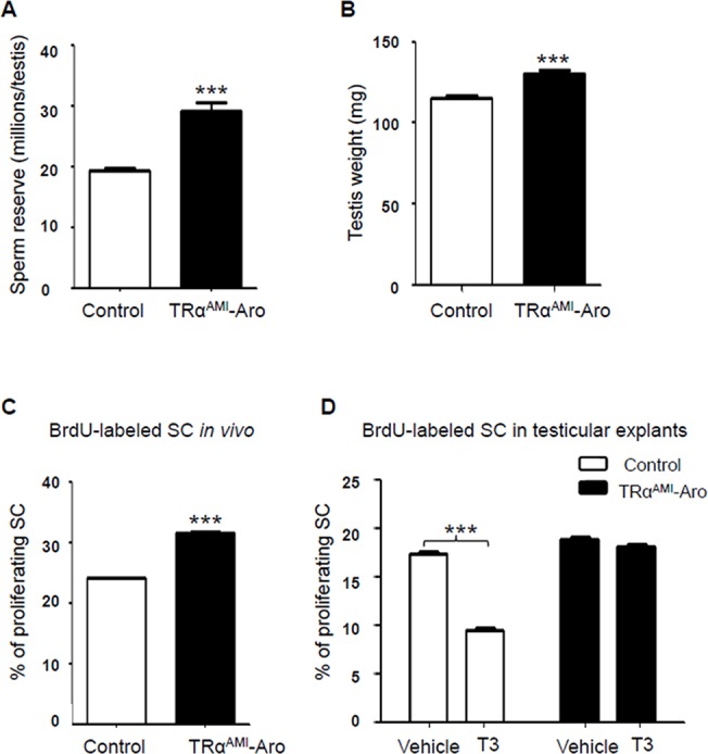
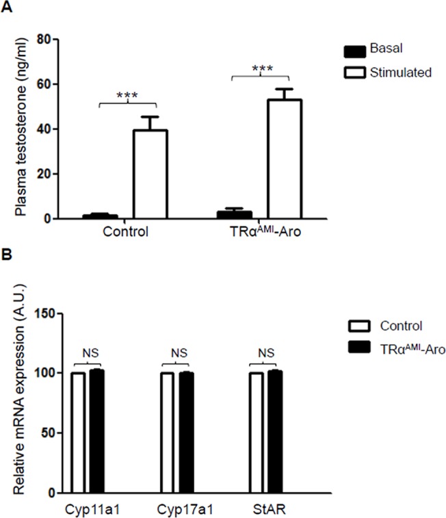
Findings: We showed that loxP recombination induced by this ARO-iCre is restricted to male and female gonads, and is effective in Sertoli and Leydig cells, but not in germ cells. We compared this model with the previous introduction of TRαAMI specifically in Sertoli cells in order to investigate T3 regulation of steroidogenesis. We demonstrated that TRαAMI-ARO males exhibited increased testis weight, increased sperm reserve in adulthood correlated to an increased proliferative index at P3 in vivo, and a loss of T3-response in vitro. Nevertheless, TRαAMI-ARO males showed normal fertility. This phenotype is similar to TRαAMI-SC males. Importantly, plasma testosterone and luteinizing hormone levels, as well as mRNA levels of steroidogenesis enzymes StAR, Cyp11a1 and Cyp17a1 were not affected in TRαAMI-ARO.

Conclusions/significance: We concluded that the presence of a mutant TRαAMI allele in both Leydig and Sertoli cells does not accentuate the phenotype in comparison with its presence in Sertoli cells only. This suggests that direct T3 regulation of steroidogenesis through TRα1 is moderate in Leydig cells, and that Sertoli cells are the main target of T3 action in the testis.


Conflict of interest statement

Competing Interests: The authors have declared that no competing interests exist.

Figures

Fig 1
Fig 1. Production and characterization of ARO-iCre transgenic mouse line.
(a) Schematic diagram of the DNA construct used for producing ARO-iCre mice. The mammalian codon-improved Cre recombinase (iCre, 1051 bp; white box) followed by an SV40 poly(A) signal cassette (right grey box) is controlled by 304 bp of the human Cyp19/aromatase(IIa)-278 promoter (left grey box). Arrows indicate position and direction of primers. Oligonucleotide sequences are in Table 1. (b) Cre excision is restricted to ovaries (Ov) and testes (Te) of iCre+/0;IGF1Rflox/WT mice. The excision was only observed in gonads, not in other tissues (He, heart; Mu, muscle; Lu, lung; Sp, spleen; Br, brain; Li, liver; Pa, pancreas) from males (M) and females (F). RT-, negative control (testis cDNA without RT). PCR detected wild-type (W, 256 bp) and floxed (F, 312 bp) alleles in all tissues, and the excised allele (E, 204 bp) exclusively in gonads. (c) Cre recombinase activity detected in somatic cells of testes in adult ARO-iCre males. After crossing the ARO-iCre mice with a ROSA26 Cre reporter mouse, β-galactosidase activity was detected in both SC and LC. No activity was present in germ cells. Right micrograph, low magnification; left micrographs, details in higher magnification. ST, seminiferous tubules. In, interstitium. Arrow head points to SC. Bar represents 50 μm.
Fig 2
Fig 2. RT-PCR detection of TRα1 mRNA in testes during post-natal development in wild-type mice.
TRα1 mRNA (720 pb) was present in whole testes from birth (P0) to P22, and in SC- and LC-enriched fractions prepared from testes of wild-type mice at P10. TRα1-specific primers were described previously [27] (Table 1). Actin mRNA (411 pb) was detected to check RNA quality. RT-, control without RT.
Fig 3
Fig 3. Histological sections of adult TRαAMI-ARO testes at low (a) and high (b) magnification.
Seminiferous tubules were fully developed in TRαAMI-ARO. Their epithelium exhibited normal structure and organization. Hematoxylin staining. Bar represents 100 μm.
Fig 4
Fig 4. Whole testicular sperm reserve and testis weight in adult TRαAMI-ARO, and percentage of proliferating SC in TRαAMI-ARO at P3 in vivo and in testicular explants using organotypic in vitro cultures with or without exogenous T3.
We observed a significant increase in whole testicular sperm reserve (a) and in testis weight (b) (***P < 0.001; n = 15 for controls and n = 16 for TRαAMI-ARO group). Data are shown as mean ± SEM. Statistical analyses were performed using Student’s t-test. White bars, control; black bars, TRαAMI-ARO (a, b). After BrdU immunohistochemical labeling, BrdU negative and BrdU positive SC were counted and the SC proliferation index was calculated. (c) When BrdU was injected in vivo 3 h before sacrifice, proliferation of Sertoli cells increased in P3 testes of TRαAMI-ARO mice in comparison with the control (***P < 0.001; n = 5 animals for each genotype). (d) When BrdU was added in vitro 3 h before the end of the organotypic cultures of P3 testicular explants, the SC proliferation index significantly decreased in the control in presence of T3 compared with vehicle (***P < 0.001, n = 6). In contrast, T3 had no effect on SC proliferative index in TRαAMI-ARO mice (P > 0.05, n = 5). Data are shown as mean ± SEM. Statistical analyses: two-way ANOVA followed by Bonferroni’s post-hoc test. White bars, control; black bars, TRαAMI-ARO.
Fig 5
Fig 5. Plasma testosterone levels in adult TRαAMI-ARO males, in basal conditions and after hCG stimulation, compared with control mice and mRNA levels of Cyp11a1/P450ssc, Cyp17a1/P450c17 and StAR in TRαAMI-ARO testes.
(a) In both TRαAMI-ARO (11 males) and controls (9 males), hCG induced a significant increase (***P < 0.001) in testosterone secretion, as expected. Plasma testosterone levels were unchanged in TRαAMI-ARO compared with the control. Data are shown as mean ± SEM. Two-way ANOVA followed by Bonferroni’s post-hoc test. Black bars, basal levels; white bars, hCG stimulated. (b) The amount of cDNA of 3 steroidogenic genes (Cyp11a1/P450ssc, Cyp17a1/P450c17 and StAR) was compared in TRαAMI-ARO (black bars) and control males (white bars). For each genotype, 6 mice were tested in triplicate using TaqMan quantitative real-time PCR, as described in materials and methods. Normalization was performed with two housekeeping genes, β-actin and GAPDH, that both exhibited similar expression among samples. mRNA expression is presented as percent of mean control level. A non-parametric test (Mann-Whitney) was used for statistical analysis. NS: not significant.

Similar articles

Cited by

References

    1. Jannini EA, Ulisse S, D'Armiento M (1995) Thyroid hormone and male gonadal function. Endocr Rev 16: 443–459. - PubMed
    1. Choksi NY, Jahnke GD, St Hilaire C, Shelby M (2003) Role of thyroid hormones in human and laboratory animal reproductive health. Birth Defects Res B Dev Reprod Toxicol 68: 479–491. - PubMed
    1. Joyce KL, Porcelli J, Cooke PS (1993) Neonatal goitrogen treatment increases adult testis size and sperm production in the mouse. J Androl 14: 448–455. - PubMed
    1. Cooke PS, Meisami E (1991) Early hypothyroidism in rats causes increased adult testis and reproductive organ size but does not change testosterone levels. Endocrinology 129: 237–243. - PubMed
    1. van Haaster LH, de Jong FH, Docter R, de Rooij DG (1993) High neonatal triiodothyronine levels reduce the period of Sertoli cell proliferation and accelerate tubular lumen formation in the rat testis, and increase serum inhibin levels. Endocrinology 133: 755–760. - PubMed

Publication types

LinkOut - more resources