. 2015 Mar 20;10(3):e0121823.
doi: 10.1371/journal.pone.0121823. eCollection 2015.

RNA interference of myocyte enhancer factor 2A accelerates atherosclerosis in apolipoprotein E-deficient mice

Affiliations

RNA interference of myocyte enhancer factor 2A accelerates atherosclerosis in apolipoprotein E-deficient mice

Wen-ping Zhou et al. PLoS One. .

Abstract

Objective: Myocyte enhancer factor-2A (MEF 2A) has been shown to be involved in atherosclerotic lesion development, but its role in preexisting lesions is still unclear. In the present study we aim to assess the role of MEF 2A in the progression of pre-existing atherosclerosis.

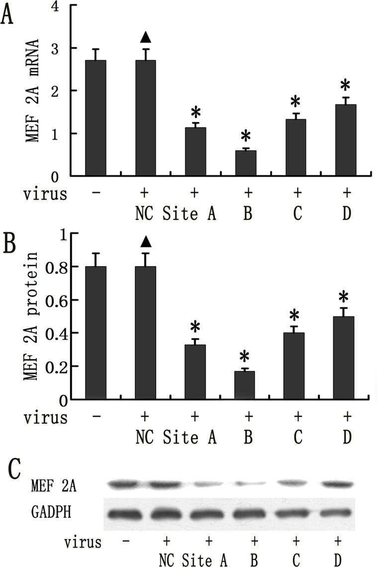
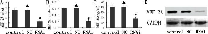
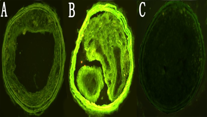
Methods: Eighty apolipoprotein E-deficient mice (APOE KO) were randomly allocated to control, scramble and MEF 2A RNA interference (RNAi) groups, and constrictive collars were used to induce plaque formation. Six weeks after surgery, lentiviral shRNA construct was used to silence the expression of MEF 2A. Carotid plaques were harvested for analysis 4 weeks after viral vector transduction. Inflammatory gene expression in the plasma and carotid plaques was determined by using ELISAs and real-time RT-PCR.

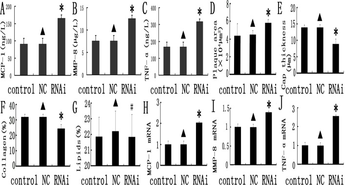
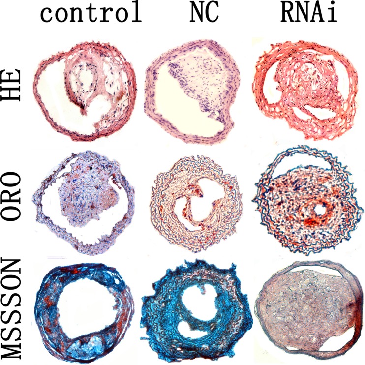
Results: The expression level of MEF 2A was significantly reduced in plasma and plaque in the RNAi group, compared to the control and NC groups, whereas the expression level of pro-inflammatory cytokines was markedly increased. Silencing MEF 2A using lentiviral shRNA significantly reduced the plaque collagen content and fibrous cap thickness, as well as increased plaque area. However, silencing MEF 2A had no obvious effect on plaque lipid content.

Conclusions: Lentivirus-mediated MEF 2A shRNA accelerates inflammation and atherosclerosis in APOE KO mice, but has no effect on lipoprotein levels in plasma.


Conflict of interest statement

Competing Interests: The authors have declared that no competing interests exist.

Figures

Fig 1
Fig 1. Lentiviral vector mediated knockdown of MEF 2A in mice aortic endothelial cells (MAECs).
MAECs were transduced with 50 MOI of 4 different shRNA vectors and MEF 2A expression was measured on day 4 by real time RT-PCR following transduction. The GAPDH was used as an internal control. (A). MEF 2A mRNA expression detected by realtime RT-PCR (*P < 0.05) (B) MEF 2A protein expression in lentivral shRNA transduced MAECs was quantified using Image J. (C) MEF2A expression in lentiviral shRNA vector transduced MAECs was determined by Western blots. Data were presented in mean±SD. *P < 0.05. (n = 8)
Fig 2
Fig 2. Knockdown of MEF 2A in vivo.
(A) mRNA expression of MEF 2A in the plaques of the control, NC, and MEF 2A shRNA groups at week 10; (B) Protein expression of MEF 2A in the plaques of the control, NC, and shRNA groups; (C) The concentration MEF 2A in the plasma; (D) Representative Western blots used for quantification. Data are presented in the mean ± SD. * P < 0.05
Fig 3
Fig 3. Efficiency of lentviral shRNA vector transduction in the carotid plaques.
A, B, C. GFP expression in sections of the carotid plaques was imaged from NC group. Carotid plaques at 1, 2 and 4 weeks following transduction were visualized under fluorescent microscope. (200×).
Fig 4
Fig 4. Inflammation gene expression and comparison of relative composition of plaques in the control, NC and MEF 2A RNAi groups.
A, B, and C: inflammatory markers (MCP-1, MMP-8 and TNF-α) in the control, NC, and MEF 2A shRNA groups; D, E,F and G plaque morphology in the control, NC, and MEF 2A shRNA groups; Plaque area (D), Fibirious cap thickness (E),collage content (F) and lipid content (G) were shown for the control, NC and RNAi groups. H, I and J: MCP-1 (H), MMP-8 (I) and TNF-α (J) mRNA expression in carotid plaques of the control, NC and shRNA groups at week 10 was detected by real-time PCR. Data were expressed as the mean ± SD. * P < 0.05.
Fig 5
Fig 5. Carotid plaques in the control, NC and MEF 2A RNAi groups.
Cross-sections of plaques in the control, NC and RNAi groups were stained with HE, ORO and masson’s trichrome. magnification 200×.

References

    1. Black BL, Olson EN. Transcriptional control of muscle development by myocyte enhancer factor-2 (MEF2) proteins. Annu Rev Cell Dev Biol. 1998;14:167–196. - PubMed
    1. McKinsey TA, Zhang ChL, Olson EN. MEF2: a calcium-dependent regulator of cell division differentiation and death. Trends Biochem Sci. 2002;27:40–47. - PubMed
    1. Wang L, Fan C, Topol SE, Topol EJ, Wang Q. Mutation of MEF2A in an inherited disorder with features of coronary artery disease. Science. 2003; 302: 1578–1581. - PMC - PubMed
    1. Bhagavatula MR, Fan C, Shen GQ, Cassano J, Plow EF, Topol EJ, et al. Transcription factor MEF2A mutations in patients with coronary artery disease. Hum Mol Genet. 2004; 13: 3181–3188. - PMC - PubMed
    1. González P, García-Castro M, Reguero JR, Batalla A, Ordóñez AG, Palop RL, et al. The Pro279Leu variant in the transcription factor MEF2A is associated with myocardial infarction. J Med Genet. 2006; 43: 167–169. - PMC - PubMed

Publication types

MeSH terms