Review
. 2015;9(1-2):154-65.
doi: 10.4161/19336918.2014.994893.

Tenascin-X: beyond the architectural function

Affiliations
Review

Tenascin-X: beyond the architectural function

Ulrich Valcourt et al. Cell Adh Migr. 2015.

Abstract

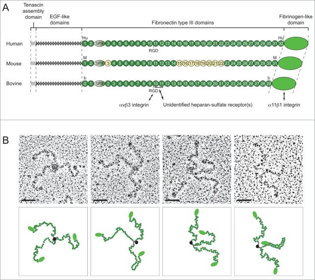
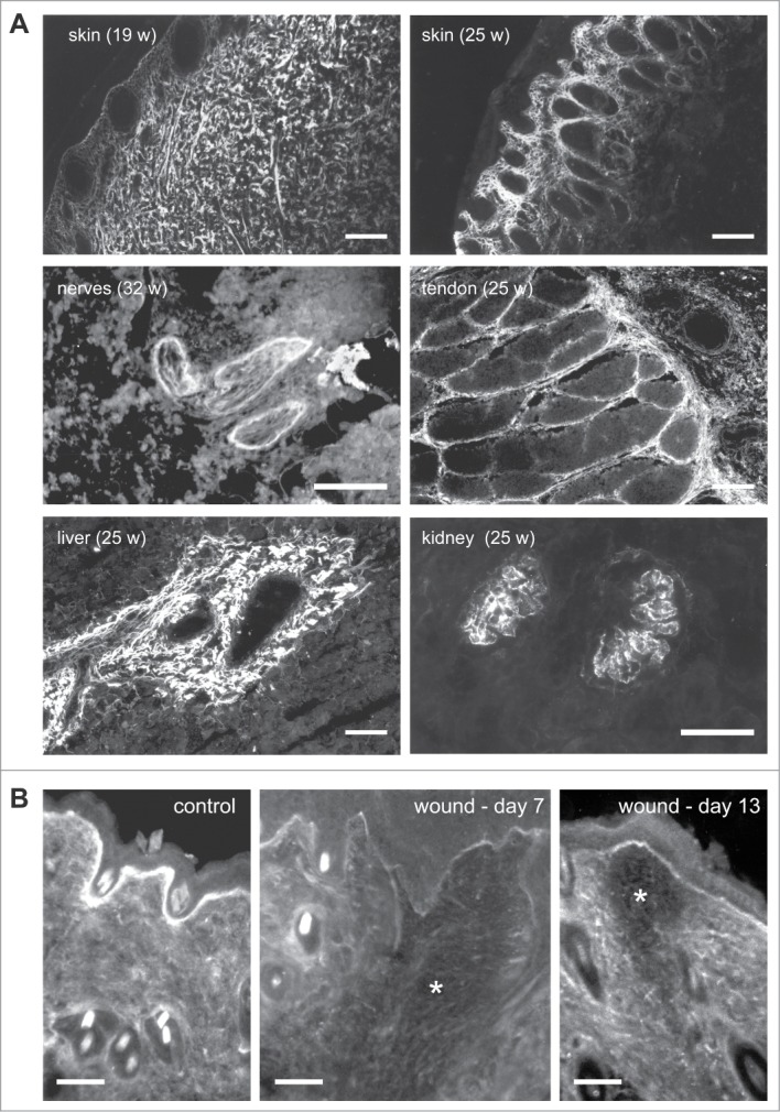
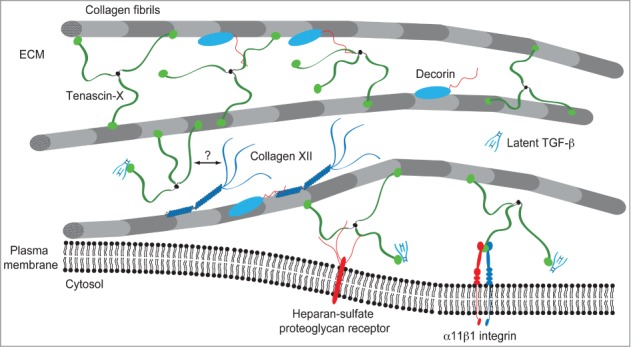
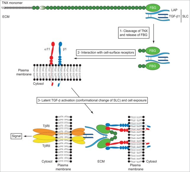
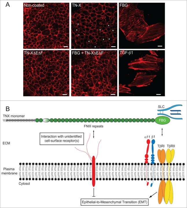
Tenascin-X is the largest member of the tenascin (TN) family of evolutionary conserved extracellular matrix glycoproteins, which also comprises TN-C, TN-R and TN-W. Among this family, TN-X is the only member described so far to exert a crucial architectural function as evidenced by a connective tissue disorder (a recessive form of Ehlers-Danlos syndrome) resulting from a loss-of-function of this glycoprotein in humans and mice. However, TN-X is more than an architectural protein, as it displays features of a matricellular protein by modulating cell adhesion. However, the cellular functions associated with the anti-adhesive properties of TN-X have not yet been revealed. Recent findings indicate that TN-X is also an extracellular regulator of signaling pathways. Indeed, TN-X has been shown to regulate the bioavailability of the Transforming Growth Factor (TGF)-β and to modulate epithelial cell plasticity. The next challenges will be to unravel whether the signaling functions of TN-X are functionally linked to its matricellular properties.

Keywords: ECM, extracellular matrix; EDS, Ehlers-Danlos syndrome; EGF, epidermal growth factor; EMT, epithelial-to-mesenchymal transition; Ehlers-Danlos syndrome (EDS); FAK, focal adhesion kinase; FBG, fibrinogen-like domain; FNIII, fibronectin type III module; LAP, latency associated peptide; MMP, matrix metalloproteinase; SLC, small latent complex; TGF-β; TGF-β activation; TN, tenascin; TSP-1, thrombospondin-1; VEGF, vascular endothelial growth factor; cell signaling; epithelial-to-mesenchymal transition (EMT); integrin α11β1; matricellular protein; tenascin-X; transforming growth factor-β.


Figures

Figure 1.
Figure 1.
TN-X is a tribrachion. (A) Schematic representation of human, mouse and bovine TN-X monomers. Each molecule is composed of the N-terminal oligomerization domain (tenascin assembly domain), followed by 18.5 epidermal growth factor (EGF)-like repeats, between 30 to 32 fibronectin type III (FNIII) repeats and with a fibrinogen (FBG)-like domain at the C-terminus. The FNIII repeats are potentially interrupted by a serine/proline-rich (SPX) region that was identified in the TN-Y molecule, the avian TN-X ortholog. For each TN-X ortholog, FNIII repeats (Hu1-32, M1-31 and b0-29) are numbered according to the nomenclature used in the original publications. The alternatively spliced FNIII repeats found in the mouse TN-X are in yellow (M3, and M15-M22). Cell-surface receptors of the bovine TN-X are depicted under the corresponding monomer. The heparin-binding site is underlined. Note that the human and bovine TN-X glycoproteins, but not the mouse ortholog, have a RGD sequence in a conserved FNIII module (Hu11 and b10, respectively). (B) Electron Micrographs of rotary shadowed purified recombinant bovine TN-X molecules (upper panels) and their respective putative schematic representation (lower panels). Bars, 50 nm.
Figure 2.
Figure 2.
Tissue distribution of TN-X (A) in fetal bovine tissues and (B) during wound healing in mice. (A) Indirect immunofluorescence of TN-X performed on cryostat sections of fetal bovine tissues (at the indicated weeks of gestation) using a monoclonal antibody (8F2) recognizing the FNIII b10 domain of the bovine glycoprotein. (B) Indirect immunofluorescence of TN-X performed on cryostat sections of mouse skin after incisional wound (*). TN-X was detected using polyclonal antibodies directed against the bovine TN-X FNIII b9-b10 repeats. Note that TN-X is hardly detected in the wound (*) even after the completion of the re-epithelialization process (day 7). TN-X staining is re-observed in the deep layers of the wound 13 days after incision. Bars, 50 μm.
Figure 3.
Figure 3.
Model of TN-X integration within the collagenous network. TN-X regulates the spacing and cohesiveness between collagen fibrils through direct interaction with collagen molecules and/or through molecular associations with several ECM components interacting with collagen fibrils. Type XII collagen interacts with TN-X through its non-collagenous N-terminal NC3 domain. The binding site of this NC3 domain has not been identified in the full-length TN-X. The dermatan sulfate chains of decorin ensure the binding to the heparin-binding site included within the FNIII b10 and b11 domains of bovine TN-X. TN-X might also anchor collagen fibrils at the vicinity of cell surface through its interaction with (not yet identified) heparin-sulfate proteoglycan receptor(s) and/or the α11β1 integrin.
Figure 4.
Figure 4.
Model of TGF-β activation by the FBG-like domain of TN-X. The small latent TGF-β complex (SLC) interacts with the FBG-like domain of TN-X. In this full-length glycoprotein, the LAP·TGF-β thus constitutes a reservoir of signaling molecule within the ECM. During physiological or pathological events of ECM remodeling, we hypothesized that some proteases would cleave the FBG-like domain of TN-X from the full-length molecule. At the vicinity of cell membrane, the FBG-like domain interacts with the α11β1 integrin receptor, allowing a conformational change of the latent complex, facilitating the presentation of mature TGF-β to TβRII and TβRI receptors. Latent TGF-β activation by the FBG-like domain results in the induction of Smad signaling and subsequent TGF-β response, such as EMT.
Figure 5.
Figure 5.
The FNIII region of TN-X antagonizes the induction of EMT by the FBG-like domain. (A) Actin direct fluorescence in normal murine mammary epithelial (NMuMG) cells seeded onto non-coated dishes or dishes containing equimolar quantities of immobilized recombinant full-length TN-X, FBG-like domain, FNIII repeats (TN-XΔEΔF) or both FNIII repeats and FBG-like domain. Note that compared to the uncoated condition, where almost all cells exhibited a cortical actin staining, the full-length TN-X induced a mild EMT, visualized by a partial delocalization of actin cytoskeleton from cell junctions (*). In contrast, the FBG-like domain caused a full EMT, as illustrated by the acquisition of elongated cell morphology and the organization of actin cytoskeleton into stress fibers. Recombinant human TGF-β1, used here as a positive control, gave similar results to those obtained with the FBG-like domain. However, the FNIII region of TN-X fully inhibited the EMT triggered by the FBG-like domain. Bars, 15 μm. (B) Model representing the regulation of epithelial cell plasticity by TN-X. When separated from the intact TN-X molecule, the FBG-like domain is able to induce a robust EMT response, through its ability to activate latent TGF-β. This response relies on the presence of TβRII/I receptors and α11β1 integrin at the cell surface. In the intact TN-X molecule, the FNIII region antagonizes the EMT induced by the FBG-like domain, most likely via distinct intracellular cues that are initiated by the interaction of certain FNIII repeats to a yet unidentified receptor.

Similar articles

  • Latent TGF-β Activation Is a Hallmark of the Tenascin Family.
    Aubert A, Mercier-Gouy P, Aguero S, Berthier L, Liot S, Prigent L, Alcaraz LB, Verrier B, Terreux R, Moali C, Lambert E, Valcourt U. Aubert A, et al. Front Immunol. 2021 May 13;12:613438. doi: 10.3389/fimmu.2021.613438. eCollection 2021. Front Immunol. 2021. PMID: 34054795 Free PMC article.
  • Tenascin-C: Form versus function.
    Giblin SP, Midwood KS. Giblin SP, et al. Cell Adh Migr. 2015;9(1-2):48-82. doi: 10.4161/19336918.2014.987587. Cell Adh Migr. 2015. PMID: 25482829 Free PMC article. Review.
  • Tenascin-X promotes epithelial-to-mesenchymal transition by activating latent TGF-β.
    Alcaraz LB, Exposito JY, Chuvin N, Pommier RM, Cluzel C, Martel S, Sentis S, Bartholin L, Lethias C, Valcourt U. Alcaraz LB, et al. J Cell Biol. 2014 May 12;205(3):409-28. doi: 10.1083/jcb.201308031. J Cell Biol. 2014. PMID: 24821840 Free PMC article.
  • Tenascin-C and integrins in cancer.
    Yoshida T, Akatsuka T, Imanaka-Yoshida K. Yoshida T, et al. Cell Adh Migr. 2015;9(1-2):96-104. doi: 10.1080/19336918.2015.1008332. Cell Adh Migr. 2015. PMID: 25793576 Free PMC article. Review.
  • Transcriptional regulation of tenascin genes.
    Chiovaro F, Chiquet-Ehrismann R, Chiquet M. Chiovaro F, et al. Cell Adh Migr. 2015;9(1-2):34-47. doi: 10.1080/19336918.2015.1008333. Cell Adh Migr. 2015. PMID: 25793574 Free PMC article. Review.

Cited by

References

    1. Morel Y, Bristow J, Gitelman SE, Miller WL. Transcript encoded on the opposite strand of the human steroid 21-hydroxylase/complement component C4 gene locus. Proc Natl Acad Sci U S A 1989; 86:6582-6; PMID:2475872; http://dx.doi.org/10.1073/pnas.86.17.6582 - DOI - PMC - PubMed
    1. Matsumoto K, Arai M, Ishihara N, Ando A, Inoko H, Ikemura T. Cluster of fibronectin type III repeats found in the human major histocompatibility complex class III region shows the highest homology with the repeats in an extracellular matrix protein, tenascin. Genomics 1992; 12:485-91; PMID:1373119; http://dx.doi.org/10.1016/0888-7543(92)90438-X - DOI - PubMed
    1. Matsumoto K, Ishihara N, Ando A, Inoko H, Ikemura T. Extracellular matrix protein tenascin-like gene found in human MHC class III region. Immunogenetics 1992; 36:400-3; PMID:1382042; http://dx.doi.org/10.1007/BF00218048 - DOI - PubMed
    1. Bristow J, Tee MK, Gitelman SE, Mellon SH, Miller WL. Tenascin-X: a novel extracellular matrix protein encoded by the human XB gene overlapping P450c21B. J Cell Biol 1993; 122:265-78; PMID:7686164; http://dx.doi.org/10.1083/jcb.122.1.265 - DOI - PMC - PubMed
    1. Chiquet-Ehrismann R, Mackie EJ, Pearson CA, Sakakura T. Tenascin: an extracellular matrix protein involved in tissue interactions during fetal development and oncogenesis. Cell 1986; 47:131-9; PMID:2428505; http://dx.doi.org/10.1016/0092-8674(86)90374-0 - DOI - PubMed

Publication types