. 2015 Mar 20;10(3):e0119225.
doi: 10.1371/journal.pone.0119225. eCollection 2015.

MicroRNA-153 inhibits osteosarcoma cells proliferation and invasion by targeting TGF-β2

Affiliations

MicroRNA-153 inhibits osteosarcoma cells proliferation and invasion by targeting TGF-β2

Guangfeng Niu et al. PLoS One. .

Retraction in

Abstract

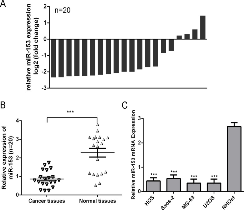
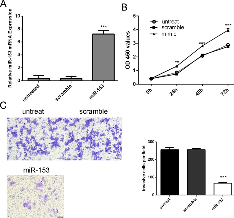
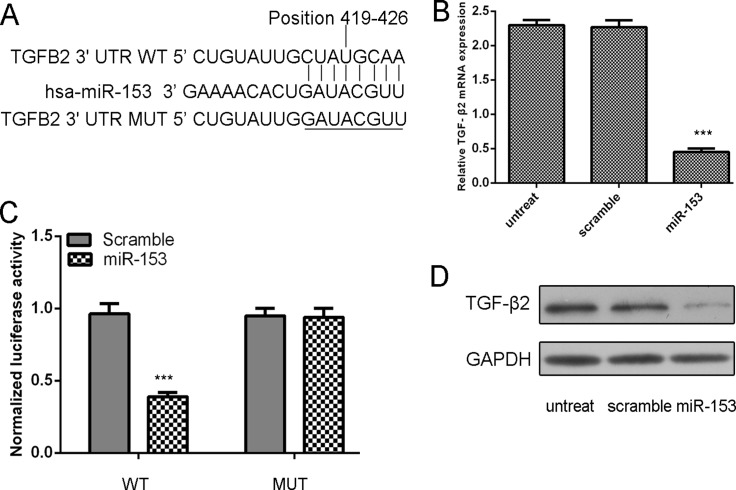
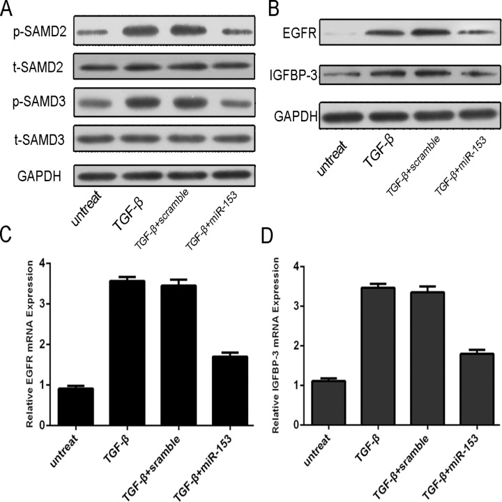
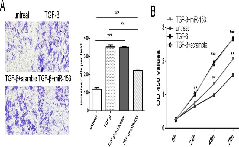
Increasing evidence indicates that microRNAs (miRNAs), a class of small noncoding RNAs, participate in almost every step of cellular processes. MiRNAs are aberrantly expressed in human cancers and contribute to cancer development and progression. Study of miRNAs may provide a new clue for understanding the mechanism of carcinogenesis and a new tool for cancer treatment. In the present study, miR-153 was downregulated in human osteosarcoma tissues and cell lines. Introduction of miR-153 mimics into the MG-63 cells inhibited cell proliferation and invasion. Our results further revealed that transforming growth factor beta 2 (TGF-β2) was negatively regulated by miR-153. Furthermore, overexpression of miR-153 decreased p-SMAD2, p-SMAD3, epidermal growth factor receptor (EGFR) and insulin-like growth factor binding protein-3 (IGFBP-3) expressions, which were the downstream signaling molecules of TGF-β. Furthermore, miRNA-153 suppressed TGF-β-mediated MG-63 proliferation and migration. Therefore, our results suggest that miR-153 may act as a tumor suppressor in osteosarcoma through targeting TGF-β2.


Conflict of interest statement

Competing Interests: The authors have declared that no competing interests exist.

Figures

Fig 1
Fig 1. miR-153 was decreased in osteosarcoma tissues and cell line.
(A) The miR-153 expression was determined by qRT-PCR in 20 paired osteosarcoma tissues and adjacent noncancerous tissues. The expression of miR-153 was normalized to U6 snRNA. Data is presented as log 2 of fold change of osteosarcoma tissues relative to non-tumor adjacent tissues. (B) Relative miR-153 expression levels in osteosarcoma tissues and their corresponding adjacent normal tissues. (C) The expression or miR-153 was analyzed in normal osteoblast cells (NHOst) and four OS cell lines (HOS, Saos-2, MG-63, and U2OS). ***p<0.001.
Fig 2
Fig 2. Overexpression of miR-153 inhibited osteosarcoma cell proliferation and invasion.
(A) qRT-PCR analysis of miR-153 expression after the transfection of miR-153 mimics or scramble or no treat. (B) The CCK8 assay used to evaluate the proliferation of the MG-63 cells after transferred with the miR-153 mimics or scramble or no treat. (C) Invasion analysis of the MG-63 cells after treatment with miRNA mimics, inhibitors or no treat; the relative ratio of invasive cells per field is shown on the right, ** p<0.01 and ***p<0.001.
Fig 3
Fig 3. TGF-β2 was a direct target of miR-153 in osteosarcoma cells.
(A) Computer prediction of miR-153 binding sites in the 3’UTR of human TGF-β2 gene. (B) Expression of TGF-β2 mRNA was examined by qRT-PCR in MG-63 cells transfected with miR-153 mimic or the scramble mimics or no treat. The expression of TGF-β2 was normalized to GAPDH. (C) MG-63cells were co-transfected with miR-153 and WT or MUT 3’UTR of TGF-β2. (D) Protein level was detected by Western blotting in MG-63 cells transfected with miR-153 mimic or the scramble mimics or no treat. GAPDH was also detected as a loading control. ***p<0.001.
Fig 4
Fig 4. MiR-153 Overexpression repressed the p-SMAD2, p-SMAD3, EGFR and IGFBP-3 expression.
The MG-63 was treated in serum-free medium in the presence and absence of TGF-β (50 ng/ml), scramble, or miRNA-153 mimic for 24 h. (A) Expression of p-SMAD2, t-SMAD2, p-SMAD3 and t-SMAD3 was detected using western blotting. GAPDH was used as a loading control. (B) Expression of EGFR and IGFBP-3 was detected using western blotting. GAPDH was used as a loading control. (C) The mRNA expression of EGFR was detected using qRT-PCR. The expression of MMP9 was normalized to GAPDH. (D) The mRNA Expression of IGFBP-3 was detected using qRT-PCR. The expression of EGFR was normalized to GAPDH.
Fig 5
Fig 5. miR-153 is involved in TGF-β-induced osteosarcoma cell proliferation and invasion.
The MG-63 was treated in serum-free medium in the presence and absence of TGF-β (50 ng/ml), scramble, or miRNA-153 mimic. (A) Invasion analysis of MG-63 cells treated with different combinations. The relative ratio of invasive cells per field is shown right. (B) Cell growth curves in MG-63 cells transfected with different combinations. ** p<0.01 and ***p<0.001.

References

    1. Zhao G, Cai C, Yang T, Qiu X, Liao B, et al. (2013) MicroRNA-221 induces cell survival and cisplatin resistance through PI3K/Akt pathway in human osteosarcoma. PLoS One 8: e53906 10.1371/journal.pone.0053906 - DOI - PMC - PubMed
    1. Namlos HM, Meza-Zepeda LA, Baroy T, Ostensen IH, Kresse SH, et al. (2012) Modulation of the osteosarcoma expression phenotype by microRNAs. PLoS One 7: e48086 10.1371/journal.pone.0048086 - DOI - PMC - PubMed
    1. Liang W, Gao B, Fu P, Xu S, Qian Y, et al. (2013) The miRNAs in the pathgenesis of osteosarcoma. Front Biosci (Landmark Ed) 18: 788–794. - PubMed
    1. Yang J, Zhang W (2013) New molecular insights into osteosarcoma targeted therapy. Curr Opin Oncol 25: 398–406. 10.1097/CCO.0b013e3283622c1b - DOI - PubMed
    1. Huang J, Gao K, Lin J, Wang Q (2014) MicroRNA-100 inhibits osteosarcoma cell proliferation by targeting Cyr61. Tumour Biol 35: 1095–1100. 10.1007/s13277-013-1146-8 - DOI - PubMed

Publication types

MeSH terms