. 2015 Mar 20;10(3):e0120627.
doi: 10.1371/journal.pone.0120627. eCollection 2015.

An S-(hydroxymethyl)glutathione dehydrogenase is involved in conidiation and full virulence in the rice blast fungus Magnaporthe oryzae

Affiliations

An S-(hydroxymethyl)glutathione dehydrogenase is involved in conidiation and full virulence in the rice blast fungus Magnaporthe oryzae

Zhen Zhang et al. PLoS One. .

Abstract

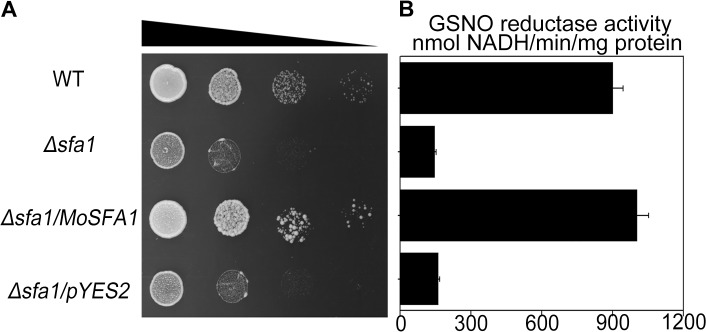
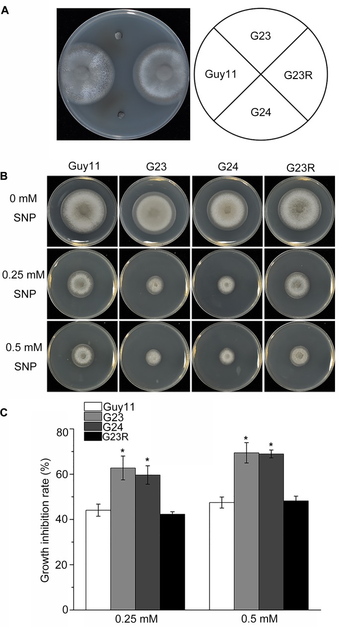
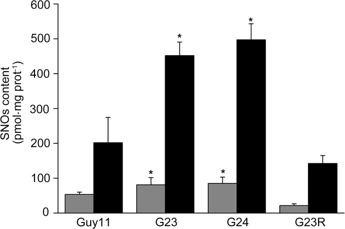
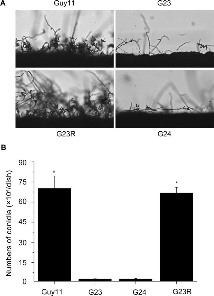
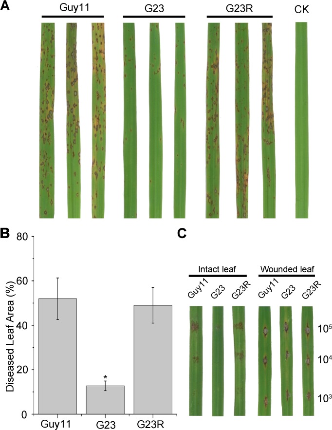
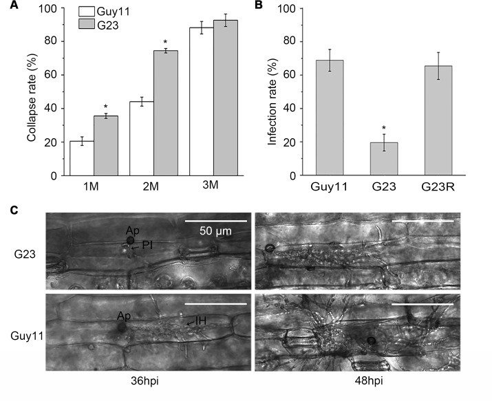
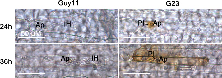
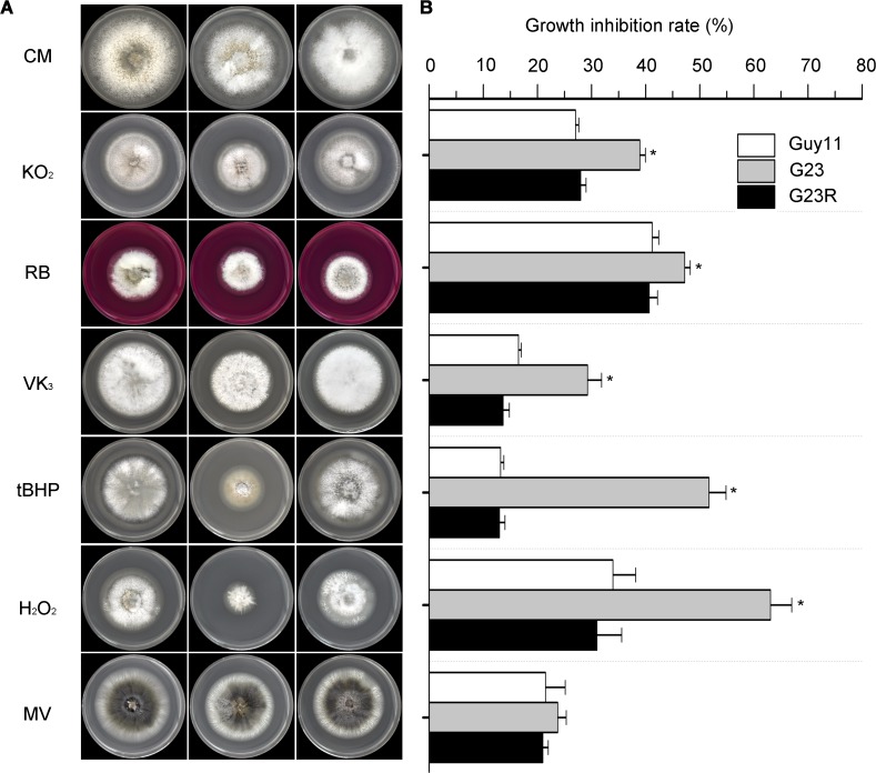
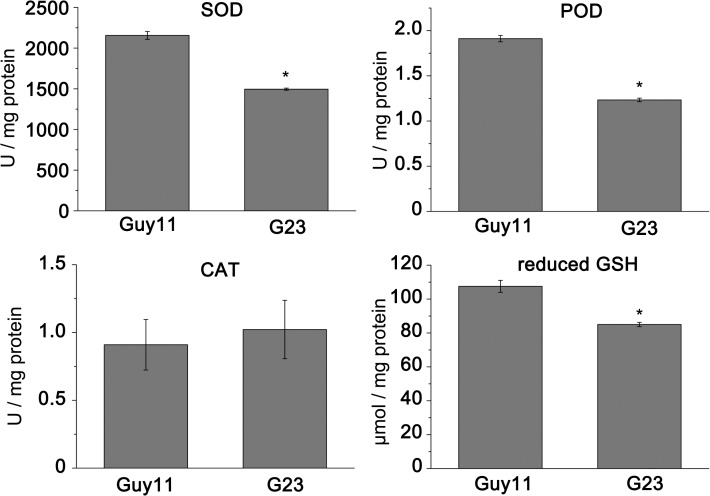
Magnaporthe oryzae is a hemibiotrophic fungal pathogen that causes rice blast disease. A compatible interaction requires overcoming plant defense responses to initiate colonization during the early infection process. Nitric oxide (NO) plays important roles in defense responses during host-pathogen interactions. Microbes generally protect themselves against NO-induced damage by using enzymes. Here, we characterized an S-(hydroxymethyl)-glutathione dehydrogenase gene in M. oryzae, MoSFA1, the homologs of which are involved in NO metabolism by specifically catalyzing the reduction of S-nitrosoglutathione (GSNO) in yeasts and plants. As expected from the activities of S-(hydroxymethyl)glutathione dehydrogenase in formaldehyde detoxification and GSNO reduction, MoSFA1 deletion mutants were lethal in formaldehyde containing medium, sensitive to exogenous NO and exhibited a higher level of S-nitrosothiols (SNOs) than that of the wild type. Notably, the mutants showed severe reduction of conidiation and appressoria turgor pressure, as well as significantly attenuated the virulence on rice cultivar CO-39. However, the virulence of MoSFA1 deletion mutants on wounded rice leaf was not affected. An infection assay on barley leaf further revealed that MoSFA1 deletion mutants exhibited a lower infection rate, and growth of infectious hyphae of the mutants was retarded not only in primary infected cells but also in expansion from cell to cell. Furthermore, barley leaf cell infected by MoSFA1 deletion mutants exhibited a stronger accumulation of H2O2 at 24 and 36 hpi. MoSFA1 deletion mutants displayed hypersensitivity to different oxidants, reduced activities of superoxide dismutases and peroxidases, and lower glutathione content in cells, compared with the wild type. These results imply that MoSFA1-mediated NO metabolism is important in redox homeostasis in response to development and host infection of M. oryzae. Taken together, this work identifies that MoSFA1 is required for conidiation and contributes to virulence in the penetration and biotrophic phases in M. oryzae.


Conflict of interest statement

Competing Interests: The authors have declared that no competing interests exist.

Figures

Fig 1
Fig 1. Complementation of the yeast Δsfa1 mutant by MoSFA1 and GSNO reductase activity in protein extracts.
(A) Yeast strains, BY4741(WT), Δsfa1, Δsfa1/MoSFA1, and Δsfa1/pYES2 were spotted onto galactose-containing medium with 1.1 mM formaldehyde at 28°C for 2 days. (B) GSNO reductase activity (mean ±SD) was determined in the tested yeast strains. Error bars represent SD.
Fig 2
Fig 2. Effect of formaldehyde and SNP on vegetative growth of the tested M. oryzae strains.
(A) M. oryzae strains were cultured on CM containing 0.3 mM formaldehyde at 28°C for 6 days. (B) Vegetative growth of Guy11, MoSFA1 deletion and reintroduction mutants were grown on CM containing 0.25 or 0.5 mM SNP at 28°C for 6 days. (C) Colony diameters of the tested strains were measured and Growth inhibition rate were evaluated. The experiments were performed in triplicate. ANOVA analysis was performed after growth inhibition rate were arcsine transformed. However, the original percentages were used for presentations. Error bars represent SD of the original percentages. Asterisks in each data column indicate significant differences at p = 0.05.
Fig 3
Fig 3. The levels of SNOs in the tested M. oryzae strains.
Total intracellular SNOs content in mycelia cultured in liquid CM in the dark at 28°C for 3 days without (gray bar) or with 100 μM SNP treatment (black bar). The values obtained were compared to a standard curve constructed using GSNO. The results were normalized to the protein content by a Bradford assay. The experiments were performed in triplicate. Error bars represent SD. Asterisks in each data column indicate significant differences at p = 0.05.
Fig 4
Fig 4. Comparison of the tested M. oryzae strains in conidiation.
(A) Development of conidia on conidiophores on CM. Conidia and conidiophores were observed under light microscopy. (B) Statistical analysis of the number of conidia in each 9-cm-diameter dish. The experiments were performed in triplicate. Error bars represent SD. Asterisks in each data column indicate significant differences at p = 0.05.
Fig 5
Fig 5. Pathogenicity assays on rice leaves.
(A) Spray inoculation with 4-week-old rice seedlings. Rice leaves (O. sativa cv. CO-39) were inoculated with conidia at a concentration of 5×10⁴ conidia ml−1. Representative leaves were photographed 6 days post-inoculation. The experiments were repeated at least three times with triple replications that yielded similar results. (B) Disease severity of each strain was assessed from the percentage of diseased leaf area as described by Fang and Dean (2000). ANOVA analysis was performed after percentages were arcsine transformed. However, the original percentages were used for presentations. Error bars represent SD of the original percentages. Asterisks in each data column indicate significant differences at p = 0.05. (C) Drop inoculation with 20 μl serial dilutions of conidia suspension on intact and wounded rice leaf segments. Representative leaves were photographed 6 days post-inoculation.
Fig 6
Fig 6. Analysis of infection-related morphogenesis of MoSFA1 deletion mutant.
(A) Cytorrhysis assay using glycerol was performed to compare the appressorial turgor pressure of MoSFA1 mutant and wild-type strain. Different glycerol solutions were given to appressoria at 48 hpi. Appressorial cytorrhysis was counted under an optical microscope. The rate of cytorrhysis was the average of three replications. (B) Infection rate of M. oryzae strains in barley leaf cells at 36 hpi. (C) Growth of infectious hyphae of MoSFA1 deletion mutant was retarded in barley leaf cells. Penetration and infectious hyphae were examined under optical microscopy. Ap, appressorium; PI, primary infectious hyphae; IH, secondary infectious hyphae. Significant difference analysis was performed after percentages were arcsine transformed. The original percentages were used for presentations. Error bars represent SD. Asterisks in each data column indicate significant differences at p = 0.05.
Fig 7
Fig 7. Detection of H2O2 accumulation at infected barley cells.
Detached barley leaf were inoculated with the tested strains, stained with DAB at 24 and 36 hpi, and observed under the light microscope. The cells infected by MoSFA1 deletion mutant were strongly stained with DAB, indicating high H2O2 accumulation at the penetration site. Bar is 50 μm. Ap, appressorium; PI, primary infectious hyphae; IH, secondary infectious hyphae.
Fig 8
Fig 8. Effect of different oxidants on vegetative growth of the tested M. oryzae strains.
(A) Vegetative growth of Guy11, MoSFA1 deletion and reintroduction mutants were grown on CM containing menadione (VK3, 100 μM), potassium superoxide (KO2, 50 mM), rose bengal (RB, 100 μM), tert-butyl-hydroxyperoxide (tBHP, 0.001%,v/v), and H2O2 (0.04%, v/v) and Methyl viologen (MV, 0.25mg/ml) at 28°C for 6 days. (B) Growth inhibition rate of tested strains exposed to different oxidants. The experiments were performed in triplicate. ANOVA analysis was performed after growth inhibition rate were arcsine transformed. However, the original percentages were used for presentations. Error bars represent SD. Aasterisks in each data column indicate significant differences at p = 0.05.
Fig 9
Fig 9. The activity of antioxidant enzymes and the content of GSH in tested M. oryzae strains.
Activities of superoxide dismutase (SOD), catalase (CAT), and peroxidase (POD) and the content of reduced GSH in the tested strains were determined using assay kits purchased from Nanjing Jiancheng Bioengineering Institute (Nanjing, China). The experiments were performed in triplicate. Error bars represent SD. Asterisks in each data column indicate significant differences at p = 0.05.

References

    1. Kankanala P, Czymmek K, Valent B. Roles for rice membrane dynamics and plasmodesmata during biotrophic invasion by the blast fungus. Plant Cell. 2007; 19: 706–724. - PMC - PubMed
    1. Mur LA, Carver TL, Prats E. NO way to live; the various roles of nitric oxide in plant-pathogen interactions. J Exp Bot. 2006; 57(3):489–505. - PubMed
    1. Arasimowicz-Jelonek M, Floryszak-Wieczorek J. Nitric oxide: an effective weapon of the plant or the pathogen? Mol Plant Pathol. 2014; 15(4):406–416. - PMC - PubMed
    1. Mur LA, Mandon J, Persijn S, Cristescu SM, Moshkov IE, Novikova GE, et al. Nitric oxide in plants: an assessment of the current state of knowledge. AoB Plants. 2013; 5:pls052. - PMC - PubMed
    1. Hu X, Neill SJ, Cai W, Tang Z. NO-mediated hypersensitive responses of rice suspension cultures induced by incompatible elicitor. Chinese Sci Bull. 2003; 48(4):358–363.

Publication types