. 2015 Mar 20;10(3):e0119402.
doi: 10.1371/journal.pone.0119402. eCollection 2015.

Alpha-catulin contributes to drug-resistance of melanoma by activating NF-κB and AP-1

Affiliations

Alpha-catulin contributes to drug-resistance of melanoma by activating NF-κB and AP-1

Birgit Kreiseder et al. PLoS One. .

Abstract

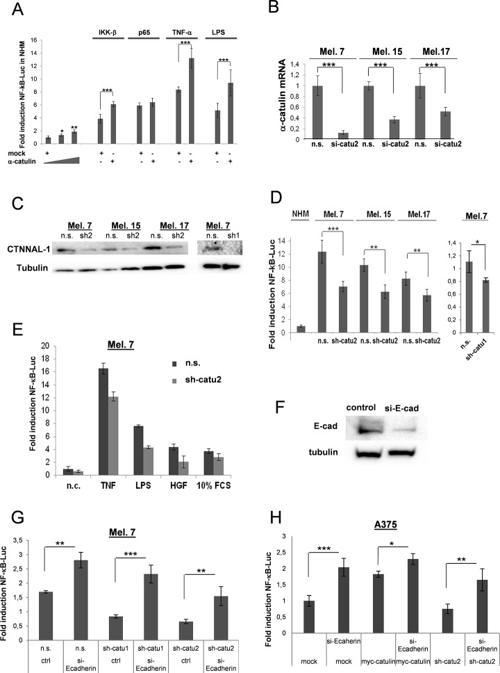
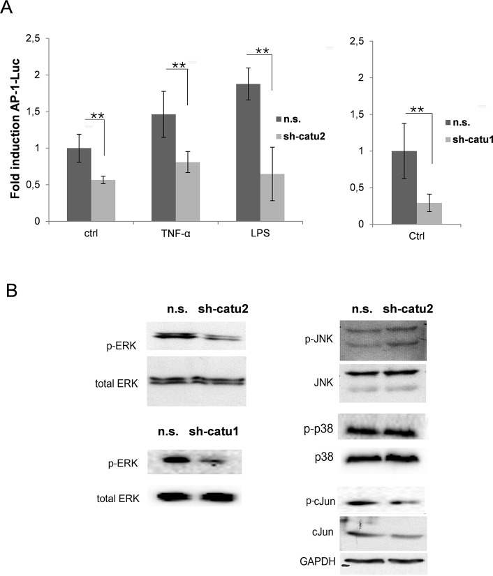
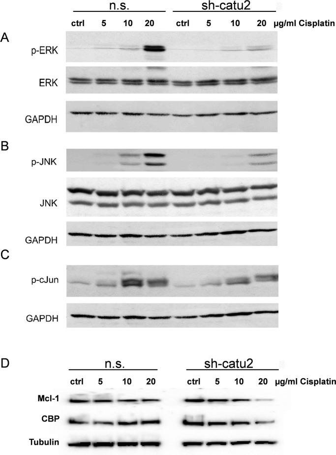
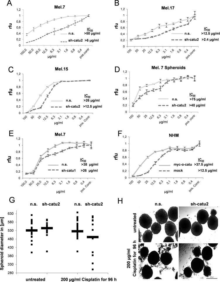
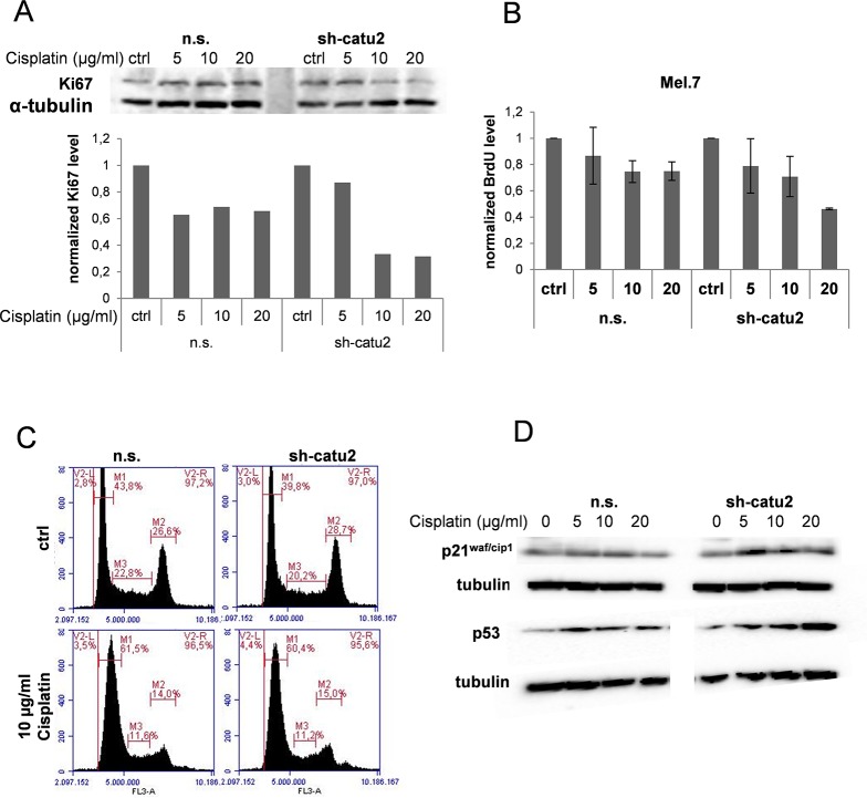
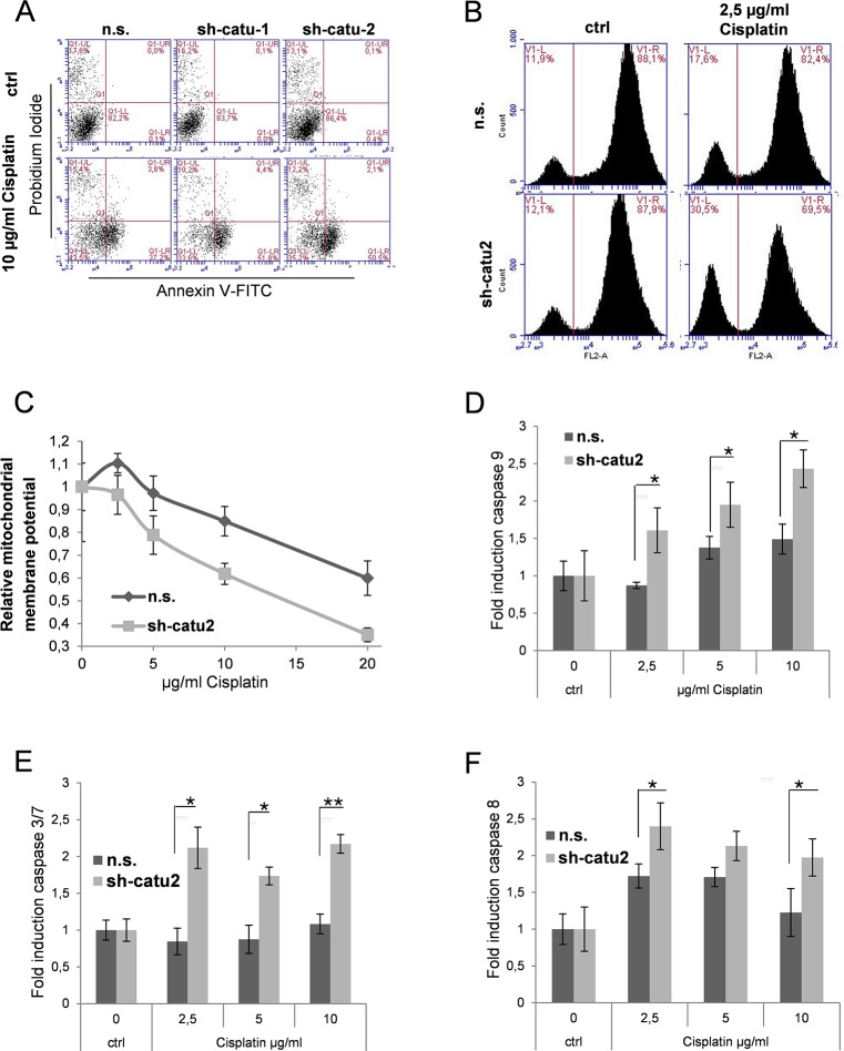
Melanoma is the most dangerous type of skin cancer accounting for 48,000 deaths worldwide each year and an average survival rate of about 6-10 months with conventional treatment. Tumor metastasis and chemoresistance of melanoma cells are reported as the main reasons for the insufficiency of currently available treatments for late stage melanoma. The cytoskeletal linker protein α-catulin (CTNNAL1) has been shown to be important in inflammation, apoptosis and cytoskeletal reorganization. Recently, we found an elevated expression of α-catulin in melanoma cells. Ectopic expression of α-catulin promoted melanoma progression and occurred concomitantly with the downregulation of E-cadherin and the upregulation of mesenchymal genes such as N-cadherin, Snail/Slug and the matrix metalloproteinases 2 and 9. In the current study we showed that α-catulin knockdown reduced NF-κB and AP-1 activity in malignant melanoma cells. Further, downregulation of α-catulin diminished ERK phosphorylation in malignant melanoma cells and sensitized them to treatment with chemotherapeutic drugs. In particular, cisplatin treatment led to decreased ERK-, JNK- and c-Jun phosphorylation in α-catulin knockdown melanoma cells, which was accompanied by enhanced apoptosis compared to control cells. Altogether, these results suggest that targeted inhibition of α-catulin may be used as a viable therapeutic strategy to chemosensitize melanoma cells to cisplatin by down-regulation of NF-κB and MAPK pathways.


Conflict of interest statement

Competing Interests: There exists no competing interests and there is no financial intent to market the results of this manuscript somehow. Most of the results were produced at the University of Applied Sciences in Krems, Austria where the first author (B. Kreiseder) was employed before she changed to SeaLife Pharma GmbH. The last author (C. Wiesner) is employed at the University in Krems and at SeaLife Pharma GmbH. Just very few results were produced at SeaLife Pharma mainly to finalize the manuscript. The research of SeaLife Pharma GmbH focuses on the production (isolation) of anti-infective molecules such as Anthraquinone-Derivatives for the treatment of MRSA. There is no interest in cancer research. Up to now, SeaLife Pharma has no products on the market. On behalf of all authors, I declare that there are no competing interests for the purposes of transparency. We confirm that this does not alter our adherence to PLOS ONE policies on sharing data and materials.

Figures

Fig 1
Fig 1. α-Catulin promotes NF-κB activation in human primary melanocytes and melanoma cells.
(A) A 5x NF-B-luc reporter gene (0.25μg) was co-transfected into melanocytes together with different concentrations of α-catulin (1 or 1.5 μg) with or without IKK-β(0.5μg) or p65 (0.5μg). 24 h later cells were non-stimulated or stimulated with TNF-α or LPS. Luciferase levels were normalized for a co-transfected RFP control (0.25μg). (B) Mel.7, Mel.15 and Mel.17 cells were stable infected with lentiviral particles containing a vector-based mirRNA construct directed against α-catulin (sh-catu 1 or sh-catu2; Materials and Methods), and α-catulin mRNA levels were analyzed by real-time PCR. (C) Mel.7, Mel.15 and Mel.17 cells were analyzed by Western blot with antibodies against α-catulin. (D) A NF-κB-luc reporter gene was tranfected into melanocytes and different melanoma cells containing stable integrated α-catulin mirRNA constructs (α-catulin-knockdown), and luciferase values were determined 24 h later and normalized for co-transfected JRED values. (E) Melanoma 7 cells as in (D) except that the cells were stimulated with TNFα, LPS, HGF and 10% FCS for further 8 h. (F) Mel.7 cells (n.s., sh-catu1/2) were transfected with NF-κB reporter plasmid and with or without si-RNA construct directed against E-cadherin and luciferase values were determined. (G) NF-κB-luc reporter assay with A375 melanoma cells transfected with mock, myc-α-catulin or sh-catu2 plasmids together with or without E-cadherin si-RNA. *Indicates P>0.005, **P>0.001, ***P>0.0001, Student´s t test.
Fig 2
Fig 2. α-Catulin knockdown in melanoma cells reduces AP-1 and ERK activation.
(A) An AP-1-luc reporter gene was transfected into non-stimulated stable infected Mel.7 cells (n.s., sh-catu1/2). The cells were non-stimulated (ctrl) or stimulated with TNF-α (10ng/ml) or LPS (5μg/ml). Luciferase levels were normalized for a co-transfected JRED control. (B) Mel.7 cells (as in A) were analyzed by Western blot with antibodies against phospho-ERK, total ERK, phospho-JNK, JNK, phospho-p38, p38, c-Jun and phospho-c-Jun. GAPDH was used as a loading control.
Fig 3
Fig 3. α-Catulin knockdown reduces phosphorylation of ERK, JNK and c-Jun in cisplatin treated melanoma cells.
Stable infected Mel.7 cells (n.s., sh-catu2) were treated with 0, 5, 10 and 20 μg/ml cisplatin for 24h and analyzed by Western blot with antibodies against (A) p-ERK, total ERK, (B) p-JNK, total JNK, (C) p-c-Jun (D) Mcl-1 and CBP. GAPDH or Tubulin were used as loading control.
Fig 4
Fig 4. α-Catulin knockdown enhances susceptibility of melanoma cells to cisplatin.
(A-F) Stable infected (n.s., sh-catu2) (A) Mel.7, (B) Mel.17, (C) Mel.15, (D) Mel.7 spheroids, (E) Mel.7 (n.s., sh-catu1) or (F) Melanocytes (mock, myc-α-catulin) were treated with different concentrations of cisplatin for 48h and cell survival normalized to untreated cells (pos. contr.). Viability was analyzed by CellTiter-Blue Assay. (G) Stable infected Mel.7 spheroids were treated with 200 μg/ml cisplatin for 96 hours and diameter of the spheroids determined before and after treatment and statistically evaluated. Observations (•) mean (-) n = 14, (H) Microscopic images from Mel.7 spheroids.
Fig 5
Fig 5. Cell proliferation is reduced in a dose- and time dependent manner in cisplatin treated melanoma cells when α-catulin is knocked down.
(A) Stable infected Mel.7 cells (n.s., sh-catu2) were treated with 0, 5, 10 and 20 μg/ml cisplatin for 24 h and analyzed by Western blot with antibody against Ki67. α-Tubulin was used as a loading control and quantification was performed with BioRad software. (B) Mel.7 cells (n.s., sh-catu2) were treated with 20, 10, 5, or 0 μg/ml cisplatin for 48 h and BrdU assay was performed. Therefore, cells were stained with BrdU solution and antibodies against BrdU and HRP conjugated secondary antibody was detected at 450 nm using a multiplate reader. (C) Mel.7 cells (n.s., sh-catu2) were treated with 0 or 10 μg/ml cisplatin for 18 hours, fixed, stained with propidium-iodide solution and analysed for cell cycle distribution using flow cytometry. (D) Cells as in (C) were analysed using western blot with antibodies against p21cip/waf and p53.
Fig 6
Fig 6. α-Catulin knockdown enhances apoptosis in cisplatin-treated melanoma cells.
(A) Stable infected Mel.7 cells (n.s., sh-catu1, sh-catu2) were treated with 0 and 10 μg/ml cisplatin for 48 h and stained with Annexin V and PI and analysed using flow cytometry (B) Stable infected Mel.7 cells (n.s., sh-catu2) were treated with 0 and 2.5μg/ml cisplatin for 6 h. For cytochrome c release assay, cells were treated with permeabilization buffer, fixed with formaldehyde, stained with antibody against cytochrome c and analyzed by Flow Cytometry. (C) Mel.7 cells (n.s., sh-catu2) were seeded in a 96 well plate and treated with 0, 2.5, 5, 10 or 20 μg/ml cisplatin for 6 h and mitochondrial membrane potential was determined using JC-1 assay (D) Mel.7 cells (n.s., sh-catu2) were treated with 0, 2.5, 5 or 10 μg/ml cisplatin for 6 h and analyzed for caspase 9 activity using caspase glo luminescence assay. (E) Cells as in (D) were analyzed for caspase 3/7 and (F) caspase 8.

References

    1. Chin L, Garraway LA, Fisher DE. Malignant melanoma: genetics and therapeutics in the genomic era. Genes Dev. 2006;20:2149–2182. - PubMed
    1. Hurst EA, Harbour JW, Cornelius LA. Ocular melanoma: a review and the relationship to cutaneous melanoma. Arch. Dermatol. 2003; 139, 1067–1073 - PubMed
    1. Chang AE, Karnell LH, Menck HR. The National Cancer Data Base Report on Cutaneous and Noncutaneous Melanoma. Cancer. 1998;83,8, 1664–1678 - PubMed
    1. Balch CM, Cascinelli N. The new melanoma staging system. Tumori. 2001; 87, S64–68 - PubMed
    1. Sun W, Schuchter LM. Metastatic Melanoma. Curr Treat Options Oncol 2001;2:139–202 - PubMed

Publication types

MeSH terms