Review
. 2015 Jan;39(1):134-54.
doi: 10.1093/femsre/fuu001. Epub 2014 Dec 4.

Exceptionally widespread nanomachines composed of type IV pilins: the prokaryotic Swiss Army knives

Affiliations
Review

Exceptionally widespread nanomachines composed of type IV pilins: the prokaryotic Swiss Army knives

Jamie-Lee Berry et al. FEMS Microbiol Rev. 2015 Jan.

Abstract

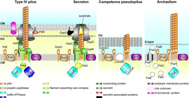
Prokaryotes have engineered sophisticated surface nanomachines that have allowed them to colonize Earth and thrive even in extreme environments. Filamentous machineries composed of type IV pilins, which are associated with an amazing array of properties ranging from motility to electric conductance, are arguably the most widespread since distinctive proteins dedicated to their biogenesis are found in most known species of prokaryotes. Several decades of investigations, starting with type IV pili and then a variety of related systems both in bacteria and archaea, have outlined common molecular and structural bases for these nanomachines. Using type IV pili as a paradigm, we will highlight in this review common aspects and key biological differences of this group of filamentous structures.

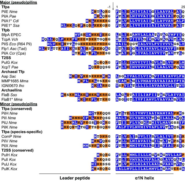
Keywords: archaellum; class III signal peptide; prepilin peptidase; type II secretion system; type IV pilus.


Figures

None
Using type IV pili as a paradigm, we review common genetic, structural and mechanistic features (many) as well as differences (few) of the exceptionally widespread and functionally versatile prokaryotic nano-machines composed of type IV pilins.
Figure 1.
Figure 1.
Conspicuous morphological features of bacterial Tfp, i.e. several μm length, 6–8 nm width, flexibility and propensity to interact laterally to form bundles. Bundle of filaments produced by N. meningitidis (individual filaments are 60 Å wide) were visualized by EM after negative staining with phosphotungstic acid.
Figure 2.
Figure 2.
Schematic representation of different Tff nanomachines: Tfp, T2SS, competence pseudopili and archaella. For Tfp, the well-characterized Tfpa system in N. meningitidis is shown. It should be noted that the protein nomenclature varies widely in other Tfpa-expressing species, although all the proteins are highly conserved. However, a major difference in Gram-positive piliated species is the absence of the proteins forming the outer membrane sub-complex (PilC, PilP, PilQ and PilW). For T2SS, the system exporting pullulanase in K. oxytoca is shown, but the proteins have been given their unifying Gsp names. For competence pseudopili, B. subtilis has been chosen. For archaeal flagella, we have chosen the representative M. maripaludis archaellum. All these systems are evolutionarily related as they are composed of proteins that show sequence and/or structural similarity and perform the same functions. To facilitate comparisons, proteins of similar function have identical colour. In brief, major (pseudo)pilins are processed by a dedicated prepilin peptidase, which removes a short hydrophilic leader peptide. For the sake of clarity, minor (pseudo)pilins that also undergo this processing are not shown. Traffic ATPases power filament extension from the inner membrane through ATP hydrolysis. The PilT ATPase which powers filament retraction has so far been identified only in Tfpa. The energy generated by ATP hydrolysis is translocated across the membrane by a multiprotein sub-complex, although the polytopic protein showing universal sequence conservation (purple) has also been proposed to play this role. In Gram-negative species, the inner membrane sub-complex is linked via a connecting protein to an outer membrane sub-complex centred on a multimeric channel known as the secretin. Several other proteins important for secretin stability/function are also part of this sub-complex. To facilitate visualization, the secretin dodecamer is shown as a vertical cross-section. OM, outer membrane; PG, peptidoglycan; CM, cytoplasmic membrane.
Figure 3.
Figure 3.
Conserved N-terminal sequence motif defining pilin subunits in a variety of Tff. This motif, known as class III signal peptide (Szabó et al., 2007), is composed of a hydrophilic leader peptide followed by a stretch of hydrophobic residues (except for a negatively charged Glu5). The 6–26 aa leader peptide contains a majority of hydrophilic (shaded in orange) and neutral (no shading) residues, and invariably ends with a Gly (except in archaea). The following tract of 21 predominantly hydrophobic residues (shaded in blue) forms an extended α-helix that is the main assembly interface of subunits within Tff. This class III signal peptide is recognized by a dedicated prepilin peptidase and cleaved (indicated by a vertical arrow) after the conserved Gly-1. *Filaments in these species might be composed of more than one major pilin. Nme, N. meningitidis; Pae, P. aeruginosa; Cdi, C. difficile; Ssa, Streptococcus sanguinis; Vch, V. cholerae; Eco, E. coli; Aac, A. actinomycetemcomitans; Ccr, C. crescentus; Kox, K. oxytoca; Sac, S. acidocaldarius; Mma, M. maripaludis; Iho, Ignicoccus hospitalis; Sso, S. solfataricus.
Figure 4.
Figure 4.
Phylogenetic distribution of the proteins involved in Tff biogenesis in archaea and bacteria. Using BioMart (Guberman et al., 2011), we have performed a global search of the InterPro database (Hunter et al., 2012) for signature motifs found only in proteins dedicated to Tff biogenesis. A black box indicates that the corresponding protein is found in the analysed phylum, while a white box indicates that it is absent. PilE, IPR007047, IPR002774 or IPR012912; PilD, IPR010627 or IPR000045; PilF, IPR007831 or IPR001482; PilG, IPR018076; PilM, IPR005883; PilN, IPR007813; PilO, IPR007445; PilP, IPR007446; PilQ, IPR001775 or IPR013355; PilC, IPR008707; PilW, IPR013360; PilT, IPR006321. In Firmicutes, the outer membrane sub-complex proteins (PilP, PilQ and PilW) are found only in the very few Gram-negative species in this phylum.
Figure 5:
Figure 5:
Common structural features of Tff and their subunits illustrated with N. gonorrhoeae Tfpa (Craig et al., 2006). (A) Structure of the full-length PilE from N. gonorrhoeae (PDB entry 2HI2) showing the conserved lollipop shape with a protruding α1N-helix (back view on the left, front view on the right). Post-translational modifications on the αβ-loop, carbohydrate at Ser63 and phosphate at Ser68, are shown in magenta. (B) Structural model of gonococcal Tfp (PDB entry 2HIL) obtained by combining X-ray crystallography and cryo-EM (side view on the left, top view on the upper right, bottom view on the lower right). In the 60 Å diameter right-handed 1-start helical assembly, the pilins are arranged in an ‘ear of wheat’ fashion (side view). The α1N-helices provide the main polymerization interface and are buried within the filament core parallel to its long axis (top and bottom views). Figures were generated using PyMOL (http://www.pymol.org).

References

    1. Aas FE, Winther-Larsen HC, Wolfgang M, et al. Substitutions in the N-terminal alpha helical spine of Neisseria gonorrhoeae pilin affect type IV pilus assembly, dynamics and associated functions. Mol Microbiol. 2007;63:69–85. - PubMed
    1. Abendroth J, Bagdasarian M, Sandkvist M, et al. The structure of the cytoplasmic domain of EpsL, an inner membrane component of the type II secretion system of Vibrio cholerae: an unusual member of the actin-like ATPase superfamily. J Mol Biol. 2004a;344:619–33. - PubMed
    1. Abendroth J, Kreger AC, Hol WG. The dimer formed by the periplasmic domain of EpsL from the type 2 secretion system of Vibrio parahaemolyticus. J Struct Biol. 2009a;168:313–22. - PMC - PubMed
    1. Abendroth J, Mitchell DD, Korotkov KV, et al. The three-dimensional structure of the cytoplasmic domains of EpsF from the type 2 secretion system of Vibrio cholerae. J Struct Biol. 2009b;166:303–15. - PMC - PubMed
    1. Abendroth J, Murphy P, Sandkvist M, et al. The X-ray structure of the type II secretion system complex formed by the N-terminal domain of EpsE and the cytoplasmic domain of EpsL of Vibrio cholerae. J Mol Biol. 2005;348:845–55. - PubMed

Publication types

Substances