. 2015 Mar 20;11(3):e1004749.
doi: 10.1371/journal.ppat.1004749. eCollection 2015 Mar.

An O antigen capsule modulates bacterial pathogenesis in Shigella sonnei

Affiliations

An O antigen capsule modulates bacterial pathogenesis in Shigella sonnei

Mariaelena Caboni et al. PLoS Pathog. .

Abstract

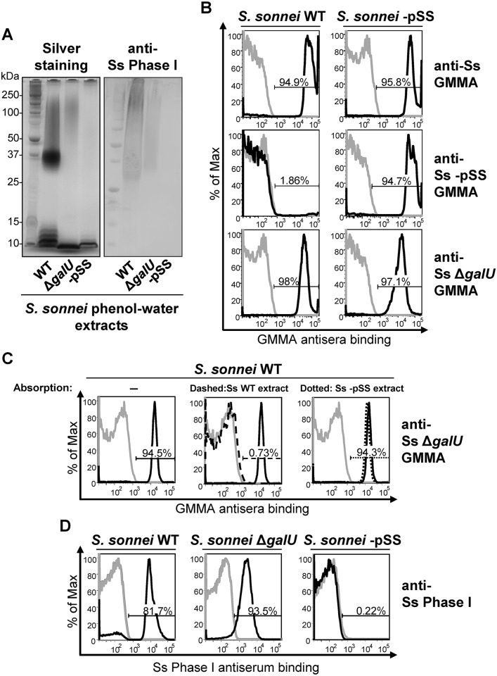
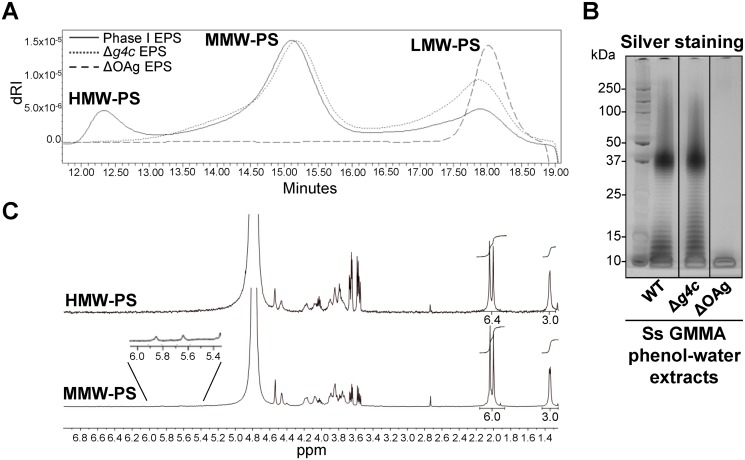
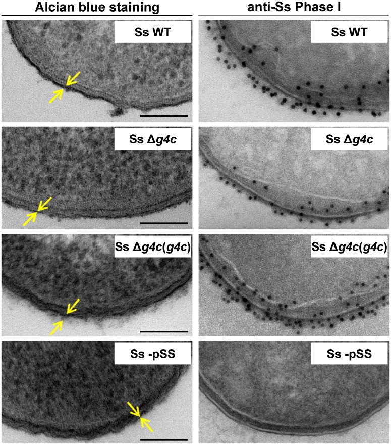
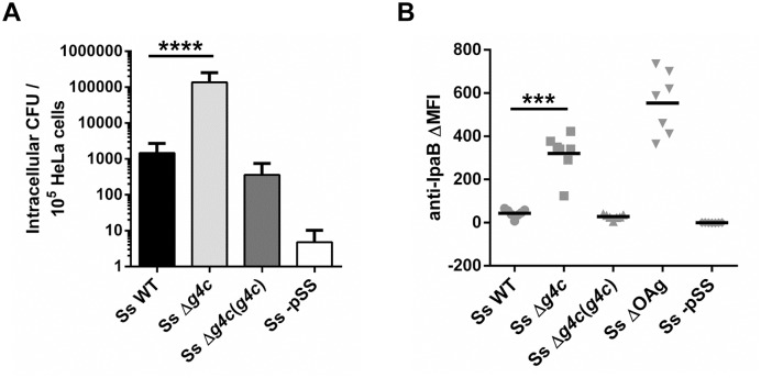
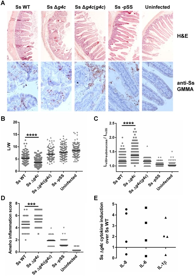
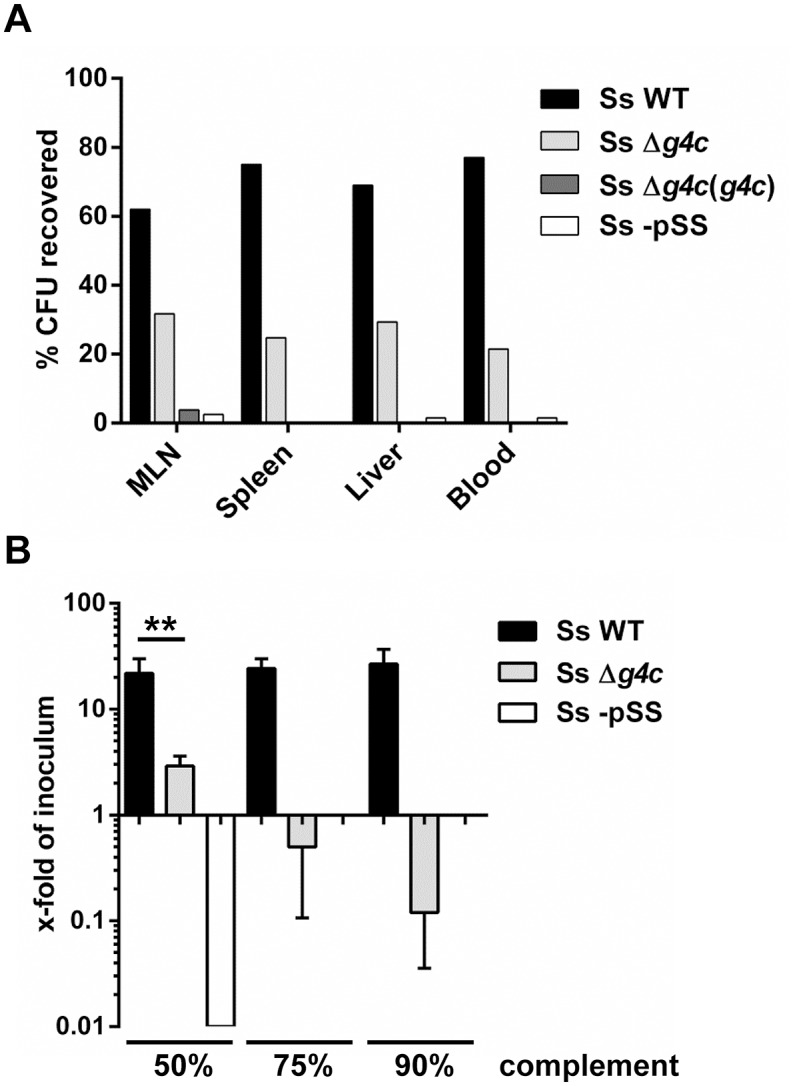
Shigella is the leading cause for dysentery worldwide. Together with several virulence factors employed for invasion, the presence and length of the O antigen (OAg) of the lipopolysaccharide (LPS) plays a key role in pathogenesis. S. flexneri 2a has a bimodal OAg chain length distribution regulated in a growth-dependent manner, whereas S. sonnei LPS comprises a monomodal OAg. Here we reveal that S. sonnei, but not S. flexneri 2a, possesses a high molecular weight, immunogenic group 4 capsule, characterized by structural similarity to LPS OAg. We found that a galU mutant of S. sonnei, that is unable to produce a complete LPS with OAg attached, can still assemble OAg material on the cell surface, but a galU mutant of S. flexneri 2a cannot. High molecular weight material not linked to the LPS was purified from S. sonnei and confirmed by NMR to contain the specific sugars of the S. sonnei OAg. Deletion of genes homologous to the group 4 capsule synthesis cluster, previously described in Escherichia coli, abolished the generation of the high molecular weight OAg material. This OAg capsule strongly affects the virulence of S. sonnei. Uncapsulated knockout bacteria were highly invasive in vitro and strongly inflammatory in the rabbit intestine. But, the lack of capsule reduced the ability of S. sonnei to resist complement-mediated killing and to spread from the gut to peripheral organs. In contrast, overexpression of the capsule decreased invasiveness in vitro and inflammation in vivo compared to the wild type. In conclusion, the data indicate that in S. sonnei expression of the capsule modulates bacterial pathogenesis resulting in balanced capabilities to invade and persist in the host environment.


Conflict of interest statement

I have read the journal's policy and the authors of this manuscript have the following competing interests: CAM, AS, and CG are employees of Novartis Vaccines Institute for Global Health, Siena, Italy. This does not alter the authors’ adherence to all the PLoS Pathogens policies on sharing data and materials.

Figures

Fig 1
Fig 1. S. sonnei ΔgalU LPS mutant possesses immunogenic and antigenic Phase I material on the surface.
(A) Silver staining and immunoblot analysis of phenol-water extracts from S. sonnei WT, S. sonnei ΔgalU and S. sonnei -pSS bacteria. Samples were run on 12% Bis-Tris SDS-PAGE, blotted, and membranes were incubated with S. sonnei Phase I monovalent antiserum (anti-Ss Phase I) at a dilution of 1:1000. (B) Flow cytometry analysis of surface staining of live S. sonnei WT and S. sonnei -pSS. Bacteria were stained with sera raised against GMMA from hyperblebbing S. sonnei with WT LPS (anti-Ss GMMA), S. sonnei -pSS with rough LPS (anti-Ss -pSS GMMA), or S. sonnei ΔgalU with deep rough LPS (anti-Ss ΔgalU GMMA). Gray profiles: staining with preimmune sera; black profiles: staining with GMMA antisera (1:1000). Representative results of three experiments are shown. (C) Competitive surface staining of S. sonnei WT live bacteria with sera raised against GMMA from hyperblebbing S. sonnei ΔgalU strain (anti-Ss ΔgalU GMMA). Gray profiles: staining with preimmune sera; black solid profiles: staining with anti-Ss ΔgalU GMMA; black dashed profile: staining with anti-Ss ΔgalU GMMA absorbed with S. sonnei WT (Ss WT) phenol-water extract; black dotted profile: staining with anti-Ss ΔgalU GMMA absorbed with S. sonnei -pSS (Ss -pSS) phenol-water extract. Sera dilution was 1:10000. (D) Surface staining of formalin-fixed S. sonnei WT, S. sonnei ΔgalU and S. sonnei -pSS with S. sonnei Phase I monovalent antiserum (anti-Ss Phase I). Gray profiles: staining with S. flexneri type II monovalent antiserum; black profiles: staining with anti-Ss Phase I. Sera dilution was 1:5000.
Fig 2
Fig 2. S. sonnei g4c cluster encodes for a high molecular weight O antigen polysaccharide.
(A) HPLC-SEC (dRI) analysis showing molecular weight distribution (high, medium, and low molecular weight polysaccharides, respectively HMW, MMW, LMW-PS) of acid-cleaved exopolysaccharide (EPS) purified from GMMA of hyperblebbing S. sonnei (Phase I EPS, solid line), S. sonnei Δg4cg4c EPS, dotted line) and S. sonnei ΔOAg (ΔOAg EPS, dashed line). Polysaccharide samples were run on TosoHaas TSK gel G3000 PWXL-CP column (distribution coefficients (Kd): KdHMW-PS = 0.09, KdMMW-PS = 0.31). Apparent average molecular weight of HMW-PS (197.49 kDa) and of MMW-PS (22.04 kDa) was estimated by running Phase I EPS with a dextran standard curve. (B) 12% Bis-Tris SDS-PAGE and silver staining of S. sonnei (Ss) phenol-water extracts from GMMA of hyperblebbing S. sonnei (WT EPS), S. sonnei Δg4c (EPS lacking HMW-PS), and S. sonnei ΔOAg (only LMW-PS). (C) 1H-NMR spectra of S. sonnei Phase I EPS populations. Integral of the signals at 2.00 and 2.04 ppm belonging to the N-acetyl groups of the FucNAc4N and the L-AltNAcA OAg residues and of the signal at 1.34–1.36 ppm of the FucNAc4N methyl group are reported. Region of anomeric signals of terminal and internal α-galactose residues from LPS outer core in MMW-PS (5.82 ppm and 5.62 ppm, respectively) is shown enlarged.
Fig 3
Fig 3. Group 4 capsule forms a dense outer layer on the surface of S. sonnei.
Surface analysis of S. sonnei WT (Ss WT), S. sonnei Δg4c (Ss Δg4c), S. sonnei Δg4c(g4c) (Ss Δg4c(g4c)) and S. sonnei -pSS (Ss -pSS) bacteria by electron microscopy. Left column: electron-dense material at the bacterial surface corresponding to exopolysaccharides is revealed by alcian blue staining. Scale bar: 100 nm. Arrows indicate the LPS/capsule layer. Right column: immunogold labelling of S. sonnei bacteria with anti-S. sonnei Phase I monovalent antiserum (anti-Ss Phase I).
Fig 4
Fig 4. Group 4 capsule impacts S. sonnei HeLa cell invasion and IpaB exposure on the surface.
(A) HeLa epithelial cells were infected with S. sonnei WT (Ss WT), S. sonnei Δg4c (Ss Δg4c), S. sonnei Δg4c(g4c) (Ss Δg4c(g4c)) or S. sonnei -pSS (Ss -pSS) at an MOI of 10. Columns show the average number and standard deviation (error bars) of intracellular bacteria (CFU) collected from 105 cells in three independent experiments in triplicate (****p<0.0001, Mann-Whitney test). (B) Flow cytometry analysis of IpaB exposure on the surface of live S. sonnei WT (Ss WT), S. sonnei Δg4c (Ss Δg4c), S. sonnei Δg4c(g4c) (Ss Δg4c(g4c)), S. sonnei ΔOAg (Ss ΔOAg) and S. sonnei -pSS (Ss -pSS). Results are expressed as differential Mean Fluorescence Intensity (ΔMFI): the difference between the MFI of the anti-IpaB immune straining and the MFI of a control staining, using only the secondary antibody. Three independent experiments were performed, twice in triplicate and once as single assay. Individual results are shown as scatter plot, the mean ΔMFI is shown as line (***p = 0.0006, Mann-Whitney test).
Fig 5
Fig 5. Group 4 capsule affects the pattern of S. sonnei pathology in the rabbit intestine.
(A) Representative results of histopathological analysis of S. sonnei WT (Ss WT), S. sonnei Δg4c (Ss Δg4c), S. sonnei Δg4c(g4c) (Ss Δg4c(g4c)) and S. sonnei -pSS (Ss -pSS) infected loops after 8 h of single strain challenge. Control sample is the uninfected tissue. Upper row: Hematoxylin and Eosin (H&E) staining of 7 μm slices of rabbit ileal loops. Lower row: immunostaining of 7 μm slices of rabbit loops with a polyclonal S. sonnei GMMA antisera (anti-Ss GMMA) to demonstrate bacteria localization (red dots) and counterstaining with Hematoxylin. (B) Histological alteration observed in the rabbit model following 8 h infection by S. sonnei strains. Severity of villi atrophy is measured according to the ratio between the length and width (L/W) of 120 villi counted on each among at least 4 different loops in 2 different animals (****p<0.0001, Mann-Whitney test). (C) Severity of submucosal edema is measured according to the ratio between the length of the entire mucosal and submucosal layer and the length of the villi (Lvilli+submucosa/Lvilli). 120 measurements were performed on each among at least 4 different loops in 2 different animals (****p<0.0001, Mann-Whitney test). (D) Evaluation of the Shigella-induced pathology for each strain according to the Ameho histopathological grading scale was performed on at least 4 image fields of 4 different loops in 2 different animals (***p = 0.0001, Mann-Whitney test). (E) Induction of IL-8, IL-6 and IL-1β in rabbit loops by S. sonnei Δg4c (Ss Δg4c) relative to S. sonnei WT (Ss WT). Expression levels were measured by RT-qPCR in the set up experiment using an infectious dose of 5x10⁹/loop (the other experiments used 3x10⁹ bacteria/loop). Two loops per strain were inoculated in 2 different animals. The scatter plot shows induction of gene expression in S. sonnei Δg4c loops relative to the adjacent S. sonnei WT loop.
Fig 6
Fig 6. Group 4 capsule affects S. sonnei peripheral spreading and sensitivity to direct complement lysis.
(A) Systemic load of S. sonnei WT (Ss WT), S. sonnei Δg4c (Ss Δg4c), S. sonnei Δg4c(g4c) (Ss Δg4c(g4c)) and S. sonnei -pSS (Ss -pSS) bacteria following 8 h of single strain infection in separate loops (3 loops per strain) in the same animal. The different strains were enumerated by plating on solid media using the distinct antibiotic resistance profiles of the strains for their differentiation. The prevalence of individual strains in mesenteric lymph nodes (MLN), spleen, liver, or blood is reported as percent of CFU recovered from the specific organ. Results present the average counts of 2 animals. (B) Sensitivity of S. sonnei WT (Ss WT), S. sonnei Δg4c (Ss Δg4c) and S. sonnei -pSS (Ss -pSS) strains to increasing concentrations of baby rabbit complement (50, 75, and 90%). Assays with 3 h incubation were performed in triplicate in three independent experiments and the results are expressed as x-fold increase/decrease compared to the number of the bacteria in the inoculum. No colonies were retrieved after incubation of S. sonnei -pSS with 75% and 90% of complement. (**p = 0.0022, Mann-Whitney test).

References

    1. Schroeder GN, Hilbi H. Molecular pathogenesis of Shigella spp.: controlling host cell signaling, invasion, and death by type III secretion. Clin Microbiol Rev. 2008;21: 134–156. 10.1128/CMR.00032-07 - DOI - PMC - PubMed
    1. Lozano R, Naghavi M, Foreman K, Lim S, Shibuya K, Aboyans V, et al. Global and regional mortality from 235 causes of death for 20 age groups in 1990 and 2010: a systematic analysis for the Global Burden of Disease Study 2010. Lancet. 2012;380: 2095–2128. 10.1016/S0140-6736(12)61728-0 - DOI - PMC - PubMed
    1. Kotloff KL, Nataro JP, Blackwelder WC, Nasrin D, Farag TH, Panchalingam S, et al. Burden and aetiology of diarrhoeal disease in infants and young children in developing countries (the Global Enteric Multicenter Study, GEMS): a prospective, case-control study. Lancet. 2013;382: 209–222. 10.1016/S0140-6736(13)60844-2 - DOI - PubMed
    1. Bennish ML. Potentially lethal complications of shigellosis. Rev Infect Dis. 1991;13 Suppl 4: S319–S324. - PubMed
    1. Struelens MJ, Patte D, Kabir I, Salam A, Nath SK, Butler T. Shigella septicemia: prevalence, presentation, risk factors, and outcome. J Infect Dis. 1985;152: 784–790. - PubMed

Publication types