. 2015 Jun;23(6):1077-1091.
doi: 10.1038/mt.2015.46. Epub 2015 Mar 20.

Polarized Macrophages Have Distinct Roles in the Differentiation and Migration of Embryonic Spinal-cord-derived Neural Stem Cells After Grafting to Injured Sites of Spinal Cord

Affiliations

Polarized Macrophages Have Distinct Roles in the Differentiation and Migration of Embryonic Spinal-cord-derived Neural Stem Cells After Grafting to Injured Sites of Spinal Cord

Kun Zhang et al. Mol Ther. 2015 Jun.

Abstract

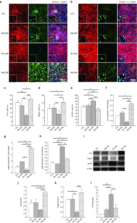
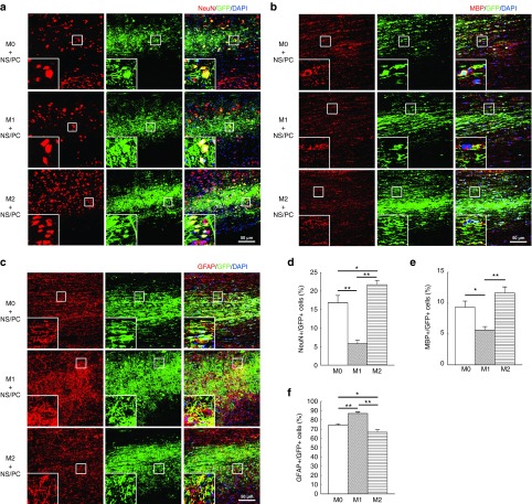
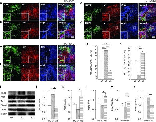
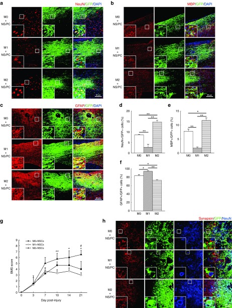
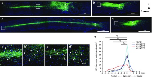
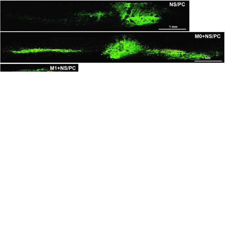
Spinal cord injury (SCI) frequently provokes serious detrimental outcomes because neuronal regeneration is limited in the central nervous system (CNS). Thus, the creation of a permissive environment for transplantation therapy with neural stem/progenitor cells (NS/PCs) is a promising strategy to replace lost neuronal cells, promote repair, and stimulate functional plasticity after SCI. Macrophages are important SCI-associated inflammatory cells and a major source of secreted factors that modify the lesion milieu. Here, we used conditional medium (CM) from bone marrow-derived M1 or M2 polarized macrophages to culture murine NS/PCs. The NS/PCs showed enhanced astrocytic versus neuronal/oligodendrocytic differentiation in the presence of M1- versus M2-CM. Similarly, cotransplantation of NS/PCs with M1 and M2 macrophages into intact or injured murine spinal cord increased the number of engrafted NS/PC-derived astrocytes and neurons/oligodendrocytes, respectively. Furthermore, when cotransplantated with M2 macrophages, the NS/PC-derived neurons integrated into the local circuitry and enhanced locomotor recovery following SCI. Interesting, engrafted M1 macrophages promoted long-distance rostral migration of NS/PC-derived cells in a chemokine (C-X-C motif) receptor 4 (CXCR4)-dependent manner, while engrafted M2 macrophages resulted in limited cell migration of NS/PC-derived cells. Altogether, these findings suggest that the cotransplantation of NS/PCs together with polarized macrophages could constitute a promising therapeutic approach for SCI repair.


Figures

Figure 1
Figure 1
Differentiation of neural stem/progenitor cells (NS/PCs) following stimulation with M0, M1- or M2-CM in vitro. (a,b) Representative immunofluorescence images of NS/PCs differentiated into Tuj1-, Oligo2-, and glial fibrillary acidic protein (GFAP)-positive (+) cells without (control, CTL) or with the assorted CM samples. The enlarged cells in the lower left-hand corner of each panel show the typical morphology for each culture. Space bars = 200 μm. (c–e) Percentage of Tuj1-, Oligo2-, and GFAP-positive cells differentiated from NS/PCs without (control) or with the assorted CM samples. (f–h) qPCR analysis of tuj1, pdgfra, and gfap, (i) western blotting of Tuj1, Oligo2, and GFAP (j–l) a densitometric quantitative analysis in cells differentiated from NS/PCs without (CTL) or with the assorted CM samples. Data in (c–h) and (j–l) were pooled from three independent experiments (*P < 0.05, **P < 0.01, ***P < 0.001); data in (a, b, and i) are representative of three independent experiments.
Figure 2
Figure 2
Differentiation of engrafted neural stem/progenitor cells (NS/PCs) cotransplanted with M0, M1, and M2 macrophages into the intact spinal cord. (a–c) Representative images of NS/PCs derived from GFP-Tg mice and cotransplanted with M0, M1, or M2 macrophages. The NS/PCs were differentiated into (a) NeuN-, (b) MBP-, and (c) glial fibrillary acidic protein (GFAP)-positive cells and detected by IHC. The boxed area in each image is enlarged at the lower left-hand corner of the panel. Space bars = 50 μm. (d–f) The percentages of (d) NeuN-, (e) MBP-, and (f) GFAP-positive cells among the engrafted GFP-expressing NS/PCs are shown. Data in (a–c) are representative of three independent experiments; data in (d–f) were pooled from three independent experiments (*P < 0.05, **P < 0.01).
Figure 3
Figure 3
Sustainability of engrafted macrophages in the M0, M1, and M2 polarization states. (a–f) Expression of iNOS (a, c, and e) and Arg1 (b, d, and f) in engrafted neural stem/progenitor cells (NS/PCs) and (a and b) M0, (c and d) M1, and (e and f) M2 macrophages at 3 days after transplantation. The boxed area in each image is enlarged at the lower left-hand corner of the panel. Space bars = 200 μm. (g and h) The percentage of (g) RFP+iNOS+ (h) RFP+Arg1+ cells among the engrafted red fluorescent protein (RFP)-positive macrophages are shown. (i) Western blot analysis of polarized macrophage markers (iNOS and Arg1) and neural lineage markers (Tuj1, Oligo2, and glial fibrillary acidic protein (GFAP)) in tissue dissected from the injured spinal cord containing engrafted NS/PCs and M0, M1, or M2 macrophages. (j and k) Densitometric analysis of iNOS and Arg1 protein expression levels in (i). (l–n) Densitometric analysis of Tuj1, Oligo2, and GFAP protein expression levels. Data in (a–i) are representative of three independent experiments; data in (g–h and j–n) are pooled from three independent experiments (*P < 0.05, ***P < 0.001).
Figure 4
Figure 4
Differentiation of engrafted neural stem/progenitor cells (NS/PCs) cotransplanted with polarized macrophages into the acutely injured spinal cord. (a–c) Representative images of engrafted NS/PCs derived from GFP-Tg mice and cotransplanted with M0, M1, or M2 macrophages. The NS/PCs were differentiated into (a) NeuN-, (b) MBP-, and (c) glial fibrillary acidic protein (GFAP)-positive cells and detected by IHC. The boxed area in each image is enlarged at the lower left-hand corner of the panel. Space bar = 50 μm. (d–f) The percentages of (d) NeuN-, (e) MBP-, and (f) GFAP-positive cells among the engrafted GFP-positive NS/PCs are shown. Data in (a–c) are representative of three independent experiments; data in (d–f) were pooled from three independent experiments (*P < 0.05, **P < 0.01). (g) BMS scores were evaluated in SCI mice engrafted with NS/PCs and M0, M1, or M2 macrophages (n = 6 mice per group). M2 + NS/PC group versus M1 + NS/PC group, *P < 0.05, **P < 0.01; M2 + NS/PC group versus M0 + NS/PC group, #P < 0.05. (h) The expression of synapsin is shown in the injured spinal cord in the three experimental groups. Space bar = 20 μm.
Figure 5
Figure 5
Migratory patterns of engrafted neural stem/progenitor cell (NS/PC)-derived cells in the injured spinal cord after co-transplantation of NS/PCs and M0, M1, or M2 macrophages. (a–d) Migration of NS/PCs alone without cotransplanted macrophages (a) and with cotransplanted (b) M0, (c) M1, and (d) M2 macrophages. The boxed areas in (a–d) are enlarged in (a'–d'). In a–d, ** denotes the lesion epicenter. Space bars = 1 mm. (a') shows the middle region of the rostral migration stream of the NS/PC-derived cells, while (b'–d') all show end-regions of the rostral migration stream. The arrowheads in (a'–d') indicate GFP-positive NS/PC-derived cells. Space bar = 50 μm. (e) Statistical results showing the integrated optical density (IOD) and the migration distance (P1, P2, P3, and P4) of engrafted NS/PC-derived cells cotransplanted without or with polarized macrophages in injured spinal cord. M1+NS/PCs versus M2+NS/PCs, P1 < 0.001; NS/PCs versus M2+NS/PCs, P2 < 0.001; M0+NS/PCs versus M2+NS/PCs, P3 < 0.001; M0+NS/PCs versus M1+NS/PCs, P4 < 0.01. Statistical data came from three independent experiments. D, dorsal; R, rostral.
Figure 6
Figure 6
CXCR4/CXCL12 signaling contributes to the migration of neural stem/progenitor cell (NS/PC)-derived cells in response to polarized macrophages. (a–d) The mRNA expression levels of cxcr4 (a and b) and cxcl12 (c and d) are shown in (a and c) M0-, M1-, and M2-CM-treated NS/PCs and (b and d) M0, M1, and M2 macrophages in vitro. (e) Western blot analysis of CXCR4 expression in M0-, M1-, and M2-CM-treated NS/PCs. Spleen tissue was used as the positive control. (f) Quantitative densitometric analysis indicated that M1-CM-treated NS/PCs expressed elevated levels of CXCR4. (g) CXCL12 expression in NS/PCs cultured with M0, M1-, or M2-CM, as well as in M0, M1, and M2 macrophages. Space bar = 10 μm. (h) IHC images of CXCL12 expression in engrafted NS/PC-derived cells cotransplanted with macrophages into the injured spinal cord. Space bar = 10 μm. (i) NS/PC migration induced by M0, M1-, or M2-CM in a transwell assay with and without AMD3100. Space bar = 50 μm. Data in (a–d) and (f) were pooled from three independent experiments (*P < 0.05, **P < 0.01, ***P < 0.001).
Figure 7
Figure 7
Migratory patterns of engrafted neural stem/progenitor cell (NS/PC)-derived cells after cotransplantation of AMD3100-treated NS/PCs and macrophages. (a–d) Migration patterns are shown of (a) NS/PCs transplanted alone and (b–d) NS/PCs cotransplanted with (b) M0 macrophages, (c) M1 macrophages and (d) M2 macrophages. In a–d, ** denotes the lesion epicenter. (e) Statistical results showing the integrated optical density (IOD) and the migration distance (P1, P2, P3, and P4) of engrafted NS/PC-derived cells after cotransplantation of AMD3100-treated NS/PCs and macrophages in injured spinal cord. M0+NS/PCs versus M2+NS/PCs, P1 < 0.001; M0+NS/PCs versus M1+NS/PCs, P2 < 0.01; NS/PCs versus M2+NS/PCs, P3 < 0.001; M1+ NS/PCs versus M2+NS/PCs, P4 < 0.001. Space bars = 1 mm.

Similar articles

Cited by

References

    1. Han, SS and Fischer, I (2000). Neural stem cells and gene therapy: prospects for repairing the injured spinal cord. JAMA 283: 2300–2301. - PubMed
    1. Mothe, AJ and Tator, CH (2012). Advances in stem cell therapy for spinal cord injury. J Clin Invest 122: 3824–3834. - PMC - PubMed
    1. Mothe, AJ and Tator, CH (2013). Review of transplantation of neural stem/progenitor cells for spinal cord injury. Int J Dev Neurosci 31: 701–713. - PubMed
    1. Weiss, S, Dunne, C, Hewson, J, Wohl, C, Wheatley, M, Peterson, AC et al. (1996). Multipotent CNS stem cells are present in the adult mammalian spinal cord and ventricular neuroaxis. J Neurosci 16: 7599–7609. - PMC - PubMed
    1. Cao, QL, Zhang, YP, Howard, RM, Walters, WM, Tsoulfas, P and Whittemore, SR (2001). Pluripotent stem cells engrafted into the normal or lesioned adult rat spinal cord are restricted to a glial lineage. Exp Neurol 167: 48–58. - PubMed

Publication types

MeSH terms

LinkOut - more resources