

Dynamic contrast-enhanced MRI for the detection of prostate cancer: meta-analysis
- PMID: 25794093
- PMCID: PMC5152763
- DOI: 10.2214/AJR.14.13373
Dynamic contrast-enhanced MRI for the detection of prostate cancer: meta-analysis
Abstract
Objective: The purpose of this study was to systematically review and meta-analyze dynamic contrast-enhanced MRI (DCE-MRI) for the detection of prostate cancer in comparison with standard evaluation with T2-weighted imaging.
Materials and methods: A PubMed electronic database search for the terms "dynamic contrast-enhanced," "prostate," and "MRI" was completed for articles up to September 17, 2013. All included studies had histopathologic correlation. Two by two contingency data were constructed for each study. A binormal bayesian ROC model was used to estimate and compare sensitivity, specificity, and AUC among eligible modalities.
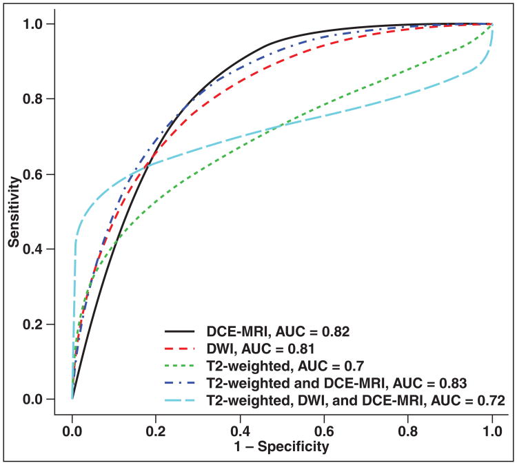
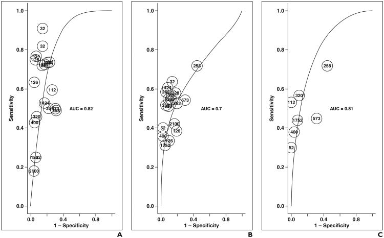
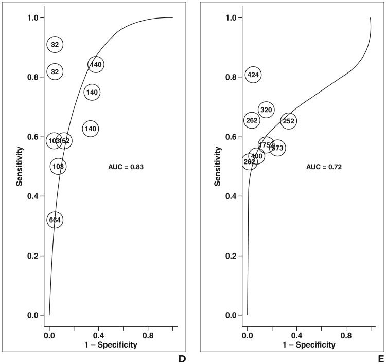
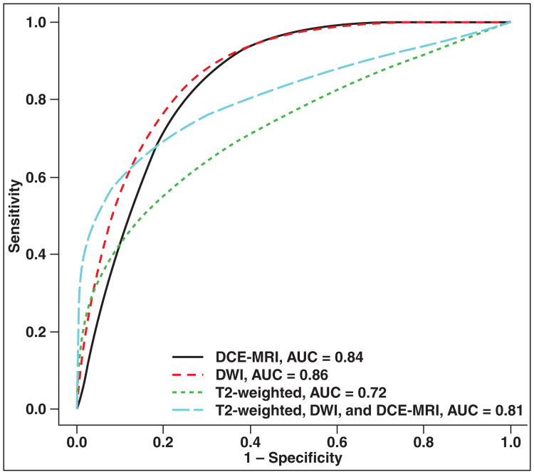
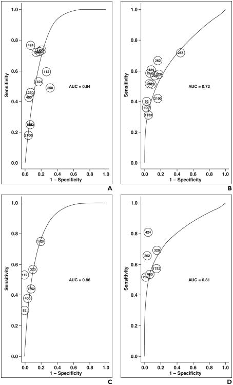
Results: Both DCE-MRI (0.82-0.86) and diffusion-weighted MRI (DWI) (0.84-0.88) yielded significantly better AUC than T2-weighted imaging (0.68-0.77). Moreover, partial AUC for the combination of DCE-MRI, DWI, and T2-weighted imaging was improved significantly (0.111; 0.103-0.119) when compared with DCE-MRI alone (0.079; 0.072-0.085) and T2-weighted imaging alone (0.079; 0.074-0.084) but not DWI alone (0.099; 0.091-0.108). Sensitivity and specificity were similar among the four modalities.
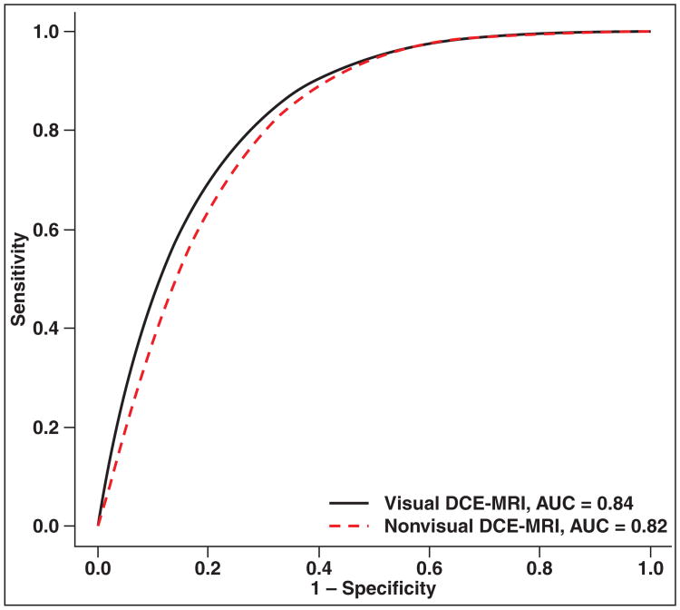
Conclusion: DCE-MRI improves AUC of tumor detection overall compared with T2-weighted imaging alone. Methods for DCE-MRI analysis require standardization, but visual analysis performs similar to semiquantitative methods. A two-parameter approach using DCE-MRI and T2-weighted imaging or DWI and T2-weighted imaging may be sufficient, and the latter may be more favorable for most routine prostate cancer imaging.
Keywords: MRI; cancer; dynamic contrast-enhanced MRI (DCE-MRI); prostate.
Figures
References
-
- Mazaheri Y, Shukla-Dave A, Muellner A, Hricak H. MR imaging of the prostate in clinical practice. MAGMA. 2008;21:379–392. - PubMed
-
- Sonnad SS, Langlotz CP, Schwartz JS. Accuracy of MR imaging for staging prostate cancer: a meta-analysis to examine the effect of technologic change. Acad Radiol. 2001;8:149–157. - PubMed
-
- Engelbrecht MR, Huisman HJ, Laheij RJ, et al. Discrimination of prostate cancer from normal peripheral zone and central gland tissue by using dynamic contrast-enhanced MR imaging. Radiology. 2003;229:248–254. - PubMed
-
- Tanimoto A, Nakashima J, Kohno H, Shinmoto H, Kuribayashi S. Prostate cancer screening: the clinical value of diffusion-weighted imaging and dynamic MR imaging in combination with T2-weighted imaging. J Magn Reson Imaging. 2007;25:146–152. - PubMed
Publication types
MeSH terms
Substances
Grants and funding
LinkOut - more resources
Full Text Sources
Medical
