. 2015 Mar 20;10(3):e0120252.
doi: 10.1371/journal.pone.0120252. eCollection 2015.

Pancreatic endoplasmic reticulum kinase activation promotes medulloblastoma cell migration and invasion through induction of vascular endothelial growth factor A

Affiliations

Pancreatic endoplasmic reticulum kinase activation promotes medulloblastoma cell migration and invasion through induction of vascular endothelial growth factor A

Stephanie Jamison et al. PLoS One. .

Abstract

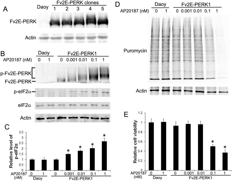
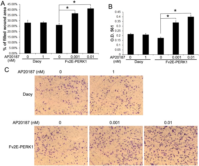
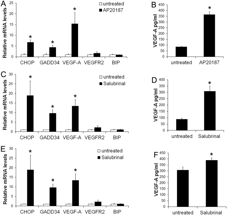
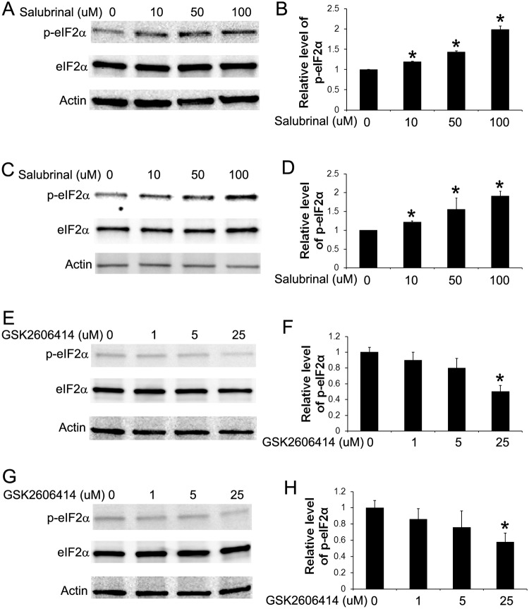
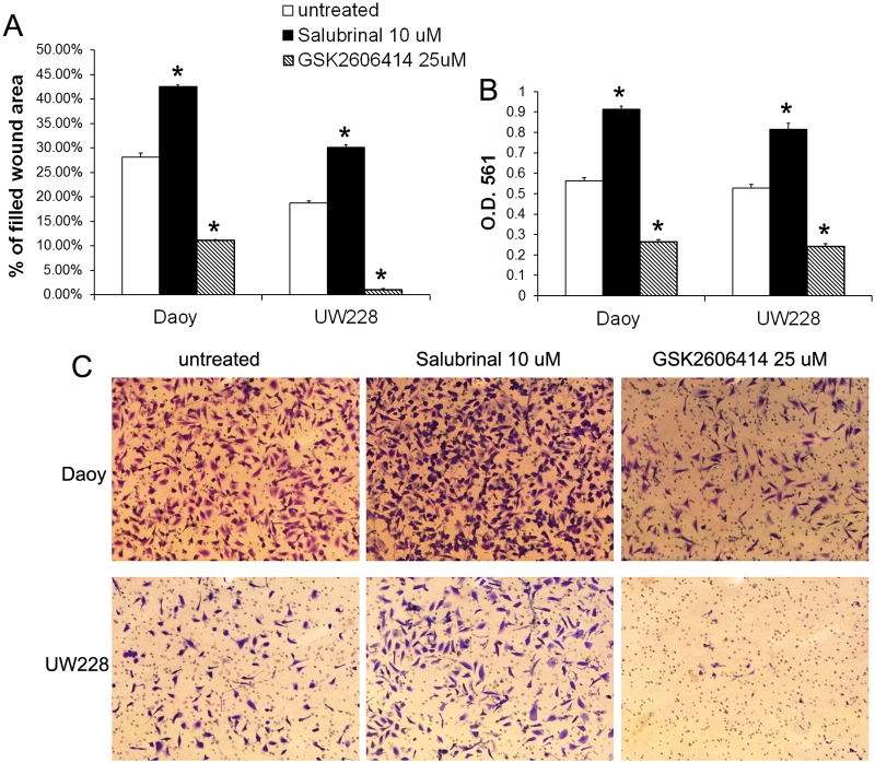
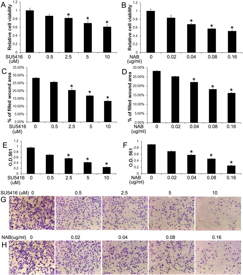
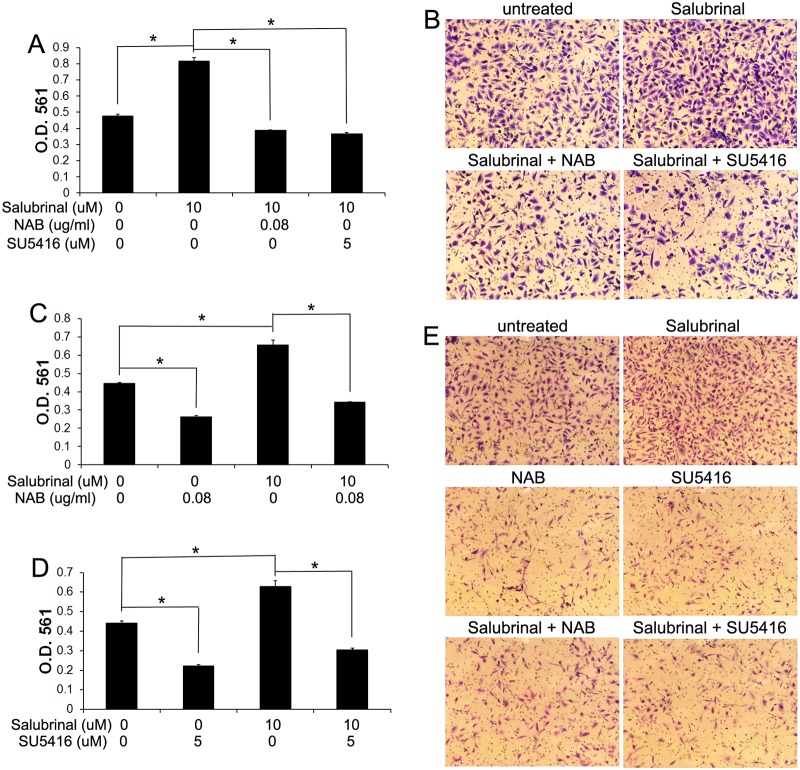
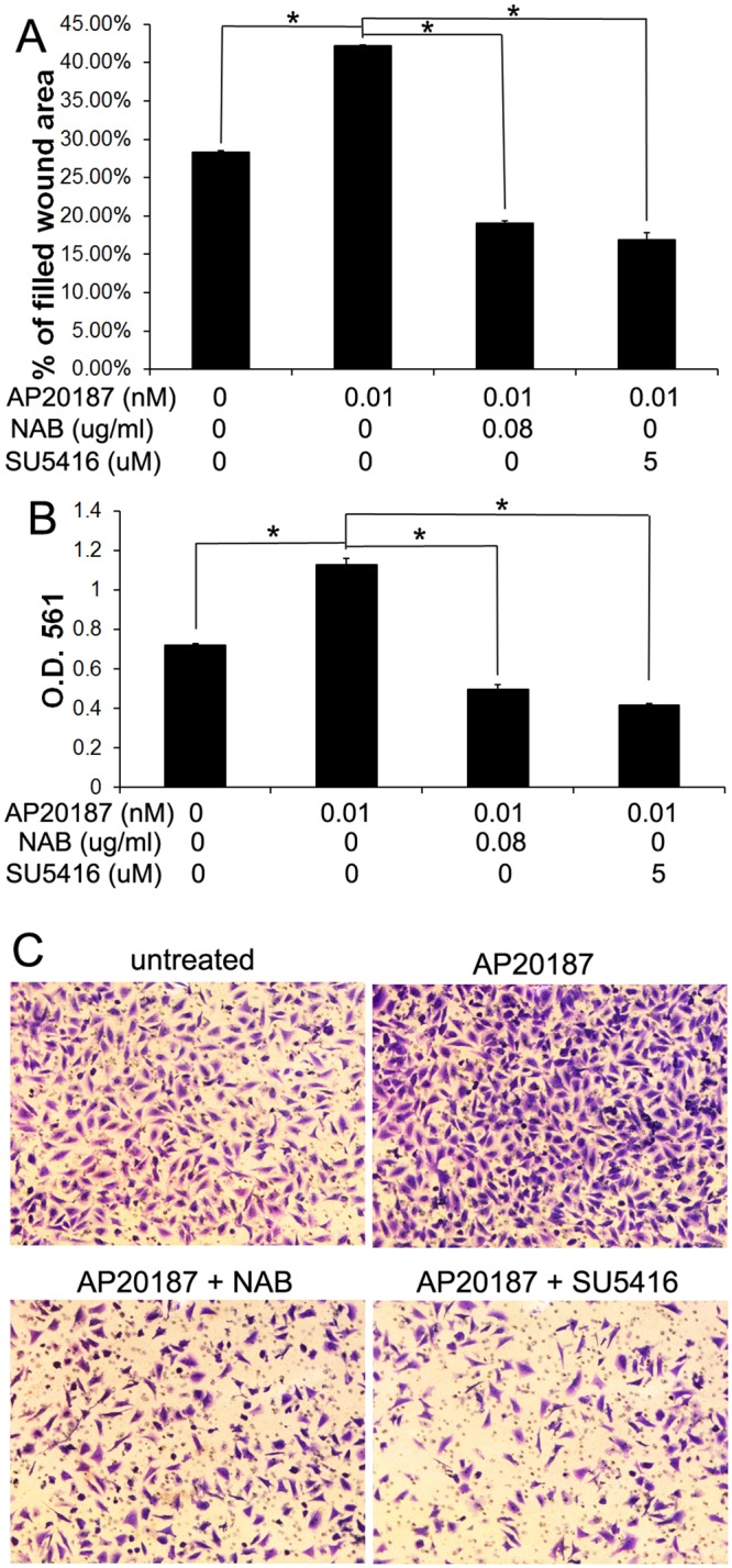
Evidence is accumulating that activation of the pancreatic endoplasmic reticulum kinase (PERK) in response to endoplasmic reticulum (ER) stress adapts tumor cells to the tumor microenvironment and enhances tumor angiogenesis by inducing vascular endothelial growth factor A (VEGF-A). Recent studies suggest that VEGF-A can act directly on certain tumor cell types in an autocrine manner, via binding to VEGF receptor 2 (VEGFR2), to promote tumor cell migration and invasion. Although several reports show that PERK activation increases VEGF-A expression in medulloblastoma, the most common solid malignancy of childhood, the role that either PERK or VEGF-A plays in medulloblastoma remains elusive. In this study, we mimicked the moderate enhancement of PERK activity observed in tumor patients using a genetic approach and a pharmacologic approach, and found that moderate activation of PERK signaling facilitated medulloblastoma cell migration and invasion and increased the production of VEGF-A. Moreover, using the VEGFR2 inhibitor SU5416 and the VEGF-A neutralizing antibody to block VEGF-A/VEGFR2 signaling, our results suggested that tumor cell-derived VEGF-A promoted medulloblastoma cell migration and invasion through VEGFR2 signaling, and that both VEGF-A and VEGFR2 were required for the promoting effects of PERK activation on medulloblastoma cell migration and invasion. Thus, these findings suggest that moderate PERK activation promotes medulloblastoma cell migration and invasion through enhancement of VEGF-A/VEGFR2 signaling.


Conflict of interest statement

Competing Interests: The authors have declared that no competing interests exist.

Figures

Fig 1
Fig 1. Characterization of stably transfected Daoy cell lines that allow for pharmacological controlled activation of PERK.
(A) Daoy cells were transfected with the plasmid pIREs-Fv2E-PERK-ZsGreen. We obtained several stably transfected cell lines that were resistant to G418 and expressed various levels of Fv2E-PERK. (B) Western blot analysis showed that AP20187 treatment activated Fv2E-PERK and led to phosphorylation of eIF2α. The positions of activated Fv2E-PERK (p-Fv2E-PERK) and inactive Fv2E-PERK proteins were indicated. (C) Densitometry analysis of western blot results showed that AP20187 treatment increased the level of p-eIF2a in Fv2E-PERK1 cells in a dose-dependent manner. The relative protein levels are relative to actin. (D) SUnSET measurement of protein biosynthesis revealed dramatic reduction of protein biosynthesis in Fv2E-PERK1 cells treated with the high dose of AP20187 (0.1–1 nM). Nevertheless, treatment with the low dose of AP20187 (0.001–0.01 nM) only slightly reduced protein biosynthesis in the cells. (E) MTT analysis showed that treatment with the high dose of AP20187 (0. 1–1 nM) significantly inhibited Fv2E-PERK1 cell growth. Nevertheless, treatment with the low dose of AP20187 (0.001–0.01 nM) had no effect on the cell growth. The experiments were repeated at least three times. Error bars represent SD, *P < 0.05.
Fig 2
Fig 2. Moderate PERK activation promoted Daoy cell migration and invasion.
(A) Wound healing assay showed that AP20187 (0.001–0.01 nM) treatment significantly increased the percentage of filled wound area of Fv2E-PERK1 cells. (B, C) Results of matrigel transwell assay. Daoy or Fv2E-PERK1 cells were seeded in the top well of a matrigel-coated chamber. After 16 h, the invaded cells at the lower surface of the chamber were stained with crystal violet and photographed (C). The stained cells were dissolved in 10% acetic acid and the absorbance was measured at 561 nm (B). AP20187 (0.001–0.01 nM) treatment significantly increased the invaded Fv2E-PERK1 cell numbers at the lower surface of the chamber. The experiments were repeated at least three times. Error bars represent SD, *P < 0.05.
Fig 3
Fig 3. PERK activation stimulated medulloblastoma cells to produce VEGF-A.
(A) Real-time PCR analysis showed that 0.01 nM AP20187 treatment significantly increased the expression of CHOP, GADD34, and VEGF-A in Fv2E-PERK1 cells, but did not affect the expression of VEGFR2 and BIP. (B) ELISA analysis showed that 0.01 nM AP20187 treatment significantly increased the production of VEGF-A in Fv2E-PERK1 cells. (C) Real-time PCR analysis showed that 10 μM Salubrinal treatment significantly increased the expression of CHOP, GADD34, and VEGF-A in Daoy cells, but did not affect the expression of VEGFR2 and BIP. (D) ELISA analysis showed that 10 μM Salubrinal treatment significantly increased the production of VEGF-A in Daoy cells. (E) Real-time PCR analysis showed that 10 μM Salubrinal treatment significantly increased the expression of CHOP, GADD34, and VEGF-A in UW228 cells, but did not affect the expression of VEGFR2 and BIP. (F) ELISA analysis showed that 10 μM Salubrinal treatment significantly increased the production of VEGF-A in UW228 cells. The experiments were repeated at least three times. Error bars represent SD, *P < 0.05.
Fig 4
Fig 4. Treatment with either Salubrinal or GSK2606414 altered the levels of p-eIF2α in medulloblastoma cells.
(A, B) Western blot analysis showed that treatment with Salubrinal (10–100 μM) moderately but significantly elevated the level of p-eIF2α in Daoy cells. (C, D) Western blot analysis showed that treatment with Salubrinal (10–100 μM) moderately but significantly elevated the level of p-eIF2α in UW228 cells. (E, F) Western blot analysis showed that treatment with GSK2606414 (25 μM) noticeably decreased the level of p-eIF2α in Daoy cells. (G, H) Western blot analysis showed that treatment with GSK2606414 (25 μM) noticeably decreased the level of p-eIF2α in UW228 cells. The relative protein levels are relative to actin. The experiments were repeated at least three times. Error bars represent SD, *P < 0.05.
Fig 5
Fig 5. Moderate PERK activation enhanced medulloblastoma cell migration and invasion.
(A) Wound healing assay showed that Salubrinal (10 μM) treatment significantly increased the percentage of filled wound area of Daoy and UW228 cells, and that GSK2606414 (25 μM) treatment significantly decreased the percentage of filled wound area of Daoy and UW228 cells. (B, C) Matrigel transwell assay showed that Salubrinal (10 μM) treatment significantly increased the numbers of the invaded Daoy and UW228 cells at the lower surface of the chamber, and that GSK2606414 (25 μM) treatment significantly decreased the numbers of the invaded Daoy and UW228 cells at the lower surface of the chamber. The experiments were repeated at least three times. Error bars represent SD, *P < 0.05.
Fig 6
Fig 6. Blockage of VEGF-A impaired medulloblastoma cell migration and invasion.
(A) MTT assay showed that SU5416 treatment significantly suppressed Daoy cell growth. (B) MTT assay showed that NAB treatment significantly suppressed Daoy cell growth. (C) Wound healing assay showed that SU5416 treatment significantly reduced the percentage of filled wound area of Daoy cells. (D) Wound-healing assay showed that NAB treatment significantly reduced the percentage of filled wound area of Daoy cells. (E, G) Matrigel transwell assay showed that SU5416 treatment significantly reduced the numbers of the invaded Daoy cells at the lower surface of the chamber. (F, H) Matrigel transwell assay showed that NAB treatment significantly reduced the numbers of the invaded Daoy cells at the lower surface of the chamber. The experiments were repeated at least three times. Error bars represent SD, *P < 0.05.
Fig 7
Fig 7. PERK activation promoted medulloblastoma cell invasion through activation of VEGF-A/VEGFR2 signaling.
(A, B) Matrigel transwell assay showed that 10 μM Salubrinal treatment significantly increased the numbers of the invaded Daoy cells at the lower surface of the chamber. Interestingly, treatment with either 0.08 μg/ml NAB or 5 μM SU5416 diminished the increased cell number at the lower surface of the chamber induced by Salubrinal treatment. (C, E) Matrigel transwell assay showed that 10 μM Salubrinal treatment significantly increased the numbers of the invaded UW228 cells at the lower surface of the chamber; however, treatment with 0.08 μg/ml NAB significantly reduced the numbers of the invaded UW228 cells at the lower surface of the chamber. Interestingly, treatment with 0.08 μg/ml NAB also diminished the increased cell number at the lower surface of the chamber induced by Salubrinal treatment. (D, E) Matrigel transwell assay showed that treatment with 5 μM SU5416 significantly reduced the numbers of the invaded UW228 cells at the lower surface of the chamber. Importantly, 5 μM SU5416 treatment also diminished the increased cell number at the lower surface of the chamber induced by Salubrinal treatment. The experiments were repeated at least three times. Error bars represent SD, *P < 0.05.
Fig 8
Fig 8. VEGF-A contributed to the promoting effects of PERK activation on Daoy cell migration and invasion.
(A) Wound-healing assay showed that 0.01 nM AP20187 treatment significantly increased the percentage of filled wound area of Fv2E-PERK1 cells. Importantly, the increased percentage of filled wound area induced by AP20187 treatment was diminished by treatment with either 0.08 μg/ml NAB or 5 μM SU5416. (B, C) Matrigel transwell assay showed that 0.01 nM AP20187 treatment significantly increased the numbers of the invaded Fv2E-PERK1 cells at the lower surface of the chamber. Interestingly, treatment with either 0.08 μg/ml NAB or 5 μM SU5416 diminished the increased cell number at the lower surface of the chamber induced by AP20187 treatment. The experiments were repeated at least three times. Error bars represent SD, *P < 0.05.

Similar articles

Cited by

References

    1. Marciniak SJ, Ron D. Endoplasmic reticulum stress signaling in disease. Physiol Rev. 2006; 86: 1133–1149. - PubMed
    1. Walter P, Ron D. The unfolded protein response: from stress pathway to homeostatic regulation. Science. 2011; 334: 1081–1086. 10.1126/science.1209038 - DOI - PubMed
    1. Pavitt GD, Ron D. New insights into translational regulation in the endoplasmic reticulum unfolded protein response. Cold Spring Harb Perspect Biol. 2012; 4: a012278 10.1101/cshperspect.a012278 - DOI - PMC - PubMed
    1. Ron D, Walter P. Signal integration in the endoplasmic reticulum unfolded protein response. Nat Rev Mol Cell Biol. 2007; 8: 519–529. - PubMed
    1. Ma Y, Hendershot LM. The role of the unfolded protein response in tumour development: friend or foe? Nat Rev Cancer. 2004; 4: 966–977. - PubMed

Publication types

MeSH terms

Substances

LinkOut - more resources