. 2015 Jun;13(6):957-68.
doi: 10.1158/1541-7786.MCR-14-0580. Epub 2015 Mar 20.

A YAP/TAZ-Regulated Molecular Signature Is Associated with Oral Squamous Cell Carcinoma

Affiliations

A YAP/TAZ-Regulated Molecular Signature Is Associated with Oral Squamous Cell Carcinoma

Samantha E Hiemer et al. Mol Cancer Res. 2015 Jun.

Abstract

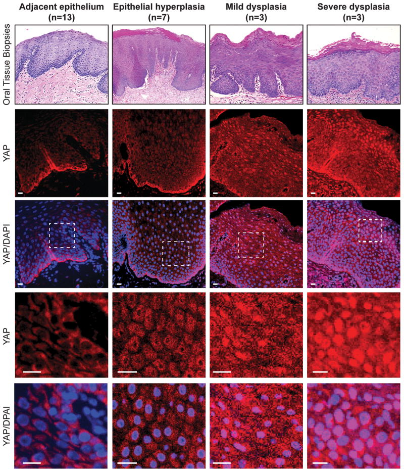
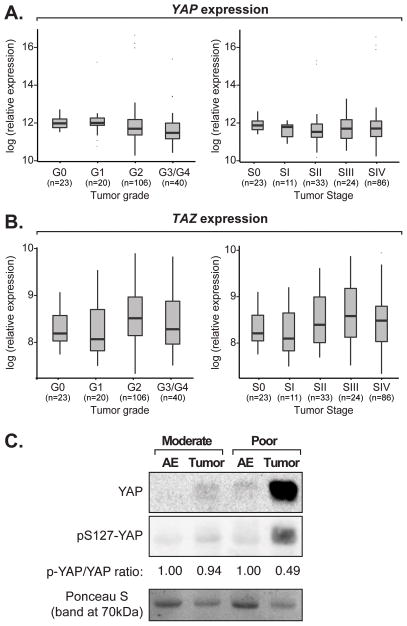
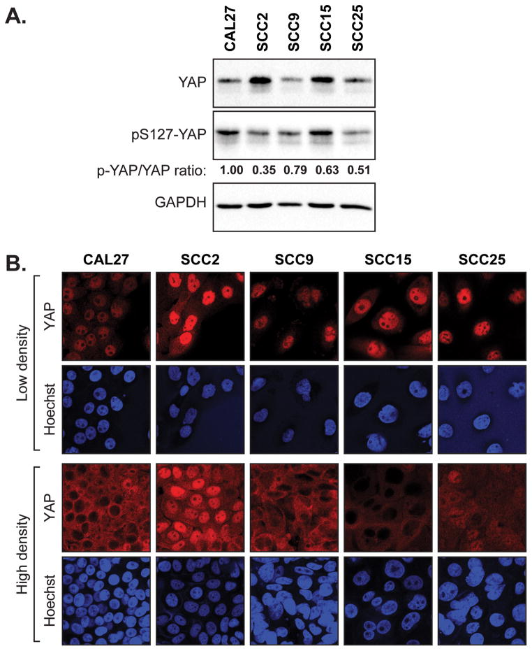
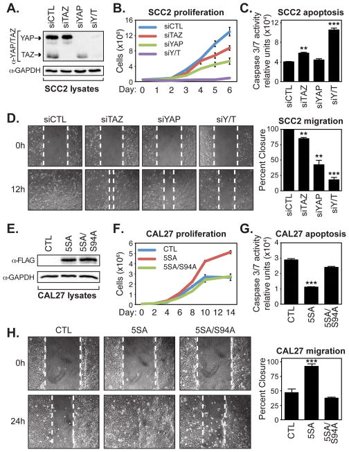
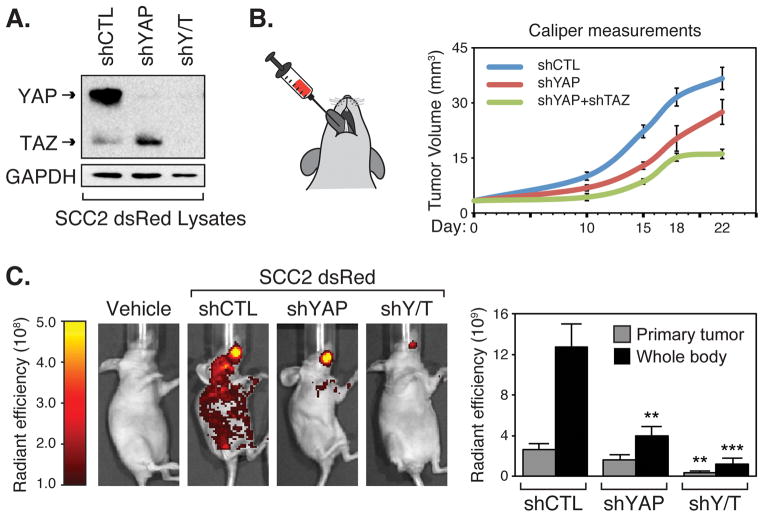
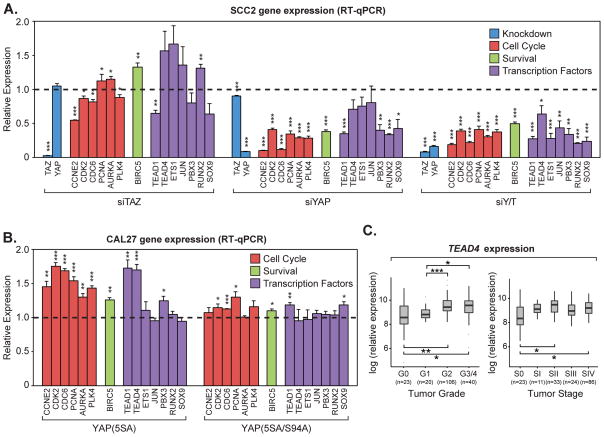
Oral squamous cell carcinoma (OSCC) is a prevalent form of cancer that develops from the epithelium of the oral cavity. OSCC is on the rise worldwide, and death rates associated with the disease are particularly high. Despite progress in understanding the mutational and expression landscape associated with OSCC, advances in deciphering these alterations for the development of therapeutic strategies have been limited. Further insight into the molecular cues that contribute to OSCC is therefore required. Here, we show that the transcriptional regulators YAP (YAP1) and TAZ (WWTR1), which are key effectors of the Hippo pathway, drive protumorigenic signals in OSCC. Regions of premalignant oral tissues exhibit aberrant nuclear YAP accumulation, suggesting that dysregulated YAP activity contributes to the onset of OSCC. Supporting this premise, we determined that nuclear YAP and TAZ activity drives OSCC cell proliferation, survival, and migration in vitro, and is required for OSCC tumor growth and metastasis in vivo. Global gene expression profiles associated with YAP and TAZ knockdown revealed changes in the control of gene expression implicated in protumorigenic signaling, including those required for cell cycle progression and survival. Notably, the transcriptional signature regulated by YAP and TAZ significantly correlates with gene expression changes occurring in human OSCCs identified by The Cancer Genome Atlas (TCGA), emphasizing a central role for YAP and TAZ in OSCC biology.

Implications: This study defines a YAP/TAZ-regulated transcriptional program in OSCC and reveals novel roles for nuclear YAP/TAZ activity in the onset and progression of this cancer.


Conflict of interest statement

The authors have no conflict of interest to disclose

Figures

Figure 1
Figure 1
Nuclear YAP accumulates in oral tissues with hyperplasia and dysplasia pathology. Tissues from patients exhibiting hyperplasia, mild dysplasia and severe dysplasia, as well as cytologically normal adjacent epithelium, were examined by H&E staining (top panels) and by immunofluorescence to detect YAP localization (the number of tissues examined is indicated). DAPI was used to stain nuclei. A zoomed in image from each sample is shown in the bottom panels, highlighting the YAP localization changes observed. Scale bars represent 20μm.
Figure 2
Figure 2
YAP and TAZ expression in OSCCs. One-way ANOVA analysis showing (A) YAP or (B) TAZ expression with respect to TCGA OSCC tumor grade or stage data. A pairwise t-test analysis comparing datasets revealed no significant expression changes. (C) Protein was extracted from poorly and moderately differentiated tumors, and associated adjacent epithelium (AE), and examined for YAP and pS127-YAP levels by immunoblotting. A representative image of our results is shown. Ponceau-S staining of the proteins on the immunoblotted membrane is shown as a loading control. Quantitation of the relative phospho-S127-YAP to total YAP is also shown.
Figure 3
Figure 3
Elevated levels of nuclear YAP are found in aggressive OSCC cells. (A) A panel of oral cancer cell lines was examined by immunoblotting for endogenous YAP, phospho-S127-YAP, and GAPDH (loading control). Quantification of phospho-S127-YAP to total YAP is shown. (B) Oral cancer cell lines were examined by immunofluorescence for endogenous YAP localization at both low and high density.
Figure 4
Figure 4
YAP/TAZ promote OSCC cell proliferation, survival, and migration. (A) SCC2 cells were transfected with control siRNA (siCTL) or siRNA targeting TAZ (siTAZ), YAP (siYAP), or YAP and TAZ (siYAP/TAZ), and lysates obtained from these cells were examined by immunoblotting with the indicated antibodies. (B) SCC2 knockdown cells were counted over 6 days to measure their proliferative capacity. Cells from three experiments were counted and the average (+S.E.) for each day is shown. (C) SCC2 knockdown cells were examined for Caspase-3 and -7 activity using a Caspase-Glo 3/7 assay. The average (+S.E.) of three experiments is shown. (D) Confluent monolayers of SCC2 knockdown cells were wounded and examined for their ability to migrate after 12 hours. Representative images are shown and the average wound healing (+S.E.) of three experiments is indicated. (E) Doxycycline-inducible CAL27 control cells, or cells expressing 3xFLAG-YAP(5SA) or 3xFLAG-YAP(5SA/S94A) were lysed and examined by immunoblotting with the indicated antibodies. (F) CAL27 expressing cells were counted over 14 days to measure their proliferative capacity. Cells from three experiments were counted and the average (+S.E.) for each day is shown. (G) CAL27 expressing cells were examined for caspase-3 and -7 activity using a Caspase-Glo 3/7 assay. The average (+S.E.) of three experiments is shown. (H) Confluent monolayers of CAL27 expressing cells were wounded and examined for their ability to migrate after 24 hours. Representative images are shown and the average wound healing (+S.E.) of three experiments is indicated. All statistics were calculated compared to the control sample using an unpaired Student’s t test and are represented as ** p-value<0.01, and *** p-value<0.001.
Figure 5
Figure 5
YAP/TAZ are required for OSCC tumor growth and metastasis in vivo. (A) SCC2-dsRED cells stably expressing control shRNA (shCTL), shRNA targeting YAP (shYAP), or shRNA targeting YAP and TAZ (shY/T) were lysed and examined by immunoblotting for the indicated proteins. (B) SCC2-dsRED cells were injected into the tongue of nude mice and primary tumor volume was determined by caliper measurements at day 10, 15, 18, and 22. (C) SCC2-dsRED cells contributing to tumor formation and metastasis were visualized by IVIS fluorescent imaging at day 22 and representative images are shown. Total radiant efficiency of cells in the primary tumor and cells that metastasized throughout the animal body were quantitated and are shown as the average (+S.E.) (n=3 for vehicle control, n=9 for shRNA-expressing cells). Statistics comparing primary tumor size or metastasis of the knockdown cells to the control cells were performed using an unpaired Student’s t test and are represented as ** p-value<0.01, and *** p-value<0.001.
Figure 6
Figure 6
YAP/TAZ-regulated transcriptional events correlate with OSCC tumor grade and stage. (A) Microarrays were performed from samples isolated from SCC2 cells transiently transfected with control siRNA (siCTL) or siRNA targeting TAZ (siTAZ), YAP (siYAP), or YAP and TAZ (siYAP/TAZ). Hierarchical clustering of the top 3000 genes with the highest median absolute deviation is shown. The number of up- and down-regulated genes (genes with adjusted p-value ≤0.05 and fold-change ≥2 compared to the control) that were identified from our microarray study is shown below. We tested for the enrichment of the YAP- and TAZ-regulated expression changes in the TCGA data by GSEA. The results for our analysis of (B) adjacent epithelium (AE) versus tumor grade and (C) tumor stages are summarized (enrichment nominal p-values reported). OSCC tumor grade and tumor stage data obtained from TCGA was projected onto (D) the entire YAP/TAZ-regulated expression signature identified from our microarray studies, or (E) only the YAP/TAZ-activated expression signature, and a heat map of the clustered data is shown. Two notable YAP/TAZ-activated gene clusters were identified, and selected data from our pathway enrichment analysis of these genes are shown on the right.
Figure 7
Figure 7
YAP/TAZ regulate the transcription of genes important for cell cycle progression and survival. (A) SCC2 cells were transiently transfected with control siRNA (siCTL) or siRNA targeting TAZ (siTAZ), YAP (siYAP), or YAP and TAZ (siYAP/TAZ). Relative expression of genes indicated in the microarray analysis was determined by qPCR. All data are relative to siCTL (dashed line) and are shown as the average of three experiments (+S.E.). Genes are grouped together by function including knockdown efficiency (blue), cell cycle regulation (red), pro-survival (green), and transcription factors (purple). (B) Doxycycline-inducible CAL27 control cells or cells expressing 3xFLAG-YAP(5SA) or 3xFLAG-YAP(5SA/S94A) were treated with doxycycline. Relative expression of genes indicated in the microarray analysis was determined by qPCR. All data are relative to control cells (dashed line) and are shown as the average of three experiments (+S.E.). Genes are grouped together by function including cell cycle regulation (red), pro-survival (green), and transcription factors (purple). Statistics comparing to the control cells (dashed line) were calculated using an unpaired Student’s t test and are represented as * p-value<0.05, ** p-value<0.01, and *** p-value<0.001. (C) Box plot of TEAD4 expression with respect to OSCC tumor grade and stage, showing TEAD4 expression is significantly induced with OSCC tumor grade (One-way ANOVA; p-value=3.51 e-05) and stage (One-way ANOVA; p-value=0.0012). Pairwise t-tests of expression between groups is also shown, and represented as * p-value<0.05, ** p-value<0.01, and *** p-value<0.001.

Similar articles

Cited by

References

    1. Varelas X, Kukuruzinska MA. Head and neck cancer: from research to therapy and cure. Annals of the New York Academy of Sciences. 2014;1333:1–32. - PubMed
    1. Harvey KF, Zhang X, Thomas DM. The Hippo pathway and human cancer. Nat Rev Cancer. 2013;13:246–57. - PubMed
    1. Vassilev A, Kaneko KJ, Shu H, Zhao Y, DePamphilis ML. TEAD/TEF transcription factors utilize the activation domain of YAP65, a Src/Yes-associated protein localized in the cytoplasm. Genes & development. 2001;15:1229–41. - PMC - PubMed
    1. Zhao B, Ye X, Yu J, Li L, Li W, Li S, et al. TEAD mediates YAP-dependent gene induction and growth control. Genes & development. 2008;22:1962–71. - PMC - PubMed
    1. Camargo FD, Gokhale S, Johnnidis JB, Fu D, Bell GW, Jaenisch R, et al. YAP1 increases organ size and expands undifferentiated progenitor cells. Curr Biol. 2007;17:2054–60. - PubMed

Publication types

MeSH terms