

Gonadotropin-releasing hormone stimulates biliary proliferation by paracrine/autocrine mechanisms
- PMID: 25794706
- PMCID: PMC4380841
- DOI: 10.1016/j.ajpath.2014.12.004
Gonadotropin-releasing hormone stimulates biliary proliferation by paracrine/autocrine mechanisms
Erratum in
-
Correction.Am J Pathol. 2023 May;193(5):654. doi: 10.1016/j.ajpath.2023.02.011. Am J Pathol. 2023. PMID: 37080663 Free PMC article. No abstract available.
Abstract
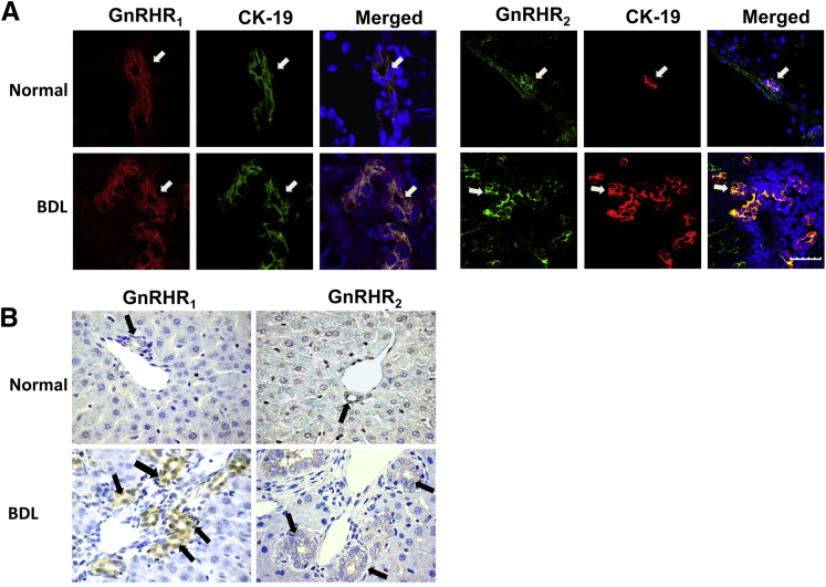
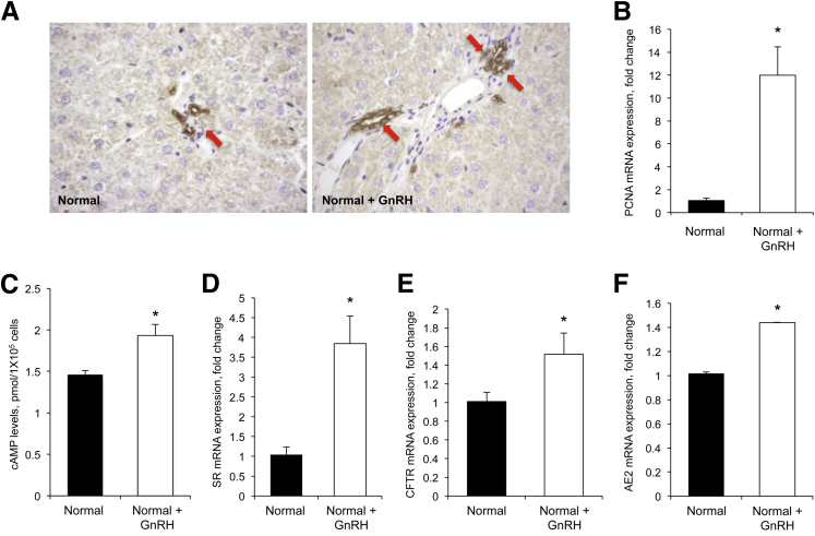
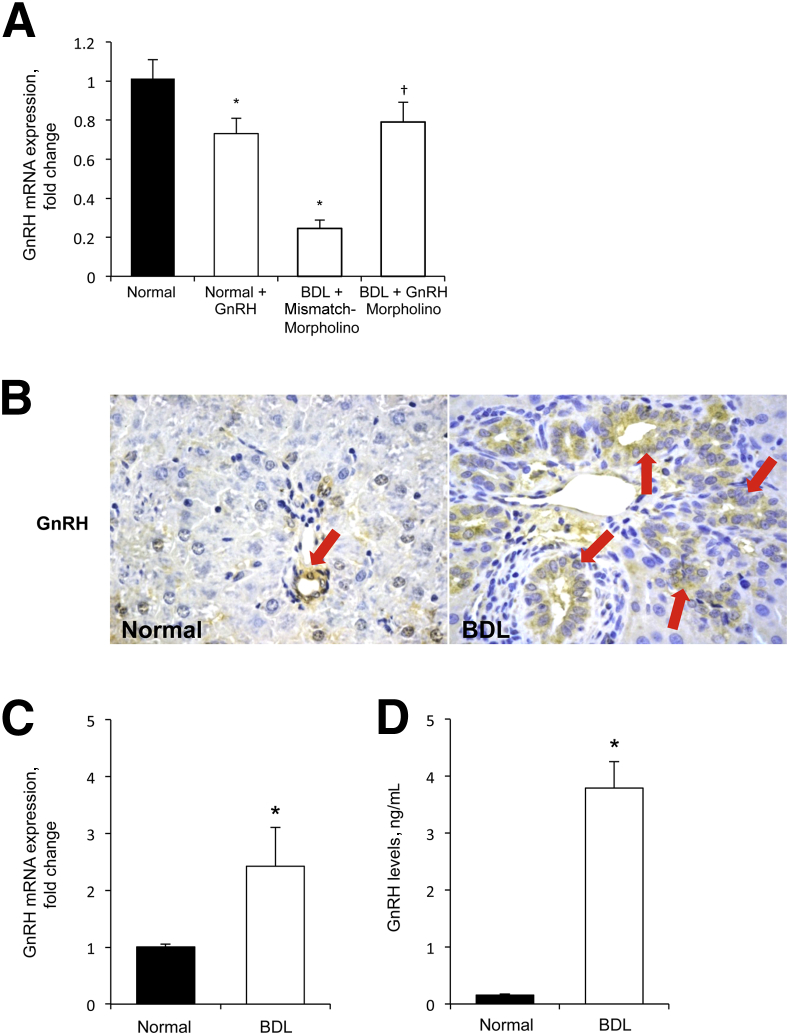
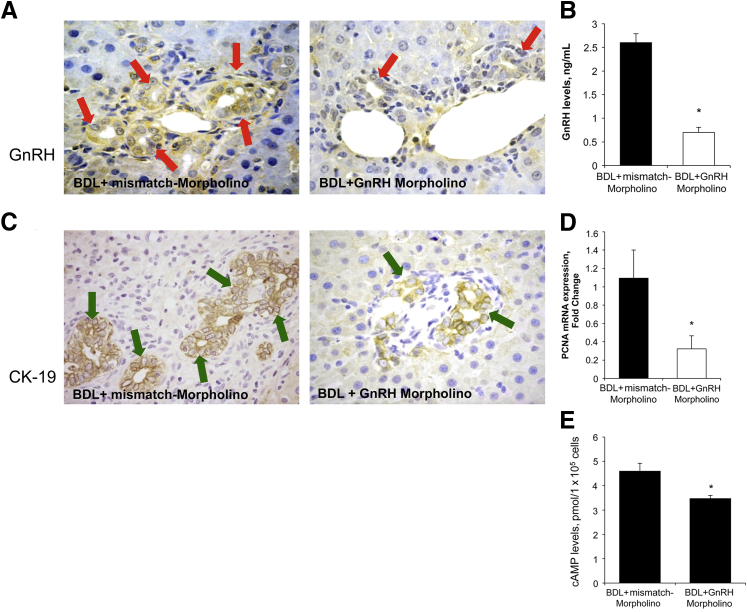
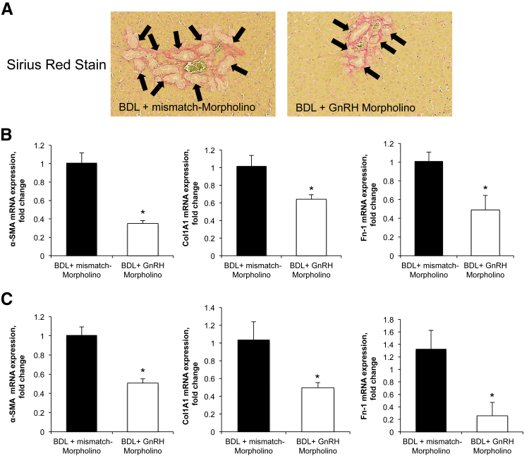
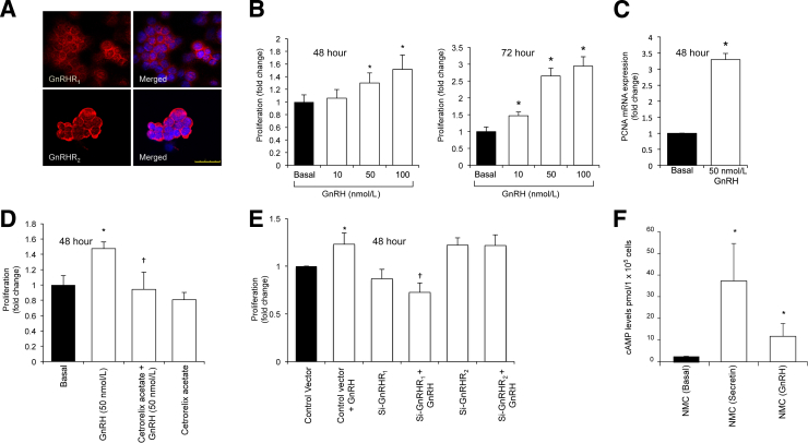
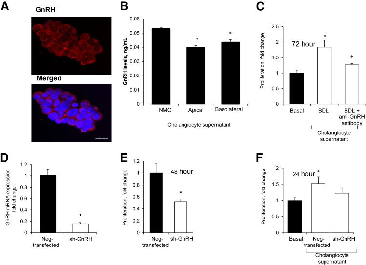
During cholestatic liver disease, there is dysregulation in the balance between biliary growth and loss in bile duct-ligated (BDL) rats modulated by neuroendocrine peptides via autocrine/paracrine pathways. Gonadotropin-releasing hormone (GnRH) is a trophic peptide hormone that modulates reproductive function and proliferation in many cell types. We evaluated the autocrine role of GnRH in the regulation of cholangiocyte proliferation. The expression of GnRH receptors was assessed in a normal mouse cholangiocyte cell line (NMC), sham, and BDL rats. The effect of GnRH administration was evaluated in normal rats and in NMC. GnRH-induced biliary proliferation was evaluated by changes in intrahepatic bile duct mass and the expression of proliferation and function markers. The expression and secretion of GnRH in NMC and isolated cholangiocytes was assessed. GnRH receptor subtypes GnRHR1 and GnRHR2 were expressed in cholangiocytes. Treatment with GnRH increased intrahepatic bile duct mass as well as proliferation and function markers in cholangiocytes. Transient knockdown and pharmacologic inhibition of GnRHR1 in NMC decreased proliferation. BDL cholangiocytes had increased expression of GnRH compared with normal rats, accompanied by increased GnRH secretion. In vivo and in vitro knockdown of GnRH decreased intrahepatic bile duct mass/cholangiocyte proliferation and fibrosis. GnRH secreted by cholangiocytes promotes biliary proliferation via an autocrine pathway. Disruption of GnRH/GnRHR signaling may be important for the management of cholestatic liver diseases.
Copyright © 2015 American Society for Investigative Pathology. Published by Elsevier Inc. All rights reserved.
Figures
Similar articles
-
Melatonin inhibits hypothalamic gonadotropin-releasing hormone release and reduces biliary hyperplasia and fibrosis in cholestatic rats.Am J Physiol Gastrointest Liver Physiol. 2017 Nov 1;313(5):G410-G418. doi: 10.1152/ajpgi.00421.2016. Epub 2017 Jul 27. Am J Physiol Gastrointest Liver Physiol. 2017. PMID: 28751425 Free PMC article.
-
Neuropeptide Y inhibits biliary hyperplasia of cholestatic rats by paracrine and autocrine mechanisms.Am J Physiol Gastrointest Liver Physiol. 2013 Aug 1;305(3):G250-7. doi: 10.1152/ajpgi.00140.2013. Epub 2013 May 23. Am J Physiol Gastrointest Liver Physiol. 2013. PMID: 23703654 Free PMC article.
-
Knockdown of Hepatic Gonadotropin-Releasing Hormone by Vivo-Morpholino Decreases Liver Fibrosis in Multidrug Resistance Gene 2 Knockout Mice by Down-Regulation of miR-200b.Am J Pathol. 2017 Jul;187(7):1551-1565. doi: 10.1016/j.ajpath.2017.03.013. Epub 2017 May 12. Am J Pathol. 2017. PMID: 28502477 Free PMC article.
-
RFamide-related peptide 3 and gonadotropin-releasing hormone-II are autocrine-paracrine regulators of testicular function in the boar.Mol Reprod Dev. 2017 Sep;84(9):994-1003. doi: 10.1002/mrd.22830. Epub 2017 Jun 21. Mol Reprod Dev. 2017. PMID: 28475264 Review.
-
Gonadotropin-releasing hormone/gonadotropin-releasing hormone receptor signaling in the placenta.Curr Opin Endocrinol Diabetes Obes. 2011 Dec;18(6):401-8. doi: 10.1097/MED.0b013e32834cd3b0. Curr Opin Endocrinol Diabetes Obes. 2011. PMID: 22024993 Review.
Cited by
-
Functional and structural features of cholangiocytes in health and disease.Cell Mol Gastroenterol Hepatol. 2015 Jul 1;1(4):368-380. doi: 10.1016/j.jcmgh.2015.05.005. Cell Mol Gastroenterol Hepatol. 2015. PMID: 26273695 Free PMC article.
-
Recent advances in understanding bile duct remodeling and fibrosis.F1000Res. 2018 Jul 31;7:F1000 Faculty Rev-1165. doi: 10.12688/f1000research.14578.1. eCollection 2018. F1000Res. 2018. PMID: 30109019 Free PMC article. Review.
-
Melatonin inhibits hypothalamic gonadotropin-releasing hormone release and reduces biliary hyperplasia and fibrosis in cholestatic rats.Am J Physiol Gastrointest Liver Physiol. 2017 Nov 1;313(5):G410-G418. doi: 10.1152/ajpgi.00421.2016. Epub 2017 Jul 27. Am J Physiol Gastrointest Liver Physiol. 2017. PMID: 28751425 Free PMC article.
-
Regulators of Cholangiocyte Proliferation.Gene Expr. 2017 Feb 10;17(2):155-171. doi: 10.3727/105221616X692568. Epub 2016 Jul 12. Gene Expr. 2017. PMID: 27412505 Free PMC article. Review.
-
Exploration of new therapeutic targets for viral hepatic fibrosis, alcoholic hepatic fibrosis, and non-alcoholic hepatic fibrosis.Ann Transl Med. 2022 Aug;10(16):886. doi: 10.21037/atm-22-3593. Ann Transl Med. 2022. PMID: 36111046 Free PMC article.
References
-
- Lazaridis K.N., Strazzabosco M., LaRusso N.F. The cholangiopathies: disorders of biliary epithelia. Gastroenterology. 2004;127:1565–1577. - PubMed
-
- Glaser S., Meng F., Han Y., Onori P., Chow B.K., Francis H., Venter J., McDaniel K., Marzioni M., Invernizzi P., Ueno Y., Lai J.M., Huang L., Standeford H., Alvaro D., Gaudio E., Franchitto A., Alpini G. Secretin stimulates biliary cell proliferation by regulating expression of MicroRNA 125b and MicroRNA let7a in mice. Gastroenterology. 2014;146:1795–1808. - PMC - PubMed
-
- Kanno N., LeSage G., Glaser S., Alpini G. Regulation of cholangiocyte bicarbonate secretion. Am J Physiol Gastrointest Liver Physiol. 2001;281:G612–G625. - PubMed
-
- Alpini G., Ulrich C.D., 2nd, Phillips J.O., Pham L.D., Miller L.J., LaRusso N.F. Upregulation of secretin receptor gene expression in rat cholangiocytes after bile duct ligation. Am J Physiol Gastrointest Liver Physiol. 1994;266:G922–G928. - PubMed
Publication types
MeSH terms
Substances
Grants and funding
LinkOut - more resources
Full Text Sources
Other Literature Sources
