Review
. 2015 May-Jun;24(3):133-40.
doi: 10.1016/j.carpath.2015.02.004. Epub 2015 Feb 19.

The winding road to regenerating the human heart

Affiliations
Review

The winding road to regenerating the human heart

Kaytlyn A Gerbin et al. Cardiovasc Pathol. 2015 May-Jun.

Abstract

Regenerating the human heart is a challenge that has engaged researchers and clinicians around the globe for nearly a century. From the repair of the first septal defect in 1953, followed by the first successful heart transplant in 1967, and later to the first infusion of bone marrow-derived cells to the human myocardium in 2002, significant progress has been made in heart repair. However, chronic heart failure remains a leading pathological burden worldwide. Why has regenerating the human heart been such a challenge, and how close are we to achieving clinically relevant regeneration? Exciting progress has been made to establish cell transplantation techniques in recent years, and new preclinical studies in large animal models have shed light on the promises and challenges that lie ahead. In this review, we will discuss the history of cell therapy approaches and provide an overview of clinical trials using cell transplantation for heart regeneration. Focusing on the delivery of human stem cell-derived cardiomyocytes, current experimental strategies in the field will be discussed as well as their clinical translation potential. Although the human heart has not been regenerated yet, decades of experimental progress have guided us onto a promising path.

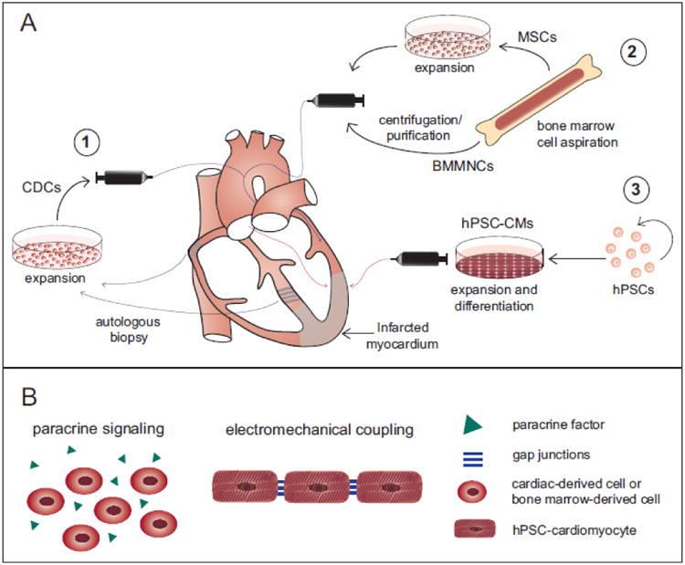
Summary: Previous work in clinical cell therapy for heart repair using bone marrow mononuclear cells, mesenchymal stem cells, and cardiac-derived cells have overall demonstrated safety and modest efficacy. Recent advancements using human stem cell-derived cardiomyocytes have established them as a next generation cell type for moving forward, however certain challenges must be overcome for this technique to be successful in the clinics.

Keywords: Cell transplantation; Heart regeneration; Myocardial infarction; Stem cell-derived cardiomyocytes.


Figures

Figure 1
Figure 1
Cell transplantation techniques and proposed mechanisms of cell therapy for heart regeneration. (A) Cell transplantation after myocardial infarction. (1) Cardiac-derived cells (CDCs) are isolated from either the atrial appendage or the septal wall, expanded in vitro, and transplanted via intracoronary catheter delivery. (2) Bone marrow mononuclear cells (BMMNCs) and mesenchymal stem cells (MSCs) are harvested from the bone marrow. BMMNCs may undergo purification steps followed by transplantation via intracoronary catheter delivery, while MSCs require in vitro expansion prior to transplantation. (3) Human cardiomyocytes are derived from human pluripotent stem cells (hPSCs) after in vitro expansion and directed cardiac differentiation. The proposed clinical delivery method for hPSC-cardiomyocytes (hPSC-CMs) is via transepicardial or transendocardial catheter-based injection. (B) Proposed mechanism of action after cell transplantation. Bone marrow-derived cells and cardiac-derived cells work primarily though paracrine signaling, in which transplanted cells secrete paracrine factors to the surrounding infarcted myocardium. HPSC-cardiomyocytes act primarily though the direct electromechanical integration with neighboring host cardiomyocytes. Paracrine factors may also be secreted by the hPSC-cardiomyocytes.
Figure 2
Figure 2
Human pluripotent stem cell-derived cardiomyocytes remuscularize the infarcted macaque heart [63]. (A) HESC-cardiomyocytes robustly engraft in the infarcted myocardium, outlined by the dashed line, as indicated by confocal immunofluorescence at day 14 after transplantation. Engrafted cardiomyocytes express GFP (human, green) and both hESC-cardiomyocytes and host cardiomyocytes express the contractile protein alpha-actinin (human and monkey, red) with nuclear DAPI counterstain (blue). Scale bar = 2 mm. (B) The in vivo maturation of engrafted hESC-cardiomyocytes is evident from 14 days to 84 days post engraftment by co-staining for alpha-actinin (human and monkey, red) and GFP (human, green). At late timepoints, engrafted hESC-cardiomyocytes display a marked increase in cell size, sarcomere alignment, and myofibril content compared to early timepoints. Scale bar = 20 μm. (C) Host vasculature perfuses the hESC-cardiomyocyte graft at 84 days post engraftment, as visualized by three-dimensional rendered microcomputed tomography. Large host arteries and veins are shown in red and blue, respectively, and small vessels perfusing the graft are shown in white. Other vessels in the myocardium are shown in gray. (D) Host-derived blood vessels are found within the hESC-cardiomyocyte grafts at 84 days post engraftment. Host endothelial cells express CD31 (human and monkey, red) and infiltrate the GFP-positive graft area (human, green). Scale bar = 20 μm. (E) Ex vivo fluorescent GCaMP3 imaging indicates that engrafted human cardiomyocytes are electrically coupled to the infarcted macaque heart at 14 days post engraftment. GCaMP3 fluorescence intensity (green) and host ECG (red) are plotted versus time and demonstrate 1:1 coupling at spontaneos rate as well as during atrial pacing at 3 Hz.

References

    1. Go AS, et al. Heart disease and stroke statistics--2013 update: a report from the American Heart Association. Circulation. 2013;127(1):e6–e245. - PMC - PubMed
    1. Murray CJL, et al. Disability-adjusted life years (DALYs) for 291 diseases and injuries in 21 regions, 1990–2010: a systematic analysis for the Global Burden of Disease Study 2010. The Lancet. 2012;380(9859):2197–2223. - PubMed
    1. Laflamme MA, Murry CE. Heart regeneration. Nature. 2011;473(7347):326–35. - PMC - PubMed
    1. Buja LM, Vela D. Cardiomyocyte death and renewal in the normal and diseased heart. Cardiovasc Pathol. 2008;17(6):349–74. - PubMed
    1. Senyo SE, et al. Mammalian heart renewal by pre-existing cardiomyocytes. Nature. 2013;493(7432):433–6. - PMC - PubMed

Publication types