. 2015 Mar 20;4(4):450-62.
doi: 10.1242/bio.201410579.

In vivo mapping of the functional regions of the DEAD-box helicase Vasa

Affiliations

In vivo mapping of the functional regions of the DEAD-box helicase Vasa

Mehrnoush Dehghani et al. Biol Open. .

Abstract

The maternally expressed Drosophila melanogaster DEAD-box helicase Vasa (Vas) is necessary for many cellular and developmental processes, including specification of primordial germ cells (pole cells), posterior patterning of the embryo, piRNA-mediated repression of transposon-encoded mRNAs, translational activation of gurken (grk) mRNA, and completion of oogenesis itself. Vas protein accumulates in the perinuclear nuage in nurse cells soon after their specification, and then at stage 10 Vas translocates to the posterior pole plasm of the oocyte. We produced a series of transgenic constructs encoding eGFP-Vas proteins carrying mutations affecting different regions of the protein, and analyzed in vivo which Vas functions each could support. We identified novel domains in the N- and C-terminal regions of the protein that are essential for localization, transposon repression, posterior patterning, and pole cell specification. One such functional region, the most C-terminal seven amino acids, is specific to Vas orthologues and is thus critical to distinguishing Vas from other closely related DEAD-box helicases. Surprisingly, we also found that many eGFP-Vas proteins carrying mutations that would be expected to abrogate DEAD-box helicase function localized to the nuage and posterior pole, and retained the capacity to support oogenesis, although they did not function in embryonic patterning, pole cell specification, grk activation, or transposon repression. We conclude from these experiments that Vas, a multifunctional protein, uses different domains and different molecular associations to carry out its various cellular and developmental roles.

Keywords: Drosophila; Embryonic patterning; Germ cells; Pole plasm; RNA helicase; piRNA biogenesis.


Conflict of interest statement

Competing interests: The authors declare no competing or financial interests.

Figures

Fig. 1.
Fig. 1.. Summary of the deletions and point mutations examined in this study.
(A) An alignment of the N-terminal ends of predicted Vas proteins from several Drosophila species. The N-terminal vas open reading frame is often incorrectly annotated in species that have not undergone extensive cDNA sequencing because of confounding factors such as poor sequence conservation, nested genes and the solo alternative splice form (Yan et al., 2010). Therefore, to produce this alignment, vas open reading frames were manually annotated from three-frame translations of genomic DNA, using the short highly-conserved amino-terminal end to identify the putative translational start site. Asterisks, colons and periods indicate full conservation, strong similarity and weak similarity, respectively. RGG motifs are shown in red. (B) Schematic representation of the N-terminal deletions used in this study. The hash box marks amino acids 141-153, which are encoded by a copy of a 39-nucleotide tandem repeat that is absent from some vas cDNA clones (Lasko and Ashburner, 1988). This segment is absent in eGFP-Vas+ and all the N-terminally deleted proteins as the constructs were built from such a cDNA clone. VasΔ17-110, 3xAGG contains a deletion of amino acids 17-110 and three mutations that convert RGG motifs to AGG. (C) The amino acid substitution mutations in conserved DEAD-box helicase motifs that were produced for this study. Motifs are identified as previously defined (Rocak and Linder, 2004). (D) Sequence alignment of the C-terminal region of Vas from D. melanogaster with orthologues from other species. The red box depicts amino acids 636-646, which are conserved among Drosophila species but not beyond. The purple letters show the conserved highly acidic residues found at the C-terminal ends of Vas orthologues from Drosophila and non-Drosophila species. A tryptophan residue (presented in blue) is also highly conserved.
Fig. 2.
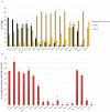
Fig. 2.. Fecundity of vasPH165 females carrying different egfp-vas constructs, and expression levels from those constructs.
(A) Fecundity. The y-axis indicates the number of eggs laid by individual females in the first three days after eclosion, and the x-axis identifies each egfp-vas construct that was tested. Data from vasPH165 and vasPH165/Df(2L)A267 controls are presented at the far right. Asterisks indicate a significant increase compared to vasPH165 (p<0.05). Error bars indicate the standard error of the mean (SEM), n (number of females tested) >50 for all genotypes. (B) Western blots (WB) comparing the expression level of eGFP-Vas in the ovaries from vas1/+ flies carrying the different constructs, using an anti-Vas antibody. α-Tubulin (α-Tub) serves as a loading control. egfp-vas+ was included in each blot for comparison. The eGFP-Vas bands migrate at different positions depending on the deletions that they carry. No Vasa protein was detected in the ovary lysate from vasPH165. The Vasa antibody raised against the full-length protein reacts with some mutant forms of Vasa such as vasΔ3-139 and vasΔ3-200 less strongly than with vas+ (see also supplementary material Fig. S1).
Fig. 3.
Fig. 3.. Dorsal-ventral patterning in eggs produced by vasPH165 females carrying different egfp-vas constructs.
(A) Dorsal appendage formation. The green bars indicate the percentage of the embryos with two separate or partly fused dorsal appendages. The beige and yellow bars represent the portion of the embryos with fused or no dorsal appendages, respectively. Data from vasPH165 controls are presented at the far right. Error bars indicate SEM, n (number of females tested) >50 for all genotypes. Asterisks indicate a significant difference compared to vas+ (p<0.05). (B) Grk expression. Red bars indicate the percentage of stage 8 egg chambers positively stained for localized Grk. Error bars represent SEM from three different replicates. n (number of stage 8 egg chambers in each replicate) >50 for all genotypes.
Fig. 4.
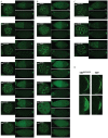
Fig. 4.. Localization of eGFP-Vas proteins in vas1 ovaries.
(A) N-terminal deletions. (B) Mutations in conserved DEAD-box helicase motifs. (C) C-terminal mutations. For each genotype the left, the top right and the bottom right images show a stage 5 egg chamber (confocal image), an early stage 10 egg chamber, and a stage 14 oocyte. Scale bars (50 µm) are included on the top set of images; all corresponding images from other genotypes are at the same magnification. (D) Higher magnification confocal images comparing distribution of eGFP-Vas at the posterior pole of vas1; egfp-vas+ and vas1;egfp-vasΔ636-646 stage 14 oocytes. eGFP-VasΔ636-646 distribution is more diffuse. For each genotype the top image shows the middle focal plane whereas the bottom image illustrates the maximum intensity projection of the z stack. Scale bars = 50 µm.
Fig. 5.
Fig. 5.. HeT-A expression in ovaries of vasPH165 females carrying different egfp-vas constructs.
Red bars indicate the expression level of HeT-A normalized to that of wild-type ovaries. Data from vasPH165 and vasPH165/Df(2L)A267 controls are presented at the far right. Asterisks indicate a significant increase compared to vas+ (p<0.05). Each bar represents the average of at least three biological replicates, error bars indicate SEM.
Fig. 6.
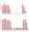
Fig. 6.. Germ cell formation in embryos from vas1 females expressing different eGFP-Vas proteins.
Red bars indicate the percentage of the embryos with germ cells. Data from the vas1 control is presented at the far right. Asterisks show a significant difference from vas+ (p<0.05). Error bars represent SEM from at least three biological replicates each with more than 50 embryos.
Fig. 7.
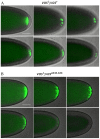
Fig. 7.. Time course of pole cell development in eGFP-Vas+ and eGFP-VasΔ636-646 expressing embryos.
(A) A series of still shots from live imaging of pole cell formation in eGFP-Vas+ expressing embryos. eGFP-Vas is tightly localized at the posterior pole and then concentrates in foci within pole buds. Posterior nuclear divisions become asynchronous with somatic nuclear divisions, and eGFP-Vas positive pole cells completely form. (B) A series of still shots from live imaging of pole cell formation in eGFP-VasΔ636-646 expressing embryos. eGFP-Vas is less tightly localized at the posterior pole and forms more foci than in wild type. Mitosis remains synchronous throughout the embryo. eGFP-Vas is then lost from the posterior region and pole cells fail to form. Scale bar = 50 µm.
Fig. 8.
Fig. 8.. The ability of various egfp-vas constructs to restore abdominal segmentation in vas1 embryos.
(A) Hatching rates: the red bars indicate the percentage of embryos hatched after 48 hours, error bars indicate SEM from at least five plates. Between 500-1000 embryos in total were scored for each genotype. (B) ftz expression in vas1 embryos containing various egfp-vas constructs: The y-axis indicates the average number of ftz stripes for each genotype. Error bars show SEM calculated from more than 50 embryos examined for each genotype. In both A and B asterisks show a significant increase or decrease from vas+ (p<0.05).

Similar articles

Cited by

References

    1. Burd C. G., Dreyfuss G. (1994). Conserved structures and diversity of functions of RNA-binding proteins. Science 265, 615–621 10.1126/science.8036511 - DOI - PubMed
    1. Carrera P., Johnstone O., Nakamura A., Casanova J., Jäckle H., Lasko P. (2000). VASA mediates translation through interaction with a Drosophila yIF2 homolog. Mol. Cell 5, 181–187 10.1016/S1097-2765(00)80414-1 - DOI - PubMed
    1. Caruthers J. M., Johnson E. R., McKay D. B. (2000). Crystal structure of yeast initiation factor 4A, a DEAD-box RNA helicase. Proc. Natl. Acad. Sci. USA 97, 13080–13085 10.1073/pnas.97.24.13080 - DOI - PMC - PubMed
    1. Castrillon D. H., Quade B. J., Wang T. Y., Quigley C., Crum C. P. (2000). The human VASA gene is specifically expressed in the germ cell lineage. Proc. Natl. Acad. Sci. USA 97, 9585–9590 10.1073/pnas.160274797 - DOI - PMC - PubMed
    1. Cordin O., Banroques J., Tanner N. K., Linder P. (2006). The DEAD-box protein family of RNA helicases. Gene 367, 17–37 10.1016/j.gene.2005.10.019 - DOI - PubMed

LinkOut - more resources