Case Reports
. 2015 Aug;131(1):63-70.
doi: 10.1007/s10633-015-9496-8. Epub 2015 Mar 22.

Rapid resolution of retinoschisis with acetazolamide

Affiliations
Case Reports

Rapid resolution of retinoschisis with acetazolamide

Lijuan Zhang et al. Doc Ophthalmol. 2015 Aug.

Abstract

Purpose: To report the results of an acetazolamide (Diamox(®)) treatment regimen in a genetically confirmed case of X-linked juvenile retinoschisis (XLRS).

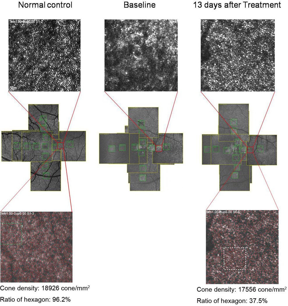
Methods: A patient with XLRS was prescribed acetazolamide (Diamox(®)) at a dose of 500 mg/day, then discontinued the treatment due to non-compliance for 4 days, and finally resumed the course of treatment. Best-corrected visual acuity, retinal structure, and function were monitored with autofluorescence, spectral domain-optical coherence tomography (SD-OCT), adaptive optics scanning laser ophthalmoscopy (AOSLO), and full-field electroretinogram (ERG). Full-field ERG was performed using DTL recording electrodes and Ganzfeld stimulation according to ISCEV standards.

Results: Serial monitoring of the cysts by SD-OCT revealed a strong association between the effects of acetazolamide administration and the size of the schisis. A reduction in foveal cyst size was significant in as rapid as 6 days after acetazolamide initiation. AOSLO data revealed that the resolution of cone cell images improves as the foveal schisis decreases in size.

Conclusions: Efficacy of acetazolamide in patients with XLRS can be apparent in as rapid as a week of therapy. AOSLO can be a good method to evaluate the cone cells after acetazolamide treatment in the early stages of XLRS.


Conflict of interest statement

Disclosures: The authors report no conflicts of interest in this work

Figures

Fig. 1
Fig. 1
An abridged timeline of events in the treatment course of a patient with X-linked juvenile retinoschisis with acetazolamide.
Fig. 2
Fig. 2
Images of the XLRS patient’s eyes at presentation: (A, B) Color fundus images of the right and left eyes, respectively, (C, D) 488nm reflectance images, (E, F) 488nm autofluorescence images. (G, H) spectral domain-optical coherence tomography scans across the fovea exhibiting small parafoveal spots seen on 488nm reflectance images, correlating with macular cysts that are less noticeable on color fundus and 488nm autofluorescence imaging.
Fig. 3
Fig. 3
Full-field electroretinogram results as compared to an age-matched control trace (bottom row). The patient exhibited electronegative maximal responses in both eyes. Scotopic rod specific ERG b-wave amplitudes were 64.5 microvolts in the right; 62.8 microvolts in the left, delayed implicit time of 120 ms was noted in both eyes. Maximal ERG a- and b-wave amplitudes were 233 microvolts and 177 microvolts in the right; 231 microvolts and 193 microvolts in the left. Photopic 30 Hz flicker ERG had implicit time and amplitudes of 28 milliseconds and 40 microvolts in the right; 31 milliseconds and 44 microvolts in the left. Transient photopic ERG b-wave amplitudes were 59 microvolts in the right; 56 microvolts in the left. Transient photopic ERG b-wave implicit times were 32 milliseconds in the right; 34 milliseconds in the left.
Fig. 4
Fig. 4
Apparent schisis resolution over time. Serial SD-OCT scans show various time points during the course of acetazolamide treatment. Between visits, the previous regions of interest were tracked and follow up SD-OCT scans were aligned at the center of the largest cyst. The initial OCT scan was taken before treatment began (Figure 4A). The follow-up examination occurred after 10 days of medication, in which the OCT revealed a decrease in the size of the foveal schisis of both eyes (Figure 4B). The next examination occured 10 days later. Regression of the schisis to baseline was noted after the patient reported discontinued use of actezolamide for four days (Figure 4C). A notable decrease in schisis was observed 6 days following the resuming of treatment (Figure 4D).
Fig. 5
Fig. 5
Parafoveal cones are visualized after acetazolamide treatment. Adaptive optics scanning laser ophthalmoloscopy (AOSLO) images of the left eye at 1mm from the central fovea in the temporal direction were shown above. Compared with the normal control, the resolution of cones before treatment is low because the schisis hinders the resolution. Our second AOSLO images were taken after 13 days of treatment when the edema was significantly smaller than baseline. The resolution of cone cells was noticeably improved. The boundaries of the cone cells are much more defined in later courses of the treatment. Compared to the AOSLO images of normal, age-matched males, the cone density and ratio of hexagons are decreased.
Fig. 6
Fig. 6
The corresponding location between AOSLO and OCT. The vertical line marking cones 1mm from the central fovea in AOSLO images corresponds to the region of schisis reduction on OCT images. Comparing the target area of the AOSLO images before and after treatment, the OCT shows that the retinoschisis has almost subsided. Note: Post-treatment autofluorescence and AOSLO images were taken on days 11 and 13, respectively.

References

    1. George ND, Yates JR, Moore AT. X linked retinoschisis. Br J Ophthalmol. 1995;79(7):697–702. - PMC - PubMed
    1. Tantri A, Vrabec TR, Cu-Unjieng A, Frost A, Annesley WH, Jr, Donoso LA. X-linked retinoschisis: a clinical and molecular genetic review. Surv Ophthalmol. 2004;49(2):214–230. - PubMed
    1. Gregori NZ, Berrocal AM, Gregori G, Murray TG, Knighton RW, Flynn HW, Jr, Dubovy S, Puliafito CA, Rosenfeld PJ. Macular spectral-domain optical coherence tomography in patients with X linked retinoschisis. Br J Ophthalmol. 2009;93(3):373–378. - PubMed
    1. Tsang SH, Vaclavik V, Bird AC, Robson AG, Holder GE. Novel phenotypic and genotypic findings in X-linked retinoschisis. Arch Ophthalmol. 2007;125(2):259–267. - PMC - PubMed
    1. Wu WW, Wong JP, Kast J, Molday RS. RS1, a discoidin domain-containing retinal cell adhesion protein associated with X-linked retinoschisis, exists as a novel disulfide-linked octamer. J Biol Chem. 2005;280(11):10721–10730. - PubMed

Publication types

Substances

LinkOut - more resources