Review
. 2015 Jul;20(4):519-32.
doi: 10.1007/s10741-015-9481-z.

Left ventricular assist devices: a kidney's perspective

Affiliations
Review

Left ventricular assist devices: a kidney's perspective

T R Tromp et al. Heart Fail Rev. 2015 Jul.

Abstract

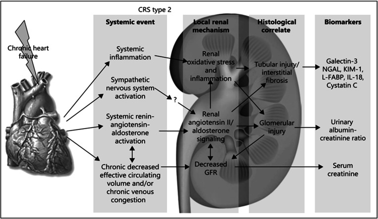
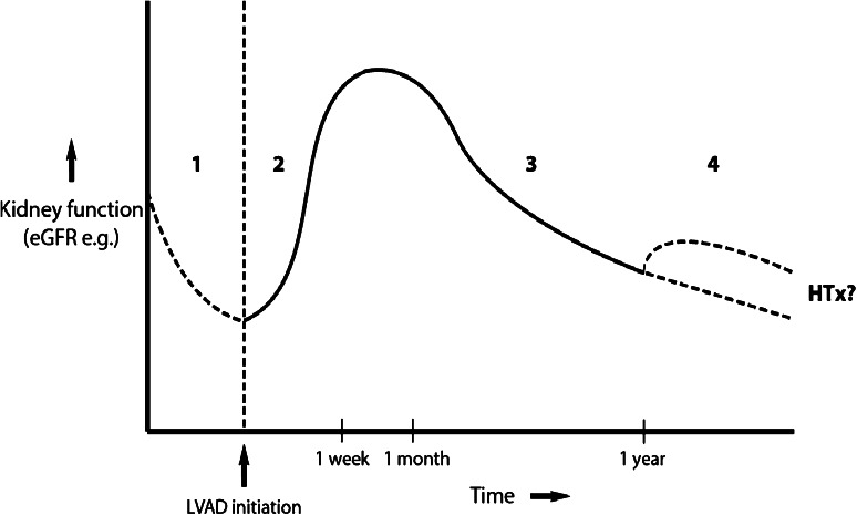
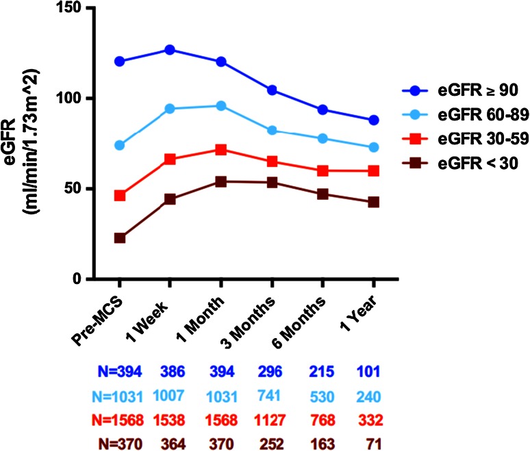
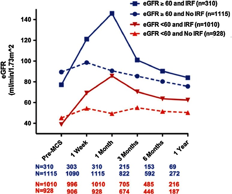
The left ventricular assist device (LVAD) has become an established treatment option for patients with refractory heart failure. Many of these patients experience chronic kidney disease (CKD) due to chronic cardiorenal syndrome type II, which is often alleviated quickly following LVAD implantation. Nevertheless, reversibility of CKD remains difficult to predict. Interestingly, initial recovery of GFR appears to be transient, being followed by gradual but significant late decline. Nevertheless, GFR often remains elevated compared to preimplant status. Larger GFR increases are followed by a proportionally larger late decline. Several explanations for this gradual decline in renal function after LVAD therapy have been proposed, yet a definitive answer remains elusive. Mortality predictors of LVAD implantation are the occurrence of either postimplantation acute kidney injury (AKI) or preimplant CKD. However, patient outcomes continue to improve as LVAD therapy becomes more widespread, and adverse events including AKI appear to decline. In light of a growing destination therapy population, it is important to understand the cumulative effects of long-term LVAD support on kidney function. Additional research and passage of time are required to further unravel the intricate relationships between the LVAD and the kidney.


Figures

Fig. 1
Fig. 1
Pathophysiology of CRS type II (reprinted with permission [25] ). NGAL, neutrophil gelatinase-associated lipocalin; KIM 1, kidney injury molecule-1; L-FABP, liver-type fatty acid binding protein; IL-18, interleukin-18
Fig. 2
Fig. 2
a (Left) Kaplan–Meier analysis of LVAD recipients grouped in changes in eGFR. Change in eGFR was taken from baseline to 1 month following surgery and represented as  % change (reprinted with permission [19] ). b (Right) schematic representation of effects of changes in early eGFR after LVAD implantation on relative mortality risk. The increased mortality risk on the left side of the U-curve is related to AKI. Surprisingly, large increases in eGFR are also associated with increased mortality risk [19]. The nadir of the U-curve lies toward a modest increase
Fig. 3
Fig. 3
Schematic representation of evolution in renal function over time. Phase 1 renal function declines with varying degrees as a result of CRS type II. Phase 2 renal function initially recovers thanks to LVAD implantation and negation of renal hypoperfusion. This effect is most notable from several weeks to up to 2 months following implantation. Phase 3 the functional improvement was only transient, and renal function continues to decline. Patients with the largest improvement consequently experience the largest deterioration, although, on average, the end-point renal function stays elevated over preimplant values, at least up to 1 year following transplantation. Phase 4 hypothetically, in the long term, renal function continues to decline and may necessitate RRT (lower dotted line). Alternatively, the patient receives a heart transplantation, which can either temporarily alleviate the downward trend (upper dotted line) or leave it unaltered (lower dotted line)
Fig. 4
Fig. 4
Change in eGFR over time, stratified by preimplant cohort, as reported by Brisco et al. [19] (reprinted with permission). Patients with low preimplant eGFR (red lines) appear to derive most benefit after MCS, with eGFR remaining notably elevated above preimplant levels up to 1-year after placement. By contrast, patients with moderate to good preimplant eGFR (blue lines) may undergo a net decrease in eGFR. Note that the fraction of patients with eGFR ≥ 90 mL/min is relatively small, and that the majority of patients have an eGFR < 60, as expected due to high prevalence of CRS type II in this population
Fig. 5
Fig. 5
Changes in eGFR, first stratified by preimplant eGFR (blue and red lines), and subsequently divided between patients who experienced improved renal function (IRF, solid lines) and those who did not (no IRF, dotted lines). IRF is defined by an increase ≥ 50 % at month 1 over baseline renal function. Although the renal function quickly declined again after 1 month in the IRF group, the eGFR remained higher compared to the non-IRF group at 1 year post-implantation [19] (reprinted with permission). Note that the dark blue solid line surpasses an eGFR of 120 mL/min at month 1 (indicated by horizontal red line), a value that is considered above the normal range of GFR maintained by autoregulation. This may hint at ongoing hyperfiltration, which can lead to renal damage

Comment in

References

    1. Seferovic PM, Stoerk S, Filippatos G, et al. Organization of heart failure management in European Society of Cardiology member countries: survey of the Heart Failure Association of the European Society of Cardiology in collaboration with the Heart Failure National Societies/Working Groups. Eur J Heart Fail. 2013;15(9):947–959. - PubMed
    1. Roger VL, Go AS, Lloyd-Jones DM, et al. Heart disease and stroke statistics-2012 update: a report from the American Heart Association. Circulation. 2012;125(1):e2–e220. - PMC - PubMed
    1. Roger VL. Epidemiology of heart failure. Circ Res. 2013;113(6):646–659. - PMC - PubMed
    1. Ammirati E, Oliva F, Cannata A, et al. Current indications for heart transplantation and left ventricular assist device: a practical point of view. Eur J Intern Med. 2014;25(5):422–429. - PubMed
    1. Jessup M, Brozena S. Heart failure. N Engl J Med. 2003;348(20):2007–2018. - PubMed

MeSH terms

LinkOut - more resources