. 2016 Jan;253(1):137-53.
doi: 10.1007/s00709-015-0798-1. Epub 2015 Mar 22.

Cell layer-specific distribution of transiently expressed barley ESCRT-III component HvVPS60 in developing barley endosperm

Affiliations

Cell layer-specific distribution of transiently expressed barley ESCRT-III component HvVPS60 in developing barley endosperm

Julia Hilscher et al. Protoplasma. 2016 Jan.

Abstract

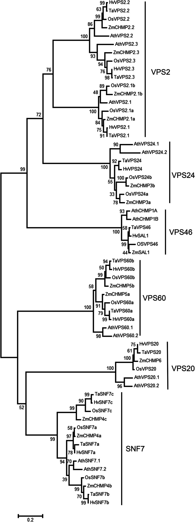
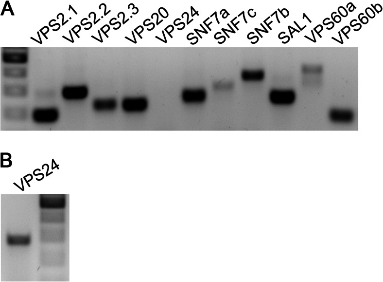
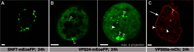
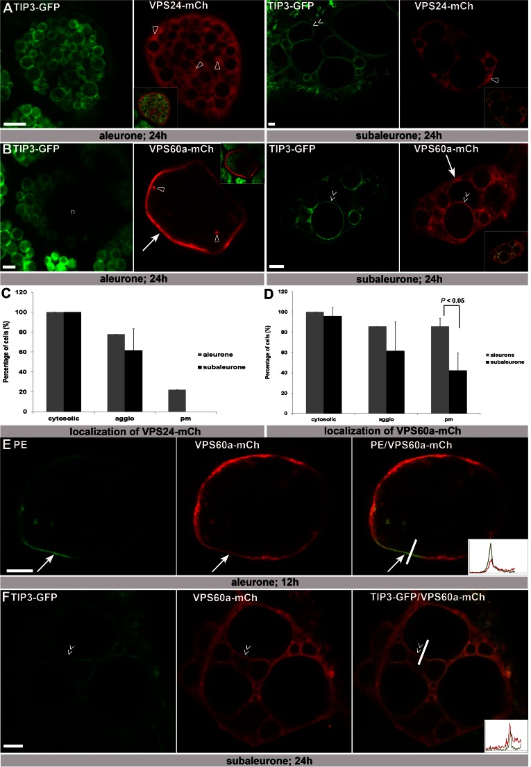
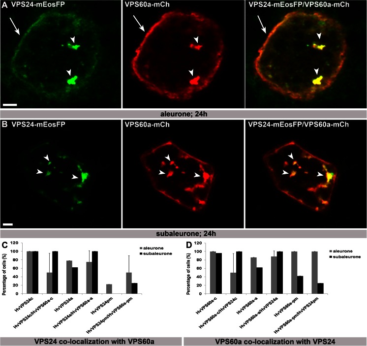
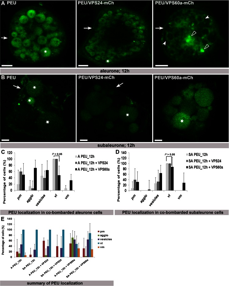
The significance of the endosomal sorting complexes required for transport (ESCRT)-III in cereal endosperm has been shown by the identification of the recessive mutant supernumerary aleurone layer1 (SAL1) in maize. ESCRT-III is indispensable in the final membrane fission step during biogenesis of multivesicular bodies (MVBs), responsible for protein sorting to vacuoles and to the cell surface. Here, we annotated barley ESCRT-III members in the (model) crop Hordeum vulgare and show that all identified members are expressed in developing barley endosperm. We used fluorescently tagged core ESCRT-III members HvSNF7a/CHMP4 and HvVPS24/CHMP3 and the associated ESCRT-III component HvVPS60a/CHMP5 for transient localization studies in barley endosperm. In vivo confocal microscopic analyses show that the localization of recombinantly expressed HvSNF7a, HvVPS24 and HvVPS60a differs within barley endosperm. Whereas HvSNF7a induces large agglomerations, HvVPS24 shows mainly cytosolic localization in aleurone and subaleurone. In contrast, HvVPS60a localizes strongly at the plasma membrane in aleurone. In subaleurone, HvVPS60a was found to a lesser extent at the plasma membrane and at vacuolar membranes. These results indicate that the steady-state association of ESCRT-III may be influenced by cell layer-specific protein deposition or trafficking and remodelling of the endomembrane system in endosperm. We show that sorting of an artificially mono-ubiquitinated Arabidopsis plasma membrane protein is inhibited by HvVPS60a in aleurone. The involvement of HvVPS60a in different cell layer-specific trafficking pathways, reflected by localization of HvVPS60a at the plasma membrane in aleurone and at the PSV membrane in subaleurone, is discussed.

Keywords: Barley; Cell layer-specific; ESCRT-III; Endosperm; Seed; VPS60.


Figures

Fig. 1
Fig. 1
Neighbour joining (NJ) tree of 61 protein sequences of ESCRT-III members of A. thaliana and barley, wheat, maize and rice presented in Table 1 and based on the alignment provided in Supplemental Figure 1. The NJ tree was built using standard configurations in MEGA6 (Tamura et al. 2013). Distances using the Poisson correction method are given in the units of the number of amino acid substitutions per site. All positions containing gaps were eliminated. Bootstrap values (1000 replicates) are shown at the branches
Fig. 2
Fig. 2
ESCRT-III members are expressed in barley endosperm. Gene-specific primers were used to detect expression of ESCRT-III members in barley endosperm cDNA (10–12 dap). a PCR on 1:10 diluted cDNA of barely endosperm. b PCR for VPS24 on undiluted cDNA of barley endosperm. DNA marker 500–200 bp
Fig. 3
Fig. 3
Localization of HvSNF7a, HvVPS24 and HvVPS60a in barley endosperm. a Actin::HvSNF7a-mEosFP induces large agglomerations (open arrowhead) within a cell in the embryo surrounding region. Note the weak signal in the nucleus (n). b Actin::HvVPS24-mEosFP localizes at vesicles (arrowhead) and to the cytoplasm concomitant with inducing agglomerations (open arrowheads). Note the maximal z-projection of 16 1-μm sections. c Actin::HvVPS60a-mCh localizes at the PM (arrow), vesicles (arrowhead), agglomerations (open arrowhead) and to a minor extent in the nucleus (n). Confocal single scans were made 24 h after bombardment. Scale = 5 μm
Fig. 4
Fig. 4
The localization of HvVPS60a-mCh differs in aleurone and subaleurone and shows plasma membrane localization in aleurone. a Single scans of barley aleurone and subaleurone cells showing transient transformation of HvVPS24-mCh in TIP3-GFP lines. In both, aleurone and subaleurone, HvVPS24-mCh localizes cytosolic and at small agglomerations (open arrowheads). Note the close ups to visualize co-localization. b Confocal single scans of HvVPS60a-mCh in TIP3-GFP lines and of co-transformation of HvVPS60a-mCh with TIP3-GFP. In aleurone, HvVPS60a-mCh shows strong signal at the pm (arrow), weak in the cytosol in parallel to small agglomeration structures (open arrowheads). Note the altered localization of HvVPS60a-mCh in subaleurone cells. HvVPS60a-mCh signal could be detected close to heterogenous, spherical PSVs (double arrow) but reduced at the PM (arrow). Note the close ups to visualize co-localization. c, d Localization of HvVPS24-mCh and HvVPS60a-mCh in aleurone and subaleurone. Relative number of cells showing localization in cytosol at agglomerations and at the PM was scored. Total number of cells: HvVPS24-mCh in aleurone (biological replicates 1; n = 18) and in subaleurone (biological replicates 3; n = 13); HvVPS60a-mCh in aleurone (biological replicates 1; n = 14) and in subaleurone (biological replicates 3; n = 24). The difference in the localization of HvVPS60a-mCh at the PM in aleurone and subaleurone is statistically significant (P < 0.05). Plus ends of the standard deviation are indicated. e Single scan of an aleurone cell showing co-localization of the plasma membrane marker PMA-EGFP (PE) and HvVPS60a-mCh at the pm (arrow). Fluorescence profile: X-axis, distance [μm] from 0 to 6 μm; Y-axis, intensity from 0 to 100. f Single confocal scan of a subaleurone cell that shows co-localization of HvVPS60a-mCh and the vacuolar membrane signal of the TIP3-GFP line. Note the double arrows indicating co-localization of HvVPS60a-mCh with the PSV membrane. Fluorescence profile: X-axis, distance [μm] from 0 to 8 μm; Y-axis, intensity from 0 to 100. Confocal single scans and z-series were made 24 h after bombardment. Scale = 5 μm
Fig 5
Fig 5
Co-bombardment of HvVPS24-mEosFP with HvVPS60a-mCh induces re-localization of both proteins to large agglomerations in aleurone and subaleurone. a, b HvVPS24m-Ch and HvVPS60a-mCh are re-localized from cytoplasm to large globulare structures (arrowheads) in aleurone and subaleurone. Note the additional localization of VPS24m-Ch and VPS60a-mCh at the PM in aleurone (arrows). c, d 8 and 4 co-transformed cells within 5 and 1 biological replicates, respectively, were analysed in aleurone and subaleurone. Relative number of cells showing localization in cytosol (-c), at agglomerations (-a) and at the plasma membrane (-pm) was scored and compared with single transformation. Plus ends of the standard deviation are indicated. Confocal single scans were made 24 h after bombardment. Scale = 5 μm.
Fig. 6
Fig. 6
Localization of PE and PEU in co-transformed aleurone and subaleurone cells. a PEU was sorted in the vacuole (asterisk) in aleurone cells. PEU is located at the PM (arrow) and sorted in vacuoles (asterisk) in co-bombarded HvVPS24-mCh cells. Note the affected sorting of PEU in co-transformed HvVPS60a-mCh cells, where PEU is localized at the pm (arrow), at agglomerations (open arrowheads) and at vacuolar membranes (arrowheads). b PEU is localized at the pm (arrowhead) and sorted in the vacuole in subaleurone cells (asterisks). In HvVPS24-mCh co-bombarded cells, PEU is localized weakly at the pm (arrowhead) and sorted in the vacuole. PEU is also sorted in the vacuole in co-transformed HvVPS60a-mCh cells (asterisk). Scale = 5 μm. c Statistical analyses of localizations of PEU in co-transformed HvVPS24-mCh and HvVPS60a-mCh aleurone cells. Biological replicates: 3 and 5 times for co-transformation with HvVPS24m-Ch and HvVPS60a-mCh, respectively; n (co-transformed cells with HvVPS24-mCh/HvVPS60a-mCh: 15/28). d Statistical analyses of localization of PEU in co-transformed HvVPS24-mCh and HvVPS60a-mCh aleurone cells. Biological replicates: 3 and 6 times for co-transformation with HvVPS24m-Ch and HvVPS60a-mCh, respectively; n (co-transformed cells with HvVPS24-mCh/HvVPS60a-mCh: 15/28). The difference in the localization of PEU in aleurone cells in the vacuolar lumen alone or in the presence of HvVPS60a-mCh is statistically significant (P < 0.05), whereas in subaleurone, the difference in the localization of PEU alone or in presence of HvVPS60a-mCh is not significant (P > 0.05). Plus ends of the standard deviation are indicated. e Summary of the statistical analyses of PEU in co-transformed HvVPS24m-Ch and HvVPS60a-mCh aleurone and subaleurone cells. Note the different localization of PEU in co-transformed HvVPS60a-mCh aleurone cells. Plus ends of the standard deviation are indicated. A aleurone, SA subaleurone, pm plasma membrane, agglo agglomeration, vl vacuolar lumen, vm vacuolar membrane.

Similar articles

Cited by

References

    1. Babst M, Katzmann DJ, Estepa-Sabal EJ, Meerloo T, Emr SD. Escrt-III: an endosome-associated heterooligomeric protein complex required for mvb sorting. Dev Cell. 2002;3:271–282. doi: 10.1016/S1534-5807(02)00220-4. - DOI - PubMed
    1. Babst M, Katzmann DJ, Snyder WB, Wendland B, Emr SD. Endosome-associated complex, ESCRT-II, recruits transport machinery for protein sorting at the multivesicular body. Dev Cell. 2002;3:283–289. doi: 10.1016/S1534-5807(02)00219-8. - DOI - PubMed
    1. Boothe J, et al. Seed-based expression systems for plant molecular farming. Plant Biotechnol J. 2010;8:588–606. doi: 10.1111/j.1467-7652.2010.00511.x. - DOI - PubMed
    1. Boura E, Ivanov V, Carlson LA, Mizuuchi K, Hurley JH. Endosomal sorting complex required for transport (ESCRT) complexes induce phase-separated microdomains in supported lipid bilayers. J Biol Chem. 2012;287:28144–28151. doi: 10.1074/jbc.M112.378646. - DOI - PMC - PubMed
    1. Boysen JH, Mitchell AP (2006) Control of Bro1-domain protein Rim20 localization by external pH, ESCRT machinery, and the Saccharomyces cerevisiae Rim101 pathway. Mol Biol Cell 17(3):1344–1353 - PMC - PubMed

Publication types

MeSH terms

Substances

LinkOut - more resources