Review
. 2015 Jul;39(4):522-40.
doi: 10.1093/femsre/fuv002. Epub 2015 Mar 21.

One cannot rule them all: Are bacterial toxins-antitoxins druggable?

Affiliations
Review

One cannot rule them all: Are bacterial toxins-antitoxins druggable?

Wai Ting Chan et al. FEMS Microbiol Rev. 2015 Jul.

Abstract

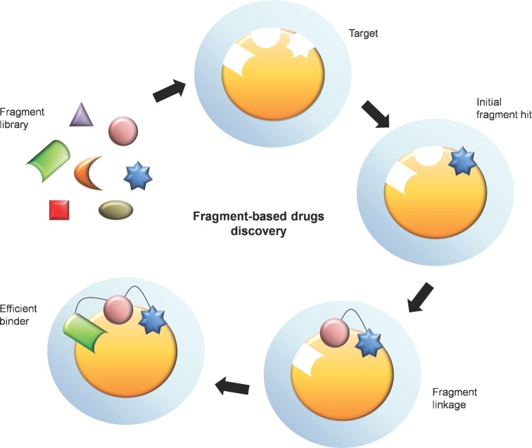
Type II (proteic) toxin-antitoxin (TA) operons are widely spread in bacteria and archaea. They are organized as operons in which, usually, the antitoxin gene precedes the cognate toxin gene. The antitoxin generally acts as a transcriptional self-repressor, whereas the toxin acts as a co-repressor, both proteins constituting a harmless complex. When bacteria encounter a stressful environment, TAs are triggered. The antitoxin protein is unstable and will be degraded by host proteases, releasing the free toxin to halt essential processes. The result is a cessation of cell growth or even death. Because of their ubiquity and the essential processes targeted, TAs have been proposed as good candidates for development of novel antimicrobials. We discuss here the possible druggability of TAs as antivirals and antibacterials, with focus on the potentials and the challenges that their use may find in the 'real' world. We present strategies to develop TAs as antibacterials in view of novel technologies, such as the use of very small molecules (fragments) as inhibitors of protein-protein interactions. Appropriate fragments could disrupt the T:A interfaces leading to the release of the targeted TA pair. Possible ways of delivery and formulation of Tas are also discussed.

Keywords: antibacterials; antivirals; drug delivery; drug discovery; inhibitors of protein–protein interactions; persistence; toxin–antitoxin operons.


Figures

Graphical Abstract Figure.
We consider various approaches to develop the toxins of the type II family as possible candidates to drug discovery; druggability of toxins-antitoxins could be possible as antivirals. As antibacterials, they might be considered as druggable but delivery and formulation may not be simple so far.
Figure 1.
Figure 1.
Features of type II TAs. Typical type II TAs consists of two genes organized as an operon. The antitoxin gene precedes the toxin one. Both genes usually overlap and are co-transcribed from one or two promoters. TA genes encode both antitoxin (oval) and toxin (crescent) proteins that bind to each other and generate a harmless complex under normal conditions. The antitoxin protein also binds to its own promoter to negatively autoregulate the TA operon. The toxin protein is not able to bind to the promoter by itself, but serves as a co-repressor upon binding of the antitoxin to the promoter, to further repress transcription of the operon. Under stressful circumstances, the antitoxin protein which is more labile, is degraded more rapidly by the host proteases and thus liberate the toxin protein to act on the cell target.
Figure 2.
Figure 2.
Conceptual models of potential approaches using TA as antivirals. (a) Activation of engineered viral promoter-MazF by early viral regulator protein. HIV-encoded Tat protein is an early viral regulator that binds to TAR sequence. A Tat-dependent MazF toxin (crescent) expression system of a retroviral vector was designed in which the mazF gene was inserted downstream the TAR sequence. MazF is an endoribonulease that cleaves free mRNA at the ACA codons. The mazF gene was engineered to avoid self-cleavage by changing the base sequences but conserving its amino acid sequence to preserve its toxicity to cleave the viral mRNAs. The vector was then transduced into human T lymphoid line CEM-SS cells. When HIV-1 attempts to enter the cell, interactions between cell surface molecules and viral envelope proteins allow the envelope to fuse with the cell membrane and subsequently viral RNA genome is released to the cell (1). The viral single-strand RNA genome is transcribed into double-strand DNA (2), and then integrated into a host chromosome (3). The proviral genome can consequently be transcribed into viral mRNA (4) for translation into HIV proteins. The early viral protein Tat will bind to the TAR sequence to induce MazF production (5) to cleave viral mRNAs (6). It is worth to mention that this system will only be triggered in the HIV-1-infected cells but not the innocent ones. (b) Cleavage of specific linker by viral protease to trigger MazF. NS3–4A is an HCV protein that has a very specific cleavage site. A recombinant vector was constructed that produced a complex in which the NS3 protease cleavage site linker was fused in between MazF (crescent) and truncated C-terminal of MazE (oval). Once the HCV enters the hepatocyte (1), HCV will take over parts of the intracellular machinery to replicate (2). NS3–4A will be produced and cleave specifically to the MazEF-linker (3) and thus liberating MazF to cleave viral mRNAs (4).
Figure 3.
Figure 3.
Detection of disruption of TA protein complex by i-PPI via BRET assays. The BRET assay technology is based on the efficient resonance energy transfer between a bioluminescent donor moiety and a fluorescent acceptor moiety. The bioluminescent Renilla luciferase (RLUC) that was fused with toxin protein catalyses the coelenterazine substrate to coelenteramide with concomitant light emission at 480 nm. When the acceptor enhanced yellow fluorescent protein (EYFP)-antitoxin is in close proximity to RLUC, EYFP will absorb the energy emitted by the RLUC/coelenterazine reaction and emit yellow light that can be measured at 530 nm. If the interactions between the toxin and antitoxin proteins are disrupted by i-PPI, RLUC and EYFP will be too far apart for resonance energy transfer and only the blue-emitting spectrum of RLUC will be detected.
Figure 4.
Figure 4.
Combination of combinatorial chemistry with high throughput has contributed to the development of large screening libraries of compounds. However, the largest imaginable collection of compounds falls short of potential chemical diversity space. As molecular size decreases, the number of possible molecules decreases exponentially. Thus, at least from a theoretical point of view, it would result easier to screen large collections of very small molecules (‘fragments’) and, later on proceed to expand, merge or link them. Fragment screening is an excellent method for the identification and validation of lead compounds that can later on be tested for development of therapeutic agents. Fragments are small (MW <300 Da) and can provide the sampling of chemical space more effectively than other screening methods. Highly ligand efficient hits have been identified for several soluble proteins and for i-PPIs purposes. Determination of the 3D structure of the target proteins in conjunction to compounds with a greater degree of 3D shape is a good method to increase the diversity of libraries. Finally, through different rounds of chemical modifications and/or combination with other molecules, fragments with increased affinity for the target protein can be developed.
Figure 5.
Figure 5.
Proposed strategies to use TA as antimicrobials. A few approaches have been suggested to make use of the toxin (crescent) of the pathogen itself for self-killing: Inhibition of TA transcription (I) or inhibition of antitoxin (oval) translation (II), thus antitoxin cannot be replenished and once the remaining antitoxin is degraded, the toxin will be free to act on the bacterial cell; Activation of host proteases (III) to rapidly degrade the labile antitoxin proteins and disruption of TA protein complex by i-PPI (IV) to liberate the toxin, as well as triggering activation of TA by quorum sensors (V).
Figure 6.
Figure 6.
What are minicells? Bacterial cells that harbour mutations in some of their genes involved in cell division (like min mutants in E. coli or divIV mutants in B. subtilis) were shown to have a septum abnormally positioned, resulting in a cell of normal size and a minicell. Because of their small size (around 400 nm), minicells can be separated and purified from normal-sized cells by employment of two successive buoyant density sucrose gradients. Purified minicells can be stored with 10% glycerol at –70 ºC without loss of their biological activity. Due to the abnormal chromosome segregation, minicells lack chromosomal DNA; however, they are metabolically active and have all the biochemical machinery to synthesize proteins. When a minicell-producing strain harbours a plasmid, these plasmids would segregate into the normal cells and into the minicells. Thus, determination of de novo protein synthesis by minicells has been successfully employed to characterize plasmid-encoded proteins, including TAs (Lacks et al., , Bravo et al., 1987, 1988).

Similar articles

Cited by

References

    1. Agarwal S, Mishra NK, Bhatnagar S, et al. PemK toxin of Bacillus anthracis is a ribonuclease. J Biol Chem. 2010;285:7254–70. - PMC - PubMed
    1. Alonso JC, Balsa D, Cherny I, et al. Bacterial toxin-antitoxin systems as targets for the development of novel antibiotics. In: Bonomo RA, Tolmasky ME, et al., editors. Enzyme-Mediated Resistance to Antibiotics: Mechanisms, Dissemination, and Prospects for Inhibition. Washington, DC: ASM Press; 2007. pp. 313–29.
    1. Allen TM, Cullis PR. Drug delivery systems: entering the mainstream. Science. 2012;303:1818–21. - PubMed
    1. Anantharaman V, Aravind L. New connections in the prokaryotic toxin-antitoxin network: relationship with the eukaryotic nonsense-mediated RNA decay system. Genome Biol. 2003;4:R81. - PMC - PubMed
    1. Arbing MA, Handelman SK, Kuzin AP, et al. Crystal structures of Phd-Doc, HigA, and YeeU establish multiple evolutionary links between microbial growth-regulating toxin-antitoxin systems. Structure. 2010;18:996–1010. - PMC - PubMed

Publication types