Review
. 2015 May;15(5):647-64.
doi: 10.1586/14737159.2015.1027194. Epub 2015 Mar 22.

Emerging technologies for studying DNA methylation for the molecular diagnosis of cancer

Affiliations
Review

Emerging technologies for studying DNA methylation for the molecular diagnosis of cancer

Diego M Marzese et al. Expert Rev Mol Diagn. 2015 May.

Abstract

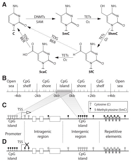
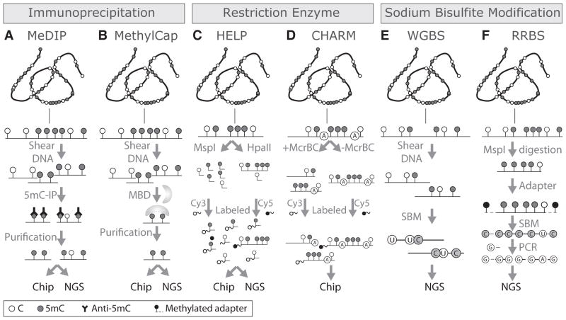
DNA methylation is an epigenetic mechanism that plays a key role in regulating gene expression and other functions. Although this modification is seen in different sequence contexts, the most frequently detected DNA methylation in mammals involves cytosine-guanine dinucleotides. Pathological alterations in DNA methylation patterns are described in a variety of human diseases, including cancer. Unlike genetic changes, DNA methylation is heavily influenced by subtle modifications in the cellular microenvironment. In all cancers, aberrant DNA methylation is involved in the alteration of a large number of oncological pathways with relevant theranostic utility. Several technologies for DNA methylation mapping have been developed recently and successfully applied in cancer studies. The scope of these technologies varies from assessing a single cytosine-guanine locus to genome-wide distribution of DNA methylation. Here, we review the strengths and weaknesses of these approaches in the context of clinical utility for the molecular diagnosis of human cancers.

Keywords: DNA methylation; cancer diagnosis; epigenomics; microarray; sequencing.


Conflict of interest statement

Conflicts of interest: The authors have declared that no competing interest exists.

The authors have no other relevant affiliations or financial involvement with any organization or entity with a financial interest in or financial conflict with the subject matter or materials discussed in the manuscript. This includes employment, consultancies, honoraria, stock ownership or options, expert testimony, grants or patents received or pending, or royalties.

Figures

Figure 1
Figure 1. Cytosine modifications and distribution of 5-methylcytosine (5mC in the human genome
A- Chemical structures of the unmodified cytosine (C), 5mC and its oxidation products 5-hydroxymethylcytosine (5hmC), 5-formylcytosine (5fC) and 5-carboxylcytosine (5caC). First, a methyl group is transferred from the s-adenosylmethionine (SAM) to the fifth carbon of cytosine by DNA methyltransferase enzymes (DNMTs). Second, the oxidation of 5mC is carried out by the ten-eleven translocation methylcytosine dioxygenases (TETs) to 5hmC, 5fC, and 5caC. Finally, 5fC and 5caC can be excised and repaired by thymine-DNA glycosylase (TDG) and base excision repair (BER) systems to generate unmethylated Cs. B- Representation of the CpG context. C–D. Distribution of 5mC in normal (C) and cancer (D) cells. CpG: cytosine – guanine
Figure 2
Figure 2. Genome-wide DNA methylation mapping technologies
Technologies based on 5mC immunoprecipitation (IP). A- Methylated DNA IP (MeDIP) is based on the use of 5mC-specific antibodies. The product is a fraction of genomic DNA enriched in methylated DNA that can be analyzed by either DNA microarrays (chip; MeDIP-chip) or next-generation sequencing (NSG; MeDIP-Seq). B- MethylCap is based on the capture of methylated DNA by methyl-CpG binding domain (MBD) proteins. The fraction of methylated DNA enriched by this approach can also be analyzed by either chip (MethylCap-chip) or NGS (MethylCap-Seq). C- HpaII tiny fragment Enrichment by Ligation-mediated PCR (HELP) is based on the differential labeling of the products of two restriction enzymes, one methylation-resistant (MspI) and other methylation-sensitive (HpaII). D- Comprehensive High-throughput Arrays for Relative Methylation (CHARM) uses the restriction enzyme McrBC which cuts 5mC that are preceded by a purine (A or G) base. The restricted (+McrBC) DNA is differentially labeled and compared with the non- McrBC restricted (-McrBC) DNA fragments in a DNA microarray. E- Whole genome bisulfite sequencing (WGBS) involves the fragmentation and sodium bisulfite modification (SBM) of genomic DNA followed by NGS. F- Reduced Representation Bisulfite Sequencing (RRBS) involves the digestion of genomic DNA by the methylation-resistant restriction enzyme MspI. Each fragment is coupled to a methylated adapter and SBM. Finally, only the converted fragments are amplified by PCR using primers that are complementary to the adaptor, and the products are analyzed by NGS. 5mC: 5-Methylcytosine; CpG: Cytosine – guanine.
Figure 3
Figure 3. Strengths and weaknesses of DNA methylation as a cancer biomarker
A–B: DNA methylation profiling can identify the tumor types. A- Heat map showing the unsupervised hierarchical cluster analysis using the DNA methylation level of the top 2,000 most variable CpG sites in four different cancer types: breast cancer (BRCA), colorectal cancer (CRC), glioblastoma (GBM), and melanoma (MEL). B- Terrain maps generated with the same DNA methylation signatures showing four separate conglomerates of samples representing the four different tumor types. C–E: Culture conditions induce genome-wide variations in DNA methylation level. C- Correlations between DNA methylation levels of three pairs of melanoma brain metastases stored as frozen and as formalin-fixed paraffin embedded tissues. D- Correlations between DNA methylation levels of the same three pairs of melanoma brain metastases stored frozen and established as cell cultures. E- Comparison of DNA methylation levels according to the CpG context of the assessed CpG sites. CpG sites located in CpG islands and in open sea regions are represented. The density plots represent the variations observed in CpG islands (upper) and open sea (lower). CpG: Cytosine – guanine.

Similar articles

Cited by

References

    1. Levin HL, Moran JV. Dynamic interactions between transposable elements and their hosts. Nat Rev Genet. 2011;12(9):615–627. - PMC - PubMed
    1. Jaenisch R, Bird A. Epigenetic regulation of gene expression: how the genome integrates intrinsic and environmental signals. Nature genetics. 2003;33 (Suppl):245–254. - PubMed
    1. Peters J. The role of genomic imprinting in biology and disease: an expanding view. Nat Rev Genet. 2014;15(8):517–530. - PubMed
    1. Smith ZD, Meissner A. DNA methylation: roles in mammalian development. Nat Rev Genet. 2013;14(3):204–220. - PubMed
    1. Lister R, Pelizzola M, Dowen RH, et al. Human DNA methylomes at base resolution show widespread epigenomic differences. Nature. 2009;462(7271):315–322. This study was the first whole-genome DNA methylation mapping of a human genome and describes the presence of non-CpG dinucleotide methylation. - PMC - PubMed

Publication types

Substances

LinkOut - more resources