Review
. 2015 Jul;34(3):173-82.
doi: 10.14366/usg.15003. Epub 2015 Feb 7.

Ultrasonography of ovarian masses using a pattern recognition approach

Affiliations
Review

Ultrasonography of ovarian masses using a pattern recognition approach

Sung Il Jung. Ultrasonography. 2015 Jul.

Abstract

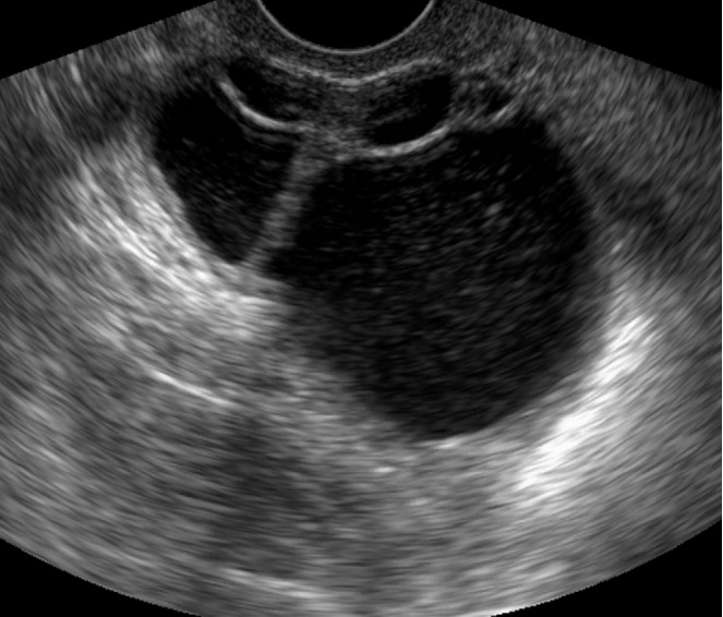
As a primary imaging modality, ultrasonography (US) can provide diagnostic information for evaluating ovarian masses. Using a pattern recognition approach through gray-scale transvaginal US, ovarian masses can be diagnosed with high specificity and sensitivity. Doppler US may allow ovarian masses to be diagnosed as benign or malignant with even greater confidence. In order to differentiate benign and malignant ovarian masses, it is necessary to categorize ovarian masses into unilocular cyst, unilocular solid cyst, multilocular cyst, multilocular solid cyst, and solid tumor, and then to detect typical US features that demonstrate malignancy based on pattern recognition approach.

Keywords: Neoplasms; Ovary; Ultrasonography.


Conflict of interest statement

No potential conflict of interest relevant to this article was reported.

Figures

Fig. 1.
Fig. 1.. Follicular cyst in a 22-year-old woman.
Transvaginal ultrasonography shows a well defined anechoic mass without a solid component.
Fig. 2.
Fig. 2.. Hemorrhagic corpus luteal cyst in a 25-year-old woman.
Transvaginal ultrasonography (US) shows a rectangular hypoechoic lesion in the cystic mass (arrows). The lesion had no demonstrable flow on Doppler US (not shown).
Fig. 3.
Fig. 3.. Hemorrhagic corpus luteal cyst in a 23-year-old woman.
Transvaginal ultrasonography shows a round complex echoic ovarian mass. A reticular pattern in the internal echo due to fibrin strands is visible (arrows).
Fig. 4.
Fig. 4.. Typical endometrioma in a 32-year-old woman.
Transvaginal ultrasonography reveals homogeneously diffuse low echoes in the cystic mass, which is known as “ground-glass” appearance.
Fig. 5.
Fig. 5.. Atypical endometrioma in a 29-year-old woman.
Transvaginal ultrasonography (US) reveals a well defined round lesion within a homogeneously hypoechoic cyst (arrows). Doppler US demonstrated no blood flow in the lesion, which proved to be a localized blood clot after surgical resection (not shown).
Fig. 6.
Fig. 6.. Endometrioid carcinoma on background of endometriosis in a 44-year-old woman.
Transvaginal ultrasonography shows several small polypoid lesions that have diffuse internal low echoes (arrows) along the wall of the cystic mass.
Fig. 7.
Fig. 7.. Mature cystic teratoma in a 37-year-old woman.
Transvaginal ultrasonography demonstrates a complex echoic mass with a hyperechoic solid portion and posterior shadowing (arrows). The mass also contains multiple hyperechoic lines and dots.
Fig. 8.
Fig. 8.. Mature cystic teratoma in a 22-year-old woman.
Transvaginal ultrasonography demonstrates a well defined hyperechoic nodule within the cystic mass, which proved to be fat after surgical resection (arrow).
Fig. 9.
Fig. 9.. Serous cystadenocarcinoma in a 59-year-old woman.
A unilocular cystic mass with internal papillary projection is seen on transvaginal ultrasonography (arrows).
Fig. 10.
Fig. 10.. Clear cell carcinoma in a 60-year-old woman.
Transvaginal ultrasonography (US) shows a well defined cystic mass with a solid nodule which has a smooth round margin and seems to be slightly different from typical papillary projections (arrows). Doppler US demonstrated blood flow in the solid nodule (not shown).
Fig. 11.
Fig. 11.. Mucinous cystadenoma in a 47-year-old woman.
Transvaginal ultrasonography shows a thin walled multilocular cystic mass with a regular septum.
Fig. 12.
Fig. 12.. Tubo-ovarian abscess in a 35-year-old woman.
Transvaginal ultrasonography shows a thick walled multilocular cystic mass with internal echogenic debris. Regular thickening of the outer wall and the septum of the cystic mass should be noted (arrows).
Fig. 13.
Fig. 13.. Ovarian hyperstimulation syndrome in a 26-year-old woman.
Transabdominal ultrasonography shows a markedly enlarged right ovary with multiple large follicles located in the ovarian cortex. The left ovary had a similar appearance (not shown).
Fig. 14.
Fig. 14.. Mucinous cystadenocarcinoma in a 65-year-old woman.
Transvaginal ultrasonography shows a multilocular cystic mass with large papillary projections (arrow).
Fig. 15.
Fig. 15.. Struma ovarii in a 37-year-old woman.
Transvaginal ultrasonography shows a multilocular cystic mass and internal solid components with various echoes (arrows).
Fig. 16.
Fig. 16.. Granulosa cell tumor in a 46-year-old woman.
Transvaginal ultrasonography reveals a multilocular solid mass. This tumor has mainly solid components, some with and some without a multilocular cystic appearance.
Fig. 17.
Fig. 17.. Fibroma in a 50-year-old woman.
Transvaginal ultrasonography shows a homogeneously hypoechoic solid mass (arrows) with strong posterior shadowing.
Fig. 18.
Fig. 18.. Brenner tumor in a 49-year-old woman.
Transvaginal ultrasonography shows a hypoechoic solid mass with punctate echogenic foci (arrows).
Fig. 19.
Fig. 19.. Dysgerminoma in a 41-year-old woman.
Transabdominal Doppler ultrasonography shows a lobulated hypoechoic solid mass with prominent blood flow in the fibrovascular septa (arrow).
Fig. 20.
Fig. 20.. Ovarian lymphoma in a 59-year-old woman.
Transvaginal ultrasonography shows a hypoechoic solid mass without necrosis or hemorrhage.
Fig. 21.
Fig. 21.. Krukenberg tumor in a 42-year-old woman with advanced gastric cancer.
Transvaginal ultrasonography shows a round hypoechoic solid mass (asterisks) with a peripheral small cyst in the right ovary (A) and left ovary (B).

References

    1. Timmerman D, Schwarzler P, Collins WP, Claerhout F, Coenen M, Amant F, et al. Subjective assessment of adnexal masses with the use of ultrasonography: an analysis of interobserver variability and experience. Ultrasound Obstet Gynecol. 1999;13:11–16. - PubMed
    1. Valentin L. Prospective cross-validation of Doppler ultrasound examination and gray-scale ultrasound imaging for discrimination of benign and malignant pelvic masses. Ultrasound Obstet Gynecol. 1999;14:273–283. - PubMed
    1. Valentin L. Pattern recognition of pelvic masses by gray-scale ultrasound imaging: the contribution of Doppler ultrasound. Ultrasound Obstet Gynecol. 1999;14:338–347. - PubMed
    1. Brown DL, Dudiak KM, Laing FC. Adnexal masses: US characterization and reporting. Radiology. 2010;254:342–354. - PubMed
    1. Geomini P, Kruitwagen R, Bremer GL, Cnossen J, Mol BW. The accuracy of risk scores in predicting ovarian malignancy: a systematic review. Obstet Gynecol. 2009;113:384–394. - PubMed

LinkOut - more resources