. 2015;16(3):307-15.
doi: 10.4142/jvs.2015.16.3.307. Epub 2015 Mar 20.

Quantitative determination of 12-hydroxyeicosatetraenoic acids by chiral liquid chromatography tandem mass spectrometry in a murine atopic dermatitis model

Affiliations

Quantitative determination of 12-hydroxyeicosatetraenoic acids by chiral liquid chromatography tandem mass spectrometry in a murine atopic dermatitis model

Seong-Ho Hong et al. J Vet Sci. 2015.

Abstract

Atopic dermatitis, one of the most important skin diseases, is characterized by both skin barrier impairment and immunological abnormalities. Although several studies have demonstrated the significant relationship between atopic dermatitis and immunological abnormalities, the role of hydroxyeicosatetraenoic acids (HETE) in atopic dermatitis remains unknown. To develop chiral methods for characterization of 12-HETE enantiomers in a 1-chloro-2,4-dinitrochlorobenzene (DNCB)-induced atopic dermatitis mouse model and evaluate the effects of 12-HETE on atopic dermatitis, BALB/c mice were treated with either DNCB or acetone/olive oil (AOO) to induce atopic dermatitis, after which 12(R)- and 12(S)-HETEs in the plasma, skin, spleen, and lymph nodes were quantified by chiral liquid chromatography-tandem mass spectrometry. 12(R)- and 12(S)-HETEs in biological samples of DNCB-induced atopic dermatitis mice increased significantly compared with the AOO group, reflecting the involvement of 12(R)- and 12(S)-HETEs in atopic dermatitis. These findings indicate that 12(R)- and 12(S)-HETEs could be a useful guide for understanding the pathogenesis of atopic dermatitis.

Keywords: (±)12-hydroxyeicosatetraenoic acid; arachidonic acid; atopic dermatitis; chiral liquid chromatography tandem mass spectrometry.


Conflict of interest statement

Conflict of Interest: There is no conflict of interest.

Figures

Fig. 1
Fig. 1. (A) Mechanism of 12 lipoxygenase (Lox) catalyzed arachidonic acid transformation and comparison of (B) skin manifestations and (C) histopathology in acetone/olive oil (AOO) or 1-chloro-2,4-dinitrochlorobenzene (DNCB)-treated mice. (A) Hydrogen is abstracted from the bisallylic methylene. Re-arrangement of radicals causes the formation of a cis-trans conjugated diene system. The molecular dioxygen is inserted sterospecifically, forming a peroxy-radical. This substance is subsequently reduced to a hydroperoxide anion. 12- hydroxyeicosatetraenoic acid (HETE) is catalyzed by glutathione peroxidase (GPx), and glutathione (GSH) is oxidized to glutathione disulfide [17]. (B) The upper row photos were taken before treatment with AOO and DNCB. No skin lesions were detected in AOO-treated mice at 49 days. Erythema, scaling, and hemorrhagic crust (yellow circle) were observed in DNCB-treated mice at 49 days. These animals were treated by application of 100 µL of 1% DNCB in AOO onto the shaved back skin at 1, 4, and 7 weeks to induce atopic dermatitis. (C) Hematoxylin and eosin (H&E) staining of dorsal skin lesions (C1), spleen (C2 and 3), lymph nodes (C4) and toluidine blue staining of dorsal skin lesions (C5) in AOO or DNCB-treated mice. (C1) H&E staining of DNCB-treated dorsal skin lesions revealed hyperkeratosis, superficial pyoderma, and infiltration of inflammatory cells (box). *Epidermis. Dermis. (C2) DNCB-treated mice spleen showed heterogeneous and indistinct white pulp (circle). *Red pulp. (C3) The number of reticular (black arrow) and giant cells (white arrow) increased in DNCB-treated mice compared with AOO-treated mice. (C4) H&E staining of lymph nodes in DNCB-treated mice showed mild lymphocyte depletion and granular lymphocytes (circle) compared with those in AOO-treated mice. (C5) Higher magnification of subcutaneous fatty layer in the DNCB-treated dorsal skin lesion showed massive infiltration of mast cells (box) under toluidine blue. Scale bars = 100 µm (C1), 200 µm (C2 and 4), 50 µm (C3 and 5).
Fig. 2
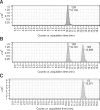
Fig. 2. Chiral separation of (±)12-HETE standards by liquid chromatography-tandem mass spectrometry analysis. 12(R)-HETE standard eluted at 10.140 min (A). Chiral separation of (±)12-HETE standards; 12(R)-HETE eluted at 10.181 min and 12(S)-HETE eluted at 12.890 min (B). 12(S)-HETE d8 standard eluted at 12.971 min (C).
Fig. 3
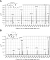
Fig. 3. Product ion mass spectra of the (A) (±)12-HETE standard and (B) internal standard (I.S.). Deprotonated molecules (m/z 319 for (±)12-HETE and m/z 327 for I.S.) were chosen as the precursor ions in the tandem mass spectrometry experiments.
Fig. 4
Fig. 4. Chiral LC/Ms/MS analysis and quantification of (±)12-HETE from AOO or DNCB-treated mice samples. Representative chromatograms of (±)12-HETE released from (A) plasma, (B) skin, (C) spleen, (D) lymph nodes of AOO-treated (top) and DNCB-treated mice (bottom). All (±)12-HETEs (A-D) were detected by electrospray negative ionization and multiple-reaction monitoring of the transition ions for the metabolites.
Fig. 5
Fig. 5. (A and B) Comparison of levels of (±)12-HETE and (C) consecutive changes in total serum IgE levels (ng/mL) in AOO-treated and DNCB-treated mice. (A and B) Brackets indicate significant differences between means (*p < 0.05, **p < 0.01, ***p < 0.001). All results are the means ± standard deviations (SD) (n = 5). Note the different scales for 12(R)-HETE and 12(S)-HETE. (C) The mean serum IgE level of AOO-treated mice remained low (327.82 ng/mL), whereas the mean serum IgE level of DNCB-treated mice increased to a very high level (1,931.7 ng/mL). Values are the means ± SD (n = 5). ***p <0.001 compared to the AOO-treated group based on a paired t-test.

References

    1. Bayer M, Mosandl A, Thaçi D. Improved enantioselective analysis of polyunsaturated hydroxy fatty acids in psoriatic skin scales using high-performance liquid chromatography. J Chromatogr B Analyt Technol Biomed Life Sci. 2005;819:323–328. - PubMed
    1. Bligh EG, Dyer WJ. A rapid method of total lipid extraction and purification. Can J Biochem Physiol. 1959;37:911–917. - PubMed
    1. De Benedetto A, Agnihothri R, McGirt LY, Bankova LG, Beck LA. Atopic dermatitis: a disease caused by innate immune defects? J Invest Dermatol. 2009;129:14–30. - PubMed
    1. de Grauw JC, van de Lest CHA, van Weeren PR. A targeted lipidomics approach to the study of eicosanoid release in synovial joints. Arthritis Res Ther. 2011;13:R123. - PMC - PubMed
    1. Dorsh W. Late Phase Allergic Reactions. Boca Raton: CRC Press; 1990.

Publication types