

Epithelial basement membrane proteins perlecan and nidogen-2 are up-regulated in stromal cells after epithelial injury in human corneas
- PMID: 25797478
- PMCID: PMC4426017
- DOI: 10.1016/j.exer.2015.03.016
Epithelial basement membrane proteins perlecan and nidogen-2 are up-regulated in stromal cells after epithelial injury in human corneas
Abstract
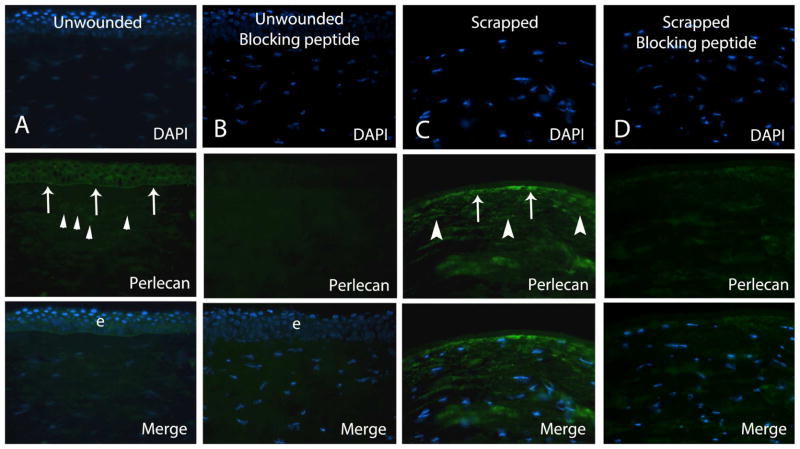
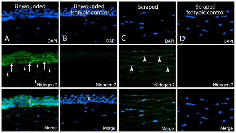
The epithelial basement membrane (BM) is a specialized extracellular matrix that has been shown to have a critical role in corneal development, wound healing, and disease. Although the epithelial BM contributes to corneal homeostasis, relatively little is know about non-epithelial production of its components that may be important in defective regeneration of the epithelial basement membrane associated with opacity after photorefractive keratectomy. The purpose of the current study was to investigate stromal production of corneal epithelial BM proteins in wounded human corneas using immunohistochemistry. A total of five unwounded control eyes and five 30-min epithelial-wounded corneas were obtained from fresh corneoscleral buttons removed from human eyes enucleated due to choroidal melanoma with normal anterior segments. In the wounded corneas, an eight mm patch of central corneal epithelium and epithelial BM was removed with a Beaver blade when the patient was under general anesthesia. Immunohistochemical analyses were performed to detect perlecan and nidogen-2 proteins-important components of the epithelial BM lamina lucida and lamina densa zones. Perlecan and nidogen-2 proteins were detected in the BM itself and at low levels in keratocytes in all unwounded corneas. After epithelial injury, both perlecan and nidogen-2 were expressed at high levels in stromal keratocytes, including superficial keratocytes in the early phases of apoptosis. Thus, after epithelial and epithelial BM injury, stromal keratocytes contribute important perlecan and nidogen-2 components to the regenerating epithelial BM.
Keywords: Apoptosis; Cornea; Epithelial basement membrane; Haze; Keratocyte; Myofibroblast; Nidogen-2; Perlecan; Stroma; Wound healing.
Copyright © 2015 Elsevier Ltd. All rights reserved.
Conflict of interest statement
Figures
References
-
- Dziadek M. Role of laminin-nidogen complexes in basement membrane formation during embryonic development. Experientia. 1995;51:901–913. - PubMed
-
- Fini ME, Stramer BM. How the cornea heals: cornea-specific repair mechanisms affecting surgical outcomes. Cornea. 2005;24:S2–S11. - PubMed
-
- Fleischmajer R, Schechter A, Bruns M, Perlish JS, Macdonald ED, Pan TC, Timpl R, Chu ML. Skin fibroblasts are the only source of nidogen during early basal lamina formation in vitro. J Invest Derm. 1995;105:597–601. - PubMed
Publication types
MeSH terms
Substances
Grants and funding
LinkOut - more resources
Full Text Sources
Other Literature Sources
Medical
