. 2015 May 14;7(18):8378-8388.
doi: 10.1039/c4nr07139e.

Multi-stimuli responsive Cu2S nanocrystals as trimodal imaging and synergistic chemo-photothermal therapy agents

Affiliations

Multi-stimuli responsive Cu2S nanocrystals as trimodal imaging and synergistic chemo-photothermal therapy agents

Aby Cheruvathoor Poulose et al. Nanoscale. .

Abstract

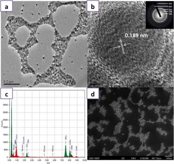
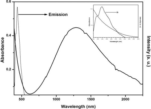
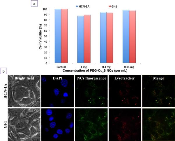
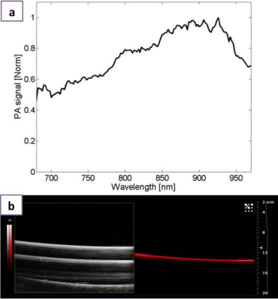
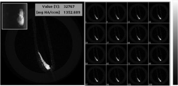
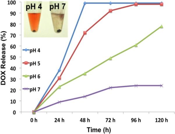
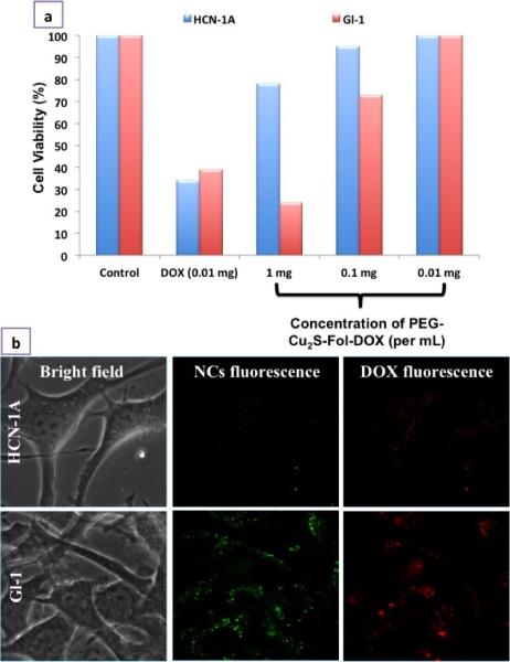
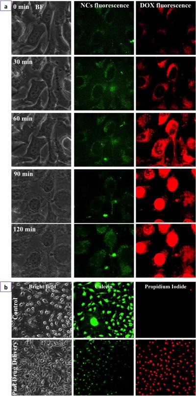
A size and shape tuned, multifunctional metal chalcogenide, Cu2S-based nanotheranostic agent is developed for trimodal imaging and multimodal therapeutics against brain cancer cells. This theranostic agent was highly efficient in optical, photoacoustic and X-ray contrast imaging systems. The folate targeted NIR-responsive photothermal ablation in synergism with the chemotherapeutic action of doxorubicin proved to be a rapid precision guided cancer-killing module. The multi-stimuli, i.e., pH-, thermo- and photo-responsive drug release behavior of the nanoconjugates opens up a wider corridor for on-demand triggered drug administration. The simple synthesis protocol, combined with the multitudes of interesting features packed into a single nanoformulation, clearly demonstrates the competing role of this Cu2S nanosystem in future cancer treatment strategies.


Figures

Figure 1
Figure 1
Structural and Elemental Characterization of the Cu2S NCs a) TEM image of Cu2S NCs, b) HR-TEM of Cu2S NCs exhibiting the crystal lattice distance with an inset presenting the SAED pattern, c) EDS analysis of Cu2S NCs and d) SEM image of Cu2S NCs.
Figure 2
Figure 2
Optical properties of the Cu2S NCs: UV-Vis spectra of Cu2S NCs exhibiting broad LSPR in the NIR. The inset shows the visible absorbance and photoluminescence spectra of Cu2S NCs.
Figure 3
Figure 3
Cellular interaction analysis of PEGylated Cu2S NCs. a) In vitro cell viability analysis of PEGylated Cu2S NCs at different concentrations (0.01 – 1 mg/mL) for 7 days b) Cellular entry analysis of PEGylated Cu2S NCs confirmed the entry of NCs into the cell's cytoplasm (NC's fluorescence) in both normal as well cancer cell line. The nuclear staining (DAPI) and lysosomal staining (Lysotracker) can be seen. The internalization of NCs was found to be endocytosis mediated which is confirmed by the co-localization of NCs signal with lysosomal mapping (Merge).
Figure 4
Figure 4
Photoacoustic imaging a) Normalized photoacoustic signal spectrum of PEGylated Cu2S and b) Ultrasound B-mode image (left) and photoacoustic image (right) of PEGylated Cu2S filled vinyl tube phantom obtained with the VisualSonics Vevo 2100-LAZR system.
Figure 5
Figure 5
X-ray microCT imaging of PEG-Cu2S NCs at tube voltage of 45 KeV. Cross sectional X-ray contrast imaging of phantom with PEG-Cu2S NCs (left). The inset in the left shows the guide view of the same. Z-stacked contrast image of the PEG-Cu2S NCs is shown in right with the intensity scale.
Figure 6
Figure 6
pH dependent DOX release from PEG FOL-Cu2S-DOX conjugate.
Figure 7
Figure 7
Selectivity and specificity of PEG-FOL-Cu2S-DOX towards cancer cells. a) In vitro selective tumoricidal activity of PEG-FOL-Cu2S-DOX at different concentrations (0.01 – 1mg/mL) in comparison to free DOX post 24 h incubation with cells. b) Cancer specific cellular entry of PEG-FOL-Cu2S-DOX NCs. The specificity of NC entry only into cancer cell (Gl-1) could be attributed to the folate targeting.
Figure 8
Figure 8
Drug delivery and Cancer cell death a) Time lapse imaging of Folate targeted PEGylated Cu2S NCs mediated intracellular trafficking of DOX. b) Gl-1 cancer cell death mediated by drug, DOX, pre and post drug delivery was confirmed with live/dead staining. Post drug delivery, after 120 min, most of the cells stained positive for propidium iodide indicating cell death.
Figure 9
Figure 9
NC photoexcitation mediated drug release a) Photoexcitation of NCs speeds up the release of DOX. The DOX was seen to localize nuclear region within 10 min of photosensitization of NCs using 488 nm excitation light. b) Cancer cell death mediated by photoexcitation mediated drug release could be visualized with positive propidium iodide staining of cancer cells.
Figure 10
Figure 10
Photothermal Efficacy of PEG-Cu2S NCs. a) Heating profile of different concentrations (1, 10, 30, 50, 100 ppm) of PEG-Cu2S NCs (suspended in water) upon NIR irradiation. b) Plot of difference in temperature attained upon 600 sec (10 min) NIR irradiation of different concentrations of PEG-Cu2S NCs. c) The heating and cooling profile of 100 ppm PEG-Cu2S NCs in terms of difference in temperature. The sample was irradiated for 600 sec, post that the laser is shut down and the gradual drop in temperature was recorded until the temperature attains initial room temperature. d) Plot of cooling period (after 600 s) versus negative natural logarithm of driving force temperature. Time constant (τs) for heat transfer is determined to be 133.20 s.
Figure 11
Figure 11
Photothermal ablation of cancer cells by dual synergistic effect of drug and heat. The culture area (10 mm in diameter) is depicted in orange in the pictorial representation of confocal dish. When treated with PEG-Cu2S and irradiated with 800 nm laser, only cells present in the zone of irradiation exhibited positive staining of propidium iodide with surrounding regions exhibiting calcein stained live cells. Whereas with PEG-FOL-Cu2S-DOX, most of the Gl-1 cells in the plate ranging from center (zone of irradiation, zone 1) to corner of the culture area of the dish (zone 4) exhibited positive propidium iodide staining with no viable cells.

References

    1. Siegel R, DeSantis C, Virgo K, Stein K, Mariotto A, Smith T, Cooper D, Gansler T, Lerro C, Fedewa S, Lin C, Leach C, Cannady RS, Cho H, Scoppa S, Hachey M, Kirch R, Jemal A, Ward E. CA Cancer J. Clin. 2012;62:220. - PubMed
    1. Siegel R, Naishadham D, Jemal A. CA Cancer J Clin. 2012;62:10. - PubMed
    1. Yin W, Yan L, Yu J, Tian G, Zhou L, Zheng X, Zhang X, Yong Y, Li J, Gu Z, Zhao Y. ACS Nano. 2014;8:6922. - PubMed
    1. Scott AM, Wolchok JD, Old LJ. Nat. Rev. Cancer. 2012;12:278. - PubMed
    1. DeVita VT, Jr., Chu E. Cancer Res. 2008;68:8643. - PubMed

Publication types

MeSH terms