Computed tomography findings of paracoccidiodomycosis in musculoskeletal system
- PMID: 25798000
- PMCID: PMC4366020
- DOI: 10.1590/0100-3984.2014.0049
Computed tomography findings of paracoccidiodomycosis in musculoskeletal system
Abstract
Objective: To evaluate musculoskeletal involvement in paracoccidioidomycosis at computed tomography.
Materials and methods: Development of a retrospective study based on a review of radiologic and pathologic reports in the institution database. Patients with histopathologically confirmed musculoskeletal paracoccidioidomycosis and submitted to computed tomography were included in the present study. The imaging findings were consensually described by two radiologists. In order to avoid bias in the analysis, one patient with uncountable bone lesions was excluded from the study.
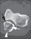
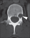
Results: A total of seven patients were included in the present study. A total of 18 bone lesions were counted. The study group consisted of 7 patients. A total number of 18 bone lesions were counted. Osteoarticular lesions were the first manifestation of the disease in four patients (57.14%). Bone lesions were multiple in 42.85% of patients. Appendicular and axial skeleton were affected in 85.71% and 42.85% of cases, respectively. Bone involvement was characterized by well-demarcated osteolytic lesions. Marginal osteosclerosis was identified in 72.22% of the lesions, while lamellar periosteal reaction and soft tissue component were present in 5.55% of them. One patient showed multiple small lesions with bone sequestra.
Conclusion: Paracoccidioidomycosis can be included in the differential diagnosis of either single or multiple osteolytic lesions in young patients even in the absence of a previous diagnosis of pulmonary or visceral paracoccidioidomycosis.
Objetivo: Avaliar o acometimento musculoesquelético da paracoccidioidomicose nas imagens de tomografia computadorizada.
Materiais e métodos: Estudo retrospectivo desenvolvido a partir de revisão de laudos radiológicos e patológicos do banco de dados da instituição. Foram selecionados pacientes com paracoccidioidomicose osteoarticular submetidos a tomografia computadorizada. Todos os casos considerados tiveram confirmação histopatológica da doença. Os achados de imagem foram descritos em consenso por dois radiologistas. Um paciente com incontáveis lesões ósseas foi excluído da contabilização das anormalidades com a finalidade de evitar viés.
Resultados: Foram incluídos 7 pacientes no presente estudo. Um total de 18 lesões ósseas foi contabilizado. Em quatro casos (57,14%) a lesão osteoarticular foi a primeira manifestação da doença. As lesões ósseas eram múltiplas em 42,85%. Os esqueletos apendicular e axial foram afetados em 85,71% e 42,85% dos pacientes, respectivamente. O envolvimento ósseo caracterizou-se por lesões osteolíticas bem delimitadas. Identificou-se osteoesclerose marginal em 72,22% das lesões contabilizadas. Reação periosteal lamelar e componente de partes moles estiveram presentes em 5,55% das anormalidades. Um paciente exibiu múltiplas lesões com sequestros ósseos.
Conclusão: A paracoccidioidomicose pode ser incluída no diagnóstico diferencial de lesões osteolíticas, únicas ou múltiplas, em pacientes jovens, mesmo sem diagnóstico prévio de paracoccidioidomicose pulmonar ou visceral.
Keywords: Computed tomography; Musculoskeletal; Osteoarticular; Osteomyelitis; Paracoccidioidomycosis.
Figures


References
-
- Shikanai-Yasuda MA, Telles FQ, Filho, Mendes RP, et al. Consenso em paracoccidioidomicose. Rev Soc Bras Med Trop. 2006;39:297–310. - PubMed
-
- Wanke B, Aidê MA. Chapter 6 - Paracoccidioidomycosis. J Bras Pneumol. 2009;35:1245–1249. - PubMed
-
- Trad HS, Trad CS, Elias J, Junior, et al. Radiological review of 173 consecutive cases of paracoccidioidomycosis. Radiol Bras. 2006;39:175–179.
-
- Campos MVS, Penna GO, Castro CN, et al. Paracoccidioidomycosis at Brasilia's University Hospital. Rev Soc Bras Med Trop. 2008;41:169–172. - PubMed
LinkOut - more resources
Full Text Sources
Other Literature Sources
