Review
. 2015 Mar 5:9:14.
doi: 10.3389/fnana.2015.00014. eCollection 2015.

The dendritic spine story: an intriguing process of discovery

Affiliations
Review

The dendritic spine story: an intriguing process of discovery

Javier DeFelipe. Front Neuroanat. .

Abstract

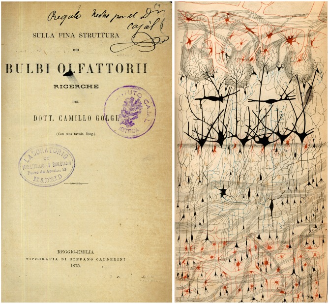
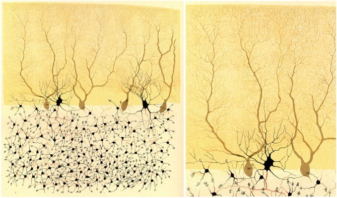
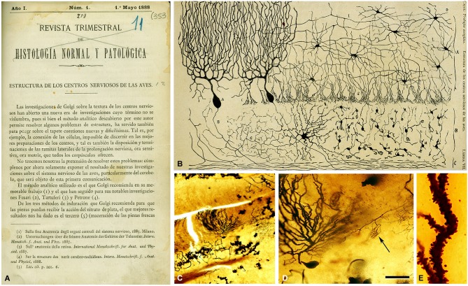
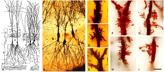
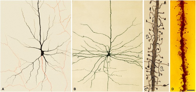
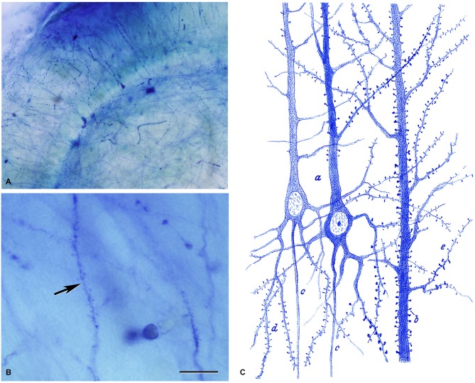
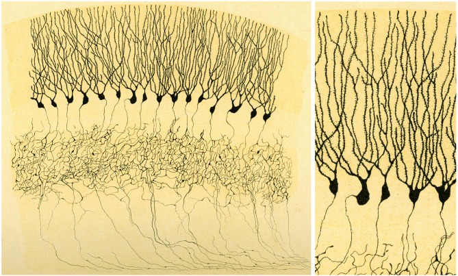
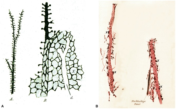
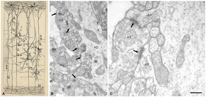
Dendritic spines are key components of a variety of microcircuits and they represent the majority of postsynaptic targets of glutamatergic axon terminals in the brain. The present article will focus on the discovery of dendritic spines, which was possible thanks to the application of the Golgi technique to the study of the nervous system, and will also explore the early interpretation of these elements. This discovery represents an interesting chapter in the history of neuroscience as it shows us that progress in the study of the structure of the nervous system is based not only on the emergence of new techniques but also on our ability to exploit the methods already available and correctly interpret their microscopic images.

Keywords: Cajal; Golgi; Purkinje cells; granule cells; history of neuroscience; neuron doctrine; pyramidal cells; reticular theory.


Figures

Figure 1
Figure 1
The first illustration by Golgi of a Golgi impregnated preparation of the nervous system. “Semi-schematic drawing of a fragment of a vertical section of the olfactory bulb of a dog” (Golgi, 1875). Taken from DeFelipe (2010).
Figure 2
Figure 2
Illustration by Golgi of a Golgi impregnated preparation of the cerebellum. “Fragment of a vertical section of a cerebellar convolution of the rabbit”. Left, panoramic view. Right, a high magnification of the drawing to illustrate that the dendrites of the Purkinje cells are smooth, without dendritic spines. Taken from Golgi (1882–1883).
Figure 3
Figure 3
First illustration by Cajal (1888) of a Golgi impregnated preparation of the nervous system. (A) First page of the article and (B) illustration whose legend states: “Vertical section of a cerebellar convolution of a hen. Impregnation by the Golgi method. A represents the molecular zone, B designates the granular layer and C the white matter”. (C) photomicrograph from one of Cajal’s preparations of the cerebellum of an adult bird stained with the Golgi method. (D) higher magnification of (C) to illustrate a Purkinje cell and a basket formation (arrow). (E) dendrite of the Purkinje cell which is covered with dendritic spines. The histological images were obtained by Pablo García-López, Virginia García-Marín, and Miguel Freire (Legado Cajal, Instituto Cajal). Scale bar: 200 μm in (C); 60 μm in (D); 8,4 μm in (E). Taken from DeFelipe (2014).
Figure 4
Figure 4
Complex dendritic spines (thorny excrescences) of CA3 pyramidal neurons. (A) Cajal’s drawing showing pyramidal cells with thorny excrescences in the CA3 (Cajal, 1893). (B–I) photomicrographs of Cajal’s original histological preparations housed at the Cajal Institute. (B) CA3 pyramidal cells of the rabbit stained by the Golgi method. (C–I) Examples of thorny excrescences on CA3 pyramidal neurons. (C–E) Dendrites from a newborn child’s CA3 pyramidal neurons stained by the Golgi method; (F–I) Dendrites of rabbit CA3 pyramidal neurons stained by Kenyon’s variant of the Golgi method. Scale bar: (B) 55 μm; (C–I), 11 μm. Taken from Blazquez-Llorca et al. (2011).
Figure 5
Figure 5
Pyramidal cells of the human cerebral cortex. (A) Drawing made by Golgi to illustrate a pyramidal cell of the human motor cortex stained with the Golgi method. The axon appears in red. Taken from Golgi (1882–1883). (B) Drawing made by Cajal to illustrate a pyramidal cell of the human motor cortex. a, initial part of the axon; b, dendrites; d, axonal collaterals. Taken from Cajal (1899). (C) Drawing by Cajal to illustrate the dendritic spines of pyramidal cells (cerebral cortex of a 2-month-old child). Taken from Cajal (1933). Note that Golgi does not draw dendritic spines. However, in the drawing of Cajal shown in (B) it can be seen that the surface of the dendrites are covered with dendrites spines. (D) photomicrograph of a preparation of Cajal of the human motor cortex (15-day-old child) stained using the Golgi method. The image illustrates an apical dendrite of a layer V pyramidal cell covered with spines. Scale bar (in D): 8 μm. The histological image was obtained by Pablo García-López, Virginia García-Marín and Miguel Freire (Legado Cajal, Instituto Cajal). Taken from DeFelipe (2014).
Figure 6
Figure 6
Dendritic spines stained with the methylene blue method. (A,B) Low and high magnification photomicrographs, respectively, of a preparation by Cajal of the hippocampus stained with the methylene blue method (preparation housed at the Cajal Institute). These photomicrographs are from unpublished material from DeFelipe and Jones (1988). Scale bar (in B): (A) 120 μm; (B) 10 μm. (C) Drawing used by Cajal to show the existence of dendritic spines on pyramidal cells with the methylene blue method. Taken from Cajal (1899–1904).
Figure 7
Figure 7
Illustration by Golgi of a Golgi impregnated preparation of the dentate gyrus. “Fascia dentata del grande piede di Hippocampo”. Left, panoramic view. Golgi used this drawing to illustrate that the axons of granule cells formed a very complex nervous network (“rete nervosa”). Right, High magnification of the drawing to illustrate that Golgi recognized the presence of dendritic spines. Taken from Golgi (1901).
Figure 8
Figure 8
Different interpretation of dendritic spines. (A) Drawings made by Bethe (1903) to illustrate dendritic spines stained with the ammonium molybdate method. A, an apical dendrite of a pyramidal cell covered with dendritic spines. B, schematic representation to illustrate his hypothesis that dendritic spines were the starting points of an interstitial network of the gray matter. d, unstained apical dendrite. (B) Drawings made by Held (1897b) to show dendrites innervated by end-feet or axon terminals (some of them are marked with an x). See text for further details.
Figure 9
Figure 9
Drawing and images showing dendritic spines as postsynaptic structures. (A) Schematic drawing by Cajal to show synaptic connections and the possible flow of information through neural circuits in the cerebral cortex. The legend states: A, small pyramid; B, and C, medium and giant pyramids, respectively; a, axon; [c], nervous collaterals that appear to cross and touch the dendrites and the trunks [apical dendrites] of the pyramids; H, white matter; [E, Martinotti cell with ascending axon]; F, special cells of the first layer of cerebral cortex; G, fiber coming from the white matter. The arrows mark the supposed direction of the nervous current”. Taken from ¿Neuronismo o reticularismo? (Cajal, 1933). (B) Electron micrograph of a typical apical dendrite showing dendritic spines with different shapes (arrows). (C) high magnification of A to illustrate an axon terminal (AX) which establishes synaptic contacts (arrows) simultaneously with two dendritic spines. AP, spine apparatus. Scale bar: (B) 0,60 μm; (C) 0,15 μm. Taken from unpublished material (Alonso-Nanclares et al., 2008).
Figure 10
Figure 10
Examples of dendritic spines forming synapses (A–F) in the adult mouse neocortex. The gold-particles allow the postsynaptic densities (PSDs; red arrows) to be clearly distinguished when present. Note the small size of the postsynaptic density (60 nm) in the head of a small spine in (B) (asterisk). (E,F) are consecutive sections of a spine head to illustrate a PSD cut tangentially. Scale bar: 560 nm in (A); 350 nm in (B,C); 300 nm in (D); 280 nm in (E,F). Taken from Arellano et al. (2007).
Figure 11
Figure 11
Reconstructions from serial electron micrographs. Serial sections of dendritic segments in the adult mouse neocortex to illustrate the distribution of some non-synaptic spines (blue) indicated by arrows. The remaining spines establish synaptic contacts (red, PSD). (A,B) Basal dendrites; (C) apical dendrite. Scale bar: 2000 nm. Taken from Arellano et al. (2007).

References

    1. Alonso-Nanclares L., Gonzalez-Soriano J., Rodriguez J. R., DeFelipe J. (2008). Gender differences in human cortical synaptic density. Proc. Natl. Acad. Sci. U S A 105, 14615–14619. 10.1073/pnas.0803652105 - DOI - PMC - PubMed
    1. Andersen P., Morris R., Amaral D., Bliss T., O’Keefe J. (eds) (2007). The Hippocampus Book. New York: Oxford University Press.
    1. Arellano J. I., Espinosa A., Fairén A., Yuste R., DeFelipe J. (2007). Non-synaptic dendritic spines in neocortex. Neuroscience 145, 464–469. 10.1016/j.neuroscience.2006.12.015 - DOI - PubMed
    1. Bethe A. (1895). Studien über das Centralnervensystem von Carcinus maenas nebst Angaben über ein neues verfahren der methylenblaufixation. Arch. Mikrosk. Anat. 44, 579–622 10.1007/bf02934031 - DOI
    1. Bethe A. (1903). Allgemeine Anatomie und Physiologie des Nervensystems. Leipzig: Thieme.

LinkOut - more resources