

Three-dimensional cone-beam computed tomography based comparison of condylar position and morphology according to the vertical skeletal pattern
- PMID: 25798412
- PMCID: PMC4367133
- DOI: 10.4041/kjod.2015.45.2.66
Three-dimensional cone-beam computed tomography based comparison of condylar position and morphology according to the vertical skeletal pattern
Abstract
Objective: To compare condylar position and morphology among different vertical skeletal patterns.
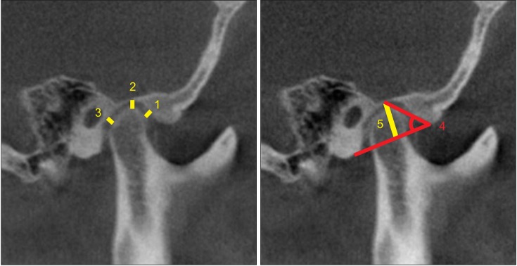
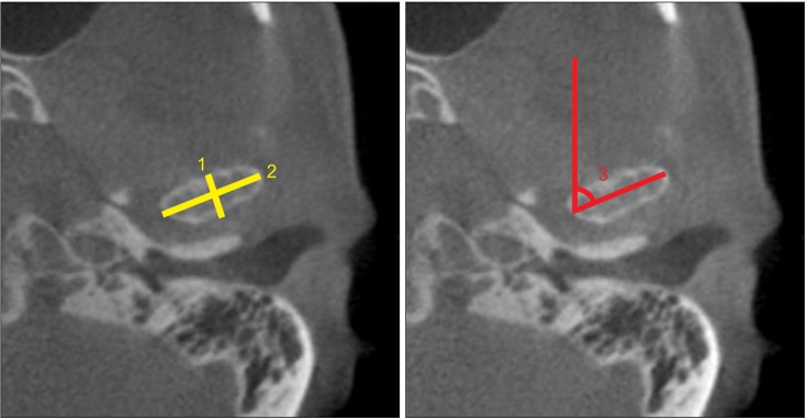
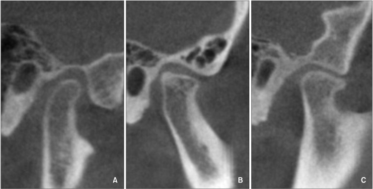
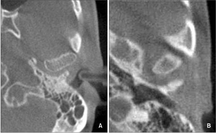
Methods: Diagnostic cone-beam computed tomography images of 60 adult patients (120 temporomandibular joints) who visited the orthodontic clinic of Hallym University Sacred Heart Hospital were reviewed. The subjects were divided into three equal groups according to the mandibular plane angle: hypodivergent, normodivergent, and hyperdivergent groups. Morphology of the condyle and mandibular fossa and condylar position were compared among the groups.
Results: The hypodivergent and hyperdivergent groups showed significant differences in superior joint spaces, antero-posterior condyle width, medio-lateral condyle width, condyle head angle, and condylar shapes.
Conclusions: Condylar position and morphology vary according to vertical facial morphology. This relationship should be considered for predicting and establishing a proper treatment plan for temporomandibular diseases during orthodontic treatment.
Keywords: Computed tomography; Condylar morphology; Condylar position; Temporomandibular joint.
Conflict of interest statement
The authors report no commercial, proprietary, or financial interest in the products or companies described in this article.
Figures
Similar articles
-
[Cone-beam CT evaluation of temporomandibular joint in skeletal class Ⅱ female adolescents with different vertical patterns].Beijing Da Xue Xue Bao Yi Xue Ban. 2020 Dec 29;53(1):109-119. doi: 10.19723/j.issn.1671-167X.2021.01.017. Beijing Da Xue Xue Bao Yi Xue Ban. 2020. PMID: 33550344 Free PMC article. Chinese.
-
Comparison of temporomandibular joints in relation to ages and vertical facial types in skeletal class II female patients: a multiple-cross-sectional study.BMC Oral Health. 2024 Apr 17;24(1):467. doi: 10.1186/s12903-024-04219-4. BMC Oral Health. 2024. PMID: 38632555 Free PMC article.
-
Comprehensive three-dimensional positional and morphological assessment of the temporomandibular joint in skeletal Class II patients with mandibular retrognathism in different vertical skeletal patterns.BMC Oral Health. 2022 Apr 28;22(1):149. doi: 10.1186/s12903-022-02174-6. BMC Oral Health. 2022. PMID: 35484618 Free PMC article.
-
Assessment of the Morphology and Degenerative Changes in the Temporomandibular Joint Using CBCT according to the Orthodontic Approach: A Scoping Review.Biomed Res Int. 2022 Feb 1;2022:6863014. doi: 10.1155/2022/6863014. eCollection 2022. Biomed Res Int. 2022. PMID: 35155678 Free PMC article.
-
Efficacy of Selective Grinding Guided by an Occlusal Splint in Management of Myofascial Pain: A Prospective Clinical Trial.Open Dent J. 2017 Jun 30;11:301-311. doi: 10.2174/1874210601711010301. eCollection 2017. Open Dent J. 2017. PMID: 28839479 Free PMC article. Review.
Cited by
-
The Role of Maxillofacial Structure and Malocclusion on Condylar Displacement in Maximum Intercuspation and Centric Relation in Patients Seeking Orthodontic Treatment-A Scoping Review.J Clin Med. 2023 Jan 15;12(2):689. doi: 10.3390/jcm12020689. J Clin Med. 2023. PMID: 36675620 Free PMC article.
-
Temporomandibular joint morphology in Korean using cone-beam computed tomography: influence of age and gender.Maxillofac Plast Reconstr Surg. 2021 Jul 5;43(1):21. doi: 10.1186/s40902-021-00307-5. Maxillofac Plast Reconstr Surg. 2021. PMID: 34224004 Free PMC article.
-
[Cone-beam CT evaluation of temporomandibular joint in skeletal class Ⅱ female adolescents with different vertical patterns].Beijing Da Xue Xue Bao Yi Xue Ban. 2020 Dec 29;53(1):109-119. doi: 10.19723/j.issn.1671-167X.2021.01.017. Beijing Da Xue Xue Bao Yi Xue Ban. 2020. PMID: 33550344 Free PMC article. Chinese.
-
Prevalence of condylar morphological changes in individuals with class II malocclusion.Braz Oral Res. 2024 Jul 12;38:e060. doi: 10.1590/1807-3107bor-2024.vol38.0060. eCollection 2024. Braz Oral Res. 2024. PMID: 39016367 Free PMC article.
-
Three-Dimensional Assessment of Temporomandibular Joint Morphology and Facial Asymmetry in Individuals with Different Vertical Skeletal Growth Patterns.Int J Environ Res Public Health. 2023 Jan 12;20(2):1437. doi: 10.3390/ijerph20021437. Int J Environ Res Public Health. 2023. PMID: 36674193 Free PMC article.
References
-
- Girardot RA., Jr Comparison of condylar position in hyperdivergent and hypodivergent facial skeletal types. Angle Orthod. 2001;71:240–246. - PubMed
-
- Vitral RW, Telles Cde S, Fraga MR, de Oliveira RS, Tanaka OM. Computed tomography evaluation of temporomandibular joint alterations in patients with class II division 1 subdivision malocclusions: condyle-fossa relationship. Am J Orthod Dentofacial Orthop. 2004;126:48–52. - PubMed
-
- Rodrigues AF, Fraga MR, Vitral RW. Computed tomography evaluation of the temporomandibular joint in Class I malocclusion patients: condylar symmetry and condyle-fossa relationship. Am J Orthod Dentofacial Orthop. 2009;136:192–198. - PubMed
-
- Schudy FF. Treatment of adult midline deviation by condylar repositioning. J Clin Orthod. 1996;30:343–347. - PubMed
LinkOut - more resources
Full Text Sources
Other Literature Sources