Clinical characteristics and perinatal outcome of fetal hydrops
- PMID: 25798421
- PMCID: PMC4366875
- DOI: 10.5468/ogs.2015.58.2.90
Clinical characteristics and perinatal outcome of fetal hydrops
Abstract
Objective: To investigate the clinical characteristics of fetal hydrops and to find the antenatal ultrasound findings predictive of adverse perinatal outcome.
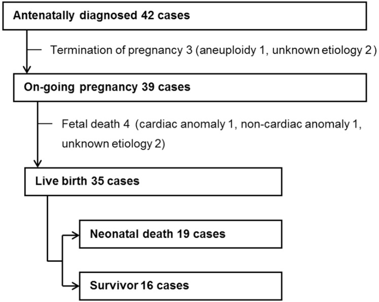
Methods: This is a retrospective study of 42 women with fetal hydrops who delivered in a tertiary-referral center from 2005 to 2013. Fetal hydrops was defined as the presence of fluid collection in ≥2 body cavities: ascites, pleural effusion, pericardial effusion, and skin edema. Predictor variables recorded included: maternal characteristics, gestational age at diagnosis, ultrasound findings, and identifiable causes. Primary outcome variables analyzed were fetal death and neonatal death.
Results: The mean gestational age at diagnosis was 29.3±5.4 weeks (range, 18 to 39 weeks). The most common identifiable causes were cardiac abnormality (10), followed by syndrome (4), aneuploidy (3), congenital infection (3), twin-to-twin transfusion syndrome (3), non-cardiac anormaly (2), chorioangioma (2), inborn errors of metabolism (1), and immune hydrops by anti-E antibody isoimmunization (1). Thirteen cases had no definite identifiable causes. Three women elected termination of pregnancy. Fetal death occurred in 4 cases. Among the 35 live-born babies, only 16 survived (54.0% neonatal mortality rate). Fetal death and neonatal mortality rate was not significantly associated with Doppler velocimetry indices or location of fluid collection, but increasing numbers of fluid collection site was significantly associated with a higher risk of neonatal death.
Conclusion: The incidence of fetal hydrops in our retrospective study was 24.4 per 10,000 deliveries and the perinatal mortality rate was 61.9% (26/42). The number of fluid collection sites was the significant antenatal risk factor to predict neonatal death.
Keywords: Fetal death in utero; Hydrops fetalis; Infant mortality; Ultrasonography.
Conflict of interest statement
No potential conflict of interest relevant to this article was reported.
Figures
Similar articles
-
Ultrasonographic severity scoring of non-immune hydrops: a predictor of perinatal mortality.J Perinat Med. 2015 Jan;43(1):53-9. doi: 10.1515/jpm-2013-0208. J Perinat Med. 2015. PMID: 24837487
-
[Clinical study on 156 cases with hydrops fetalis].Zhonghua Fu Chan Ke Za Zhi. 2011 Dec;46(12):905-10. Zhonghua Fu Chan Ke Za Zhi. 2011. PMID: 22333280 Chinese.
-
Transient hydropic signs in the donor fetus after fetoscopic laser coagulation in severe twin-twin transfusion syndrome: incidence and clinical relevance.Ultrasound Obstet Gynecol. 2002 May;19(5):449-53. doi: 10.1046/j.1469-0705.2002.00642.x. Ultrasound Obstet Gynecol. 2002. PMID: 11982976
-
Outcome of fetuses with congenital parvovirus B19 infection: systematic review and meta-analysis.Ultrasound Obstet Gynecol. 2018 Nov;52(5):569-576. doi: 10.1002/uog.19092. Ultrasound Obstet Gynecol. 2018. PMID: 29785793
-
Fetal primary pleural effusions: Prenatal diagnosis and management.Best Pract Res Clin Obstet Gynaecol. 2019 Jul;58:66-77. doi: 10.1016/j.bpobgyn.2019.01.005. Epub 2019 Jan 12. Best Pract Res Clin Obstet Gynaecol. 2019. PMID: 30737016 Review.
Cited by
-
Non-Immune Hydrops Fetalis: Do Placentomegaly and Polyhydramnios Matter?J Ultrasound Med. 2018 May;37(5):1185-1191. doi: 10.1002/jum.14462. Epub 2017 Oct 27. J Ultrasound Med. 2018. PMID: 29076544 Free PMC article.
-
Frequency and Prognosis of Hydrops Fetalis: A 10-Year Single-Center Experience.Sisli Etfal Hastan Tip Bul. 2021 Sep 24;55(3):366-373. doi: 10.14744/SEMB.2021.65632. eCollection 2021. Sisli Etfal Hastan Tip Bul. 2021. PMID: 34712079 Free PMC article.
-
[Clinical features and prognosis of neonates with nonimmune hydrops fetalis].Zhongguo Dang Dai Er Ke Za Zhi. 2019 Mar;21(3):253-258. doi: 10.7499/j.issn.1008-8830.2019.03.013. Zhongguo Dang Dai Er Ke Za Zhi. 2019. PMID: 30907350 Free PMC article. Chinese.
-
Etiology and Outcome of non-immune Hydrops Fetalis in Southern China: report of 1004 cases.Sci Rep. 2019 Jul 24;9(1):10726. doi: 10.1038/s41598-019-47050-6. Sci Rep. 2019. PMID: 31341179 Free PMC article.
-
Our Experience of Immune Fetal Hydrops: its Clinical Characteristics and Perinatal Outcome.J Obstet Gynaecol India. 2021 Jun;71(3):239-245. doi: 10.1007/s13224-020-01423-4. Epub 2021 Feb 5. J Obstet Gynaecol India. 2021. PMID: 34408342 Free PMC article.
References
-
- Bellini C, Hennekam RC. Non-immune hydrops fetalis: a short review of etiology and pathophysiology. Am J Med Genet A. 2012;158A:597–605. - PubMed
-
- Randenberg AL. Nonimmune hydrops fetalis part I: etiology and pathophysiology. Neonatal Netw. 2010;29:281–295. - PubMed
-
- Favre R, Dreux S, Dommergues M, Dumez Y, Luton D, Oury JF, et al. Nonimmune fetal ascites: a series of 79 cases. Am J Obstet Gynecol. 2004;190:407–412. - PubMed
-
- Randenberg AL. Nonimmune hydrops fetalis part II: does etiology influence mortality? Neonatal Netw. 2010;29:367–380. - PubMed
-
- Abrams ME, Meredith KS, Kinnard P, Clark RH. Hydrops fetalis: a retrospective review of cases reported to a large national database and identification of risk factors associated with death. Pediatrics. 2007;120:84–89. - PubMed
LinkOut - more resources
Full Text Sources
Other Literature Sources
Molecular Biology Databases