

Prevalence of incidental pancreatic cysts on 3 tesla magnetic resonance
- PMID: 25798910
- PMCID: PMC4370618
- DOI: 10.1371/journal.pone.0121317
Prevalence of incidental pancreatic cysts on 3 tesla magnetic resonance
Abstract
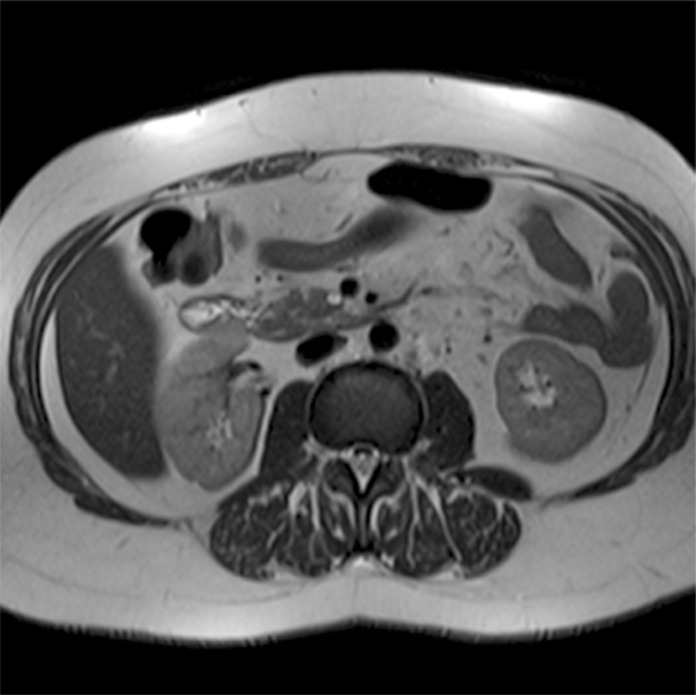
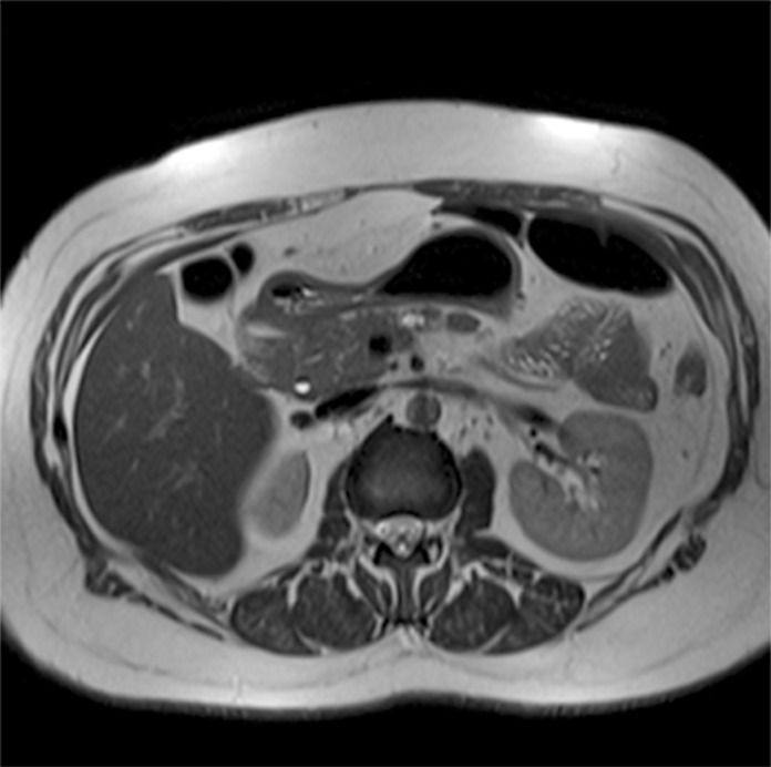
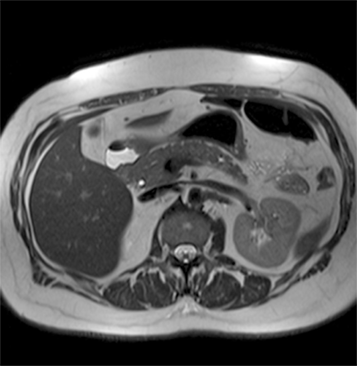
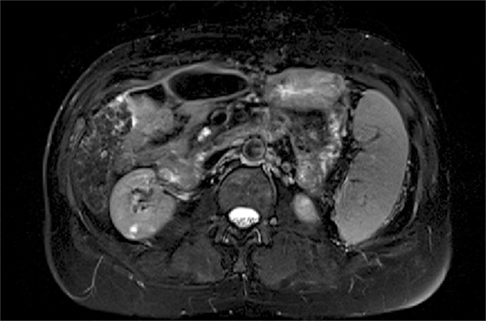
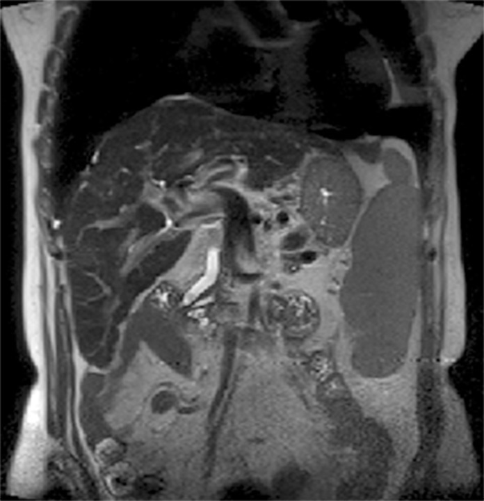
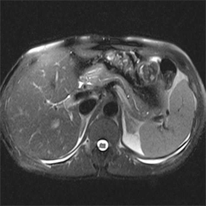
Objectives: To ascertain the prevalence of pancreatic cysts detected incidentally on 3-Tesla magnetic resonance imaging (MRI) of the abdomen and correlate this prevalence with patient age and gender; assess the number, location, and size of these lesions, as well as features suspicious for malignancy; and determine the prevalence of incidentally detected dilatation of the main pancreatic duct (MPD).
Methods: Retrospective analysis of 2,678 reports of patients who underwent abdominal MRI between January 2012 and June 2013. Patients with a known history of pancreatic conditions or surgery were excluded, and the remaining 2,583 reports were examined for the presence of pancreatic cysts, which was then correlated with patient age and gender. We also assessed whether cysts were solitary or multiple, as well as their location within the pancreatic parenchyma, size, and features suspicious for malignancy. Finally, we calculated the prevalence of incidental MPD dilatation, defined as MPD diameter ≥ 2.5 mm.
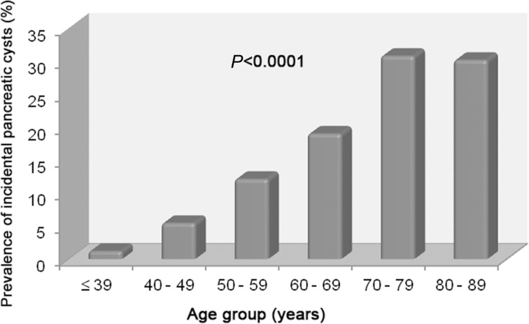
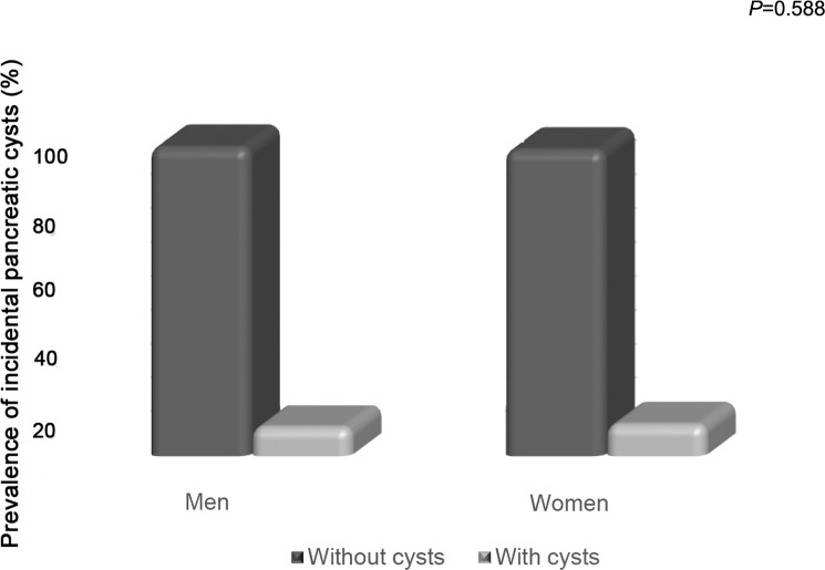
Results: Pancreatic cysts were detected incidentally in 9.3% of patients (239/2,583). The prevalence of pancreatic cysts increased significantly with age (p<0.0001). There were no significant differences in prevalence between men and women (p=0.588). Most cysts were multiple (57.3%), distributed diffusely throughout the pancreas (41.8%), and 5 mm or larger (81.6%). In 12.1% of cases, cysts exhibited features suspicious for malignancy. Overall, 2.7% of subjects exhibited incidental MPD dilatation.
Conclusions: In this sample, the prevalence of pancreatic cysts detected incidentally on 3T MRI of the abdomen was 9.3%. Prevalence increased with age and was not associated with gender. The majority of cysts were multiple, diffusely distributed through the pancreatic parenchyma, and ≥ 5 mm in size; 12.1% were suspicious for malignancy. An estimated 2.7% of subjects had a dilated MPD.
Conflict of interest statement
Figures
References
-
- Balcom IJ, Fernandez-Del Castillo C, Warshaw A. Cystic lesions in the pancreas: when to watch, when to resect. Curr Gastroenterol Rep. 2000; 2: 152–158. - PubMed
MeSH terms
LinkOut - more resources
Full Text Sources
Other Literature Sources
