. 2015 Mar 23;10(3):e0121541.
doi: 10.1371/journal.pone.0121541. eCollection 2015.

Oligodendrocyte birth and death following traumatic brain injury in adult mice

Affiliations

Oligodendrocyte birth and death following traumatic brain injury in adult mice

Krista A Dent et al. PLoS One. .

Abstract

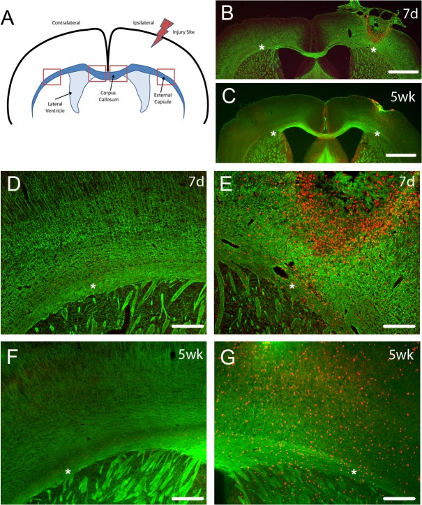
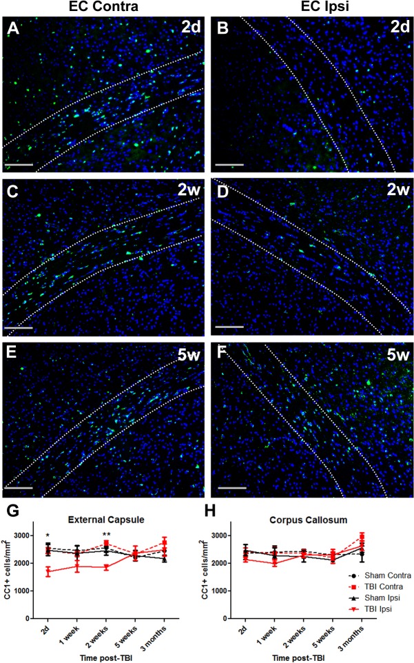
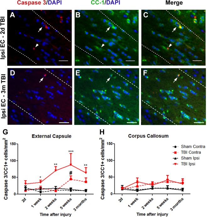
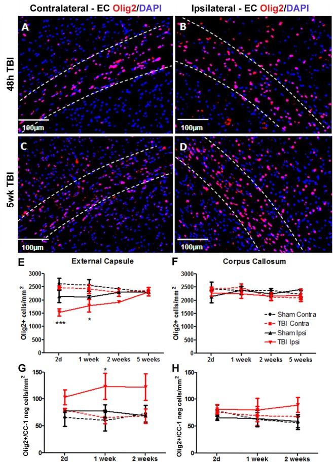
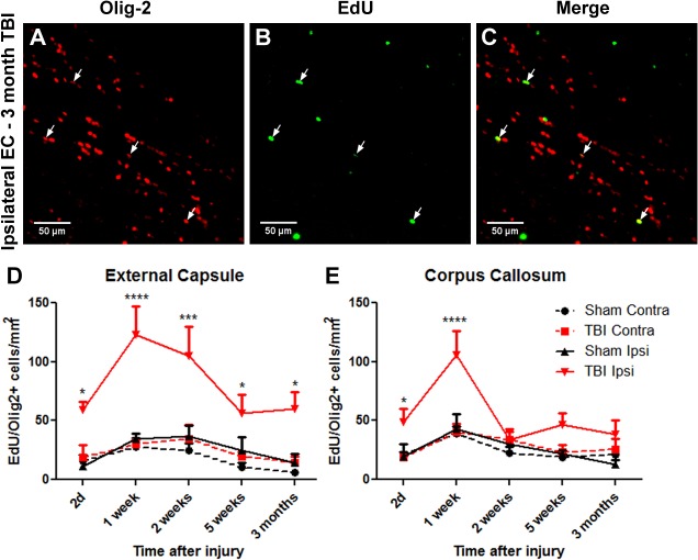
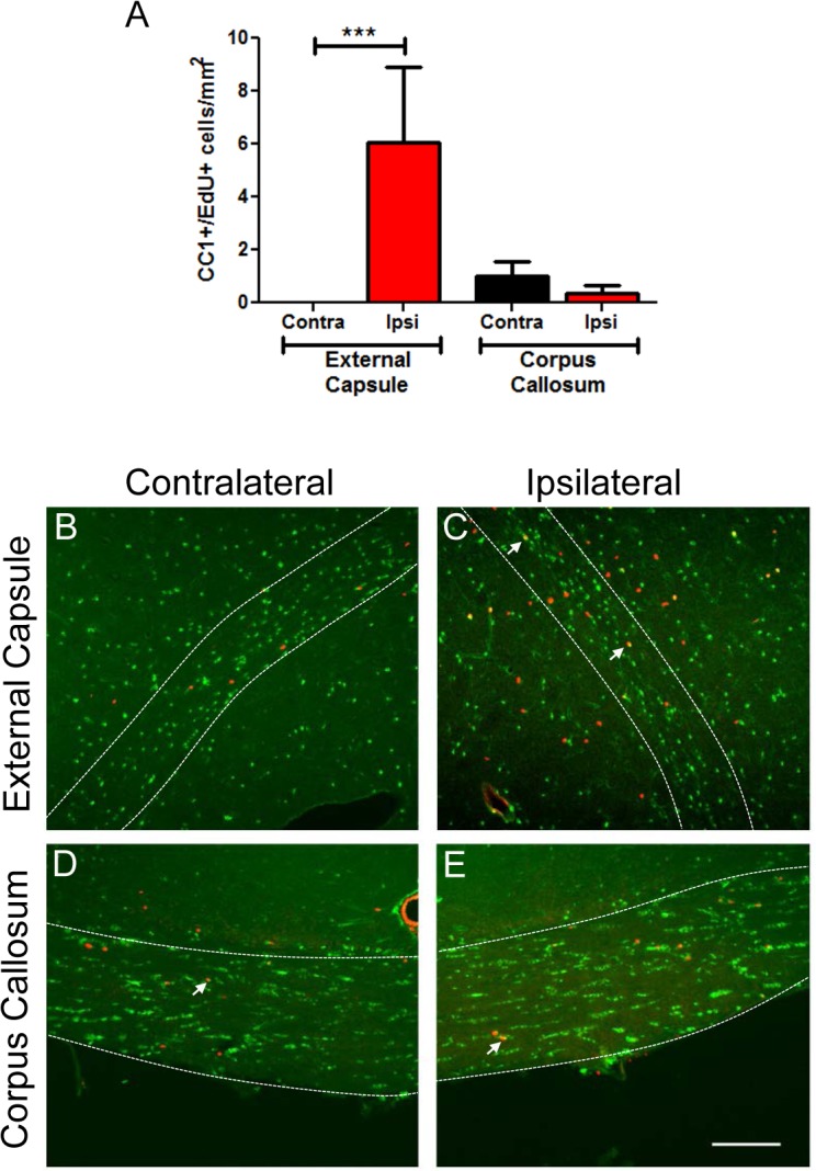
Oligodendrocytes are responsible for producing and maintaining myelin throughout the CNS. One of the pathological features observed following traumatic brain injury (TBI) is the progressive demyelination and degeneration of axons within white matter tracts. While the effect of TBI on axonal health has been well documented, there is limited information regarding the response of oligodendrocytes within these areas. The aim of this study was to characterize the response of both mature oligodendrocytes and immature proliferative oligodendrocyte lineage cells across a 3 month timecourse following TBI. A computer-controlled cortical impact model was used to produce a focal lesion in the left motor cortex of adult mice. Immunohistochemical analyses were performed at 48 hours, 7 days, 2 weeks, 5 weeks and 3 months following injury to assess the prevalence of mature CC-1+ oligodendrocyte cell death, immature Olig2+ cell proliferation and longer term survival in the corpus callosum and external capsule. Decreased CC-1 immunoreactivity was observed in white matter adjacent to the site of injury from 2 days to 2 weeks post TBI, with ongoing mature oligodendrocyte apoptosis after this time. Conversely, proliferation of Olig2+ cells was observed as early as 48 hours post TBI and significant numbers of these cells and their progeny survived and remained in the external capsule within the injured hemisphere until at least 3 months post injury. These findings demonstrate that immature oligodendrocyte lineage cells respond to TBI by replacing oligodendrocytes lost due to damage and that this process occurs for months after injury.


Conflict of interest statement

Competing Interests: The authors have declared that no competing interests exist.

Figures

Fig 1
Fig 1. Diagram of TBI location and CNPase staining of myelin fibers.
(A) The location of the TBI was in the cortex above the external capsule, with analyses performed on oligodendrocyte lineage cells in the ipsilateral and contralateral external capsule and corpus callosum (regions analyzed indicated by boxes). CNPase staining (B-D) of myelin (green) and EdU (red) labelling of proliferative cells at 7 days (7d) and 5 weeks (5wk) after TBI showed that this injury largely affected the cortex but also perturbed myelin tracts in the ipsilateral external capsule (B, C, E, G) with few EdU labeled cells on the contralateral side (D, F). This did not usually result in extensive long term myelinated fiber loss, as indicated by CNPase staining at 5 weeks (wk) after TBI, with ipsilateral external capsule showing similar levels of myelination as contralateral external capsule. Scale bars in B,C 1mm; D-G 200μm. Asterisks in B-G indicate the external capsule with panels D and E an enlargement of B and panels F and G an enlargement of C.
Fig 2
Fig 2. CC-1+ immunostained cell numbers decrease by 2 days after TBI and return to control levels by 5 weeks.
Brain sections from TBI mice were immunostained for mature CC-1 expressing oligodendrocytes and counterstained with DAPI. Panels show contralateral (Contra) and ipsilateral (Ipsi) external capsule (EC) at (A,B) 2 days (2d), (C,D) 2 weeks (2w) and (E,F) 5 weeks (5w) (G) In the external capsule numbers of CC-1+ oligodendrocytes were decreased by 2d after injury, remained decreased at 2 weeks and returned to normal levels by 5 weeks. (H) There was no significant loss of CC-1+ oligodendrocytes in the corpus callosum. Results in panels G and H show mean+/-SEM of sections from n = 3–4 mice per timepoint. Statistical comparisons were made using ANOVA (F(19,58) = 2.281, p<0.01 for external capsule in G, F(19,54) = 1.51, p = 0.12 for corpus callosum in H) and comparisons were made between ipsilateral TBI and sham at each timepoint using the Bonferroni post hoc test; *p<0.05, **p<0.01.
Fig 3
Fig 3. Activated Caspase-3+/CC-1+ cell numbers increase to a maximum at 5 weeks after TBI.
Brain sections were immunostained for activated caspase-3 and CC-1 and counterstained with DAPI. Panels show ipsilateral external capsule (EC) of brains at 2d (A-C) and 3 months (D-F) post-TBI: (A, D) Caspase-3 and DAPI, (B, E) CC-1 and DAPI, (C F) merged. Arrows show co-labelled cells, indicative of apoptotic oligodendrocytes. In panels A-C, (G) the arrows indicate an early apoptotic CC-1+ oligodendrocyte with cytoplasmic CC-1 and Caspase-3 staining and a pyknotic nucleus, while the arrowhead indicates a later apoptotic cell where Caspase-3 is present in the nucleus. In the ipsilateral external capsule, numbers of Caspase-3+/CC-1+ oligodendrocytes were significantly increased by 1 week after injury. Apoptotic oligodendrocytes were also elevated in the contralateral external capsule by 5 weeks. (H) No significant oligodendrocyte apoptosis was detected in the corpus callosum. Results in panels D and E show mean+/-SEM of sections from n = 3–4 mice per timepoint. Statistical comparisons were made using ANOVA (F(19,58) = 4.637, p<0.0001 for external capsule in D, F(3,12) = 5.638, p = 0.01 for the 1 week timepoint in D and F(19,58) = 1.569, p = 0.097 for corpus callosum in E) and comparisons were made between ipsilateral TBI and sham at each timepoint using the Bonferroni post hoc test; *p<0.05; **p<0.01, **p<0.001; # Contralateral TBI was significantly different to sham by t-test, p<0.05.
Fig 4
Fig 4. Olig2 cells in external capsule at 48h and 5 weeks after TBI.
Brain sections were immunostained for Olig2 and counterstained with DAPI. Panels show (A,C) contralateral and (B,D) ipsilateral external capsule (EC) at (A,B) 48h and (C,D) 5 weeks (5wk) post-TBI. The bordered regions indicate external capsule. (E) In the external capsule numbers of Olig2+ oligodendrocytes were decreased by 2d after injury, remained decreased at 1 week and returned to normal levels by 2 weeks. (F) There was no significant loss of Olig2+ oligodendrocytes in the corpus callosum. (G) There was an increase in numbers of Olig2+/CC-1 negative cells at 1 week post-injury but (H) no increase in these cells in the corpus callosum. Results in panels E-H show mean+/-SEM of sections from n = 3–4 mice per timepoint. Statistical comparisons were made using ANOVA (F(15,38) = 3.624, p<0.001 for external capsule in E, F(11,30) = 2.062, p = 0.05 for external capsule in G) and comparisons were made between ipsilateral TBI and sham at each timepoint using the Bonferroni post hoc test; *p<0.05, ***p<0.001.
Fig 5
Fig 5. Proliferative Olig2 cell numbers increase within 2d after TBI and remain elevated to at least 3 months.
Brain sections were immunostained for Olig2 and counterstained for EdU and DAPI. Panels show ipsilateral external capsule (EC) of a 3 month post-TBI brain: (A) Olig2, (B) EdU, (C) merged. Arrows show some co-labelled cells, indicative of oligodendrocytes generated after TBI. (D) In the ipsilateral external capsule, numbers of EdU+/Olig2+ oligodendrocytes were significantly increased within 2d after injury, were further increased at 1 and 2 weeks, with lower numbers by 3 months. (E) A significant increase in EdU+ oligodendrocytes was also detected in the corpus callosum between 2d and 1 week, decreasing to control levels by 2 weeks. Results in panels D and E show mean+/-SEM of sections from n = 3–4 mice per timepoint. Statistical comparisons were made using ANOVA (F(19,58) = 7.545, p<0.0001 for external capsule in D and F(19,58) = 4.808, p<0.0001 for corpus callosum in E) and comparisons were made between TBI and sham at each timepoint using the Bonferroni post hoc test; *p<0.05, ***p<0.001.
Fig 6
Fig 6. EdU+/CC-1+ cells are present to at least 3 months post-TBI.
Brain sections were immunostained for CC-1 and counterstained with EdU and DAPI. (A) Increased numbers of EdU+/CC-1+ cells were present in ipsilateral external capsule at 5 weeks post injury, compared to the contralateral side or corpus callosum. EdU+/CC-1+ cells were also present at 3 months post-TBI. Panels show (B,C) external capsule and (D,E) corpus callosum at 3 months post-TBI. Examples of EdU+/CC-1+ oligodendrocytes are indicated by arrows. The bordered regions indicate external capsule or corpus callosum respectively. Scale bar 50μm. Results in panel A show mean+/-SEM of sections from n = 3 mice per group. Statistical comparisons were made using ANOVA (F(3,8) = 3.784, p = 0.05) and comparisons were made between ipsilateral and contralateral sides using the Bonferroni post hoc test; *p<0.05.

References

    1. Bramlett HM, Dietrich WD. Quantitative structural changes in white and gray matter 1 year following traumatic brain injury in rats. Acta Neuropathol. 2002; 103: 607–614. - PubMed
    1. Johnson SC, Bigler EDB RB, Blatter DD. White Matter Atrophy, Ventricular Dilation, and Intellectual Functioning Following Traumatic Brain Injury. Neuropsych. 1994; 8: 307–315.
    1. Levin HS, Williams DH, Valastro M, Eisenberg HM, Crofford MJ, Handei SF. Corpus callosal atrophy following closed head injury: detection with magnetic resonance imaging. J. Neurosurg. 1990; 73: 77–81. - PubMed
    1. Johnson VE, Stewart JE, Begbie FD, Trojanowski JQ, Smith DH, Stewart W. Inflammation and white matter degeneration persist for years after a single traumatic brain injury. Brain 2013; 136: 28–42. 10.1093/brain/aws322 - DOI - PMC - PubMed
    1. Flygt J, Djupsjo A, Lenne F, Marklund N. Myelin loss and oligodendrocyte pathology in white matter tracts following traumatic brain injury in the rat. Eur. J. Neurosci. 2013; 38: 2153–2165. 10.1111/ejn.12179 - DOI - PubMed

Publication types

Substances