. 2015 Mar 23;10(3):e0119859.
doi: 10.1371/journal.pone.0119859. eCollection 2015.

Potent anti-proliferative, pro-apoptotic activity of the Maytenus royleanus extract against prostate cancer cells: evidence in in-vitro and in-vivo models

Affiliations

Potent anti-proliferative, pro-apoptotic activity of the Maytenus royleanus extract against prostate cancer cells: evidence in in-vitro and in-vivo models

Maria Shabbir et al. PLoS One. .

Retraction in

Abstract

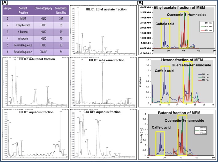
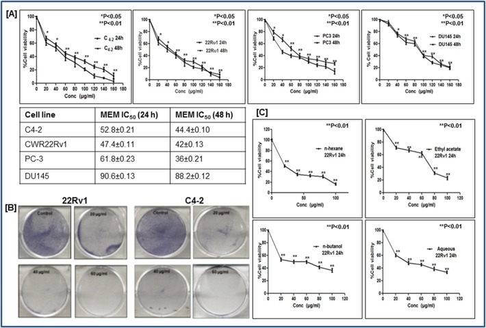
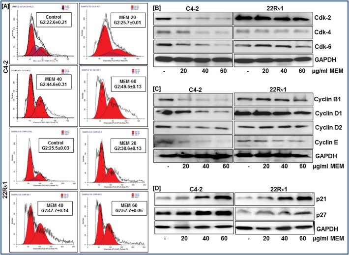
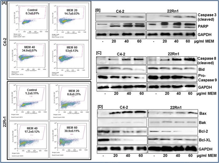
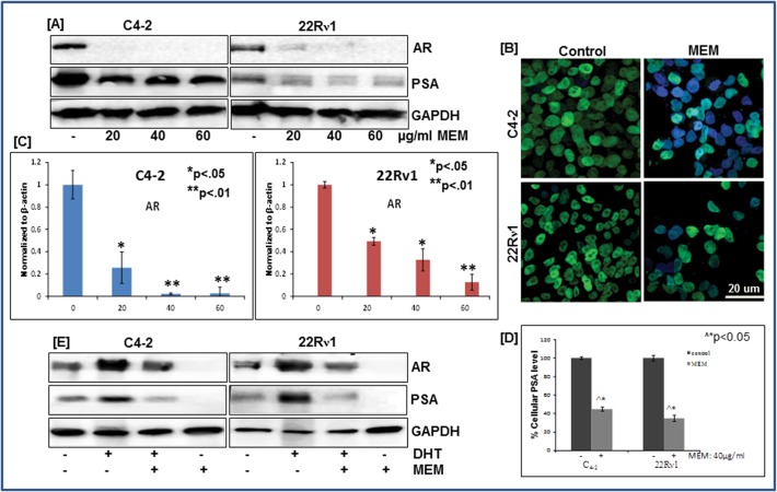
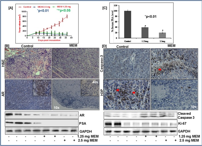
Prostate cancer is a leading of cause of cancer related death in men. Despite intensive investment in improving early diagnosis, it often escapes timely detection. Mortality remains high in advanced stage prostate cancer where palliative care remains the only option. Effective strategies are therefore needed to prevent the occurrence and progression of the disease. Plant-derived compounds have been an important source of several clinically useful anti-cancer agents and offer an attractive approach against prostate cancer. We previously showed that the methanol extract of Maytenus royleanus (MEM) leaves and its fractions possess significant antioxidant activity with therapeutic potential against free-radical associated damages. The present study evaluated the anti-proliferative activity of MEM in the prostate cancer model system. Analysis of MEM and its various fractions revealed the presence of triterpenoids, flavonoids and tannins, conjugated to one or more polar groups and carbohydrate moieties. Further studies against known standards established the existence of caffeic acid and quercetin 3-rhamnoside in varying concentration in different MEM fractions. Time course analysis of MEM treated prostate cancer cells indicated significant decrease in cell viability, assessed by MTT and clonogenic survival assays. This was accompanied by G2 phase arrest of cell cycle, downregulation of cyclin/cdk network and increase in cdk inhibitors. MEM treated cells exhibited cleavage of Caspase-3 and PARP, and modulation of apoptotic proteins, establishing apoptosis as the primary mechanism of cell death. Notably MEM suppressed AR/PSA signaling both in prostate cancer cell cultures and in the in vivo model. Intraperitoneal injection of MEM (1.25 and 2.5 mg/ animal) to athymic nude mice implanted with androgen sensitive CWR22Rν1 cells showed significant inhibition in tumor growth and decreased serum PSA levels reciprocating in vitro findings. Taken together, our data suggest that MEM may be explored further for its potential therapeutic effects against prostate cancer progression in humans.


Conflict of interest statement

Competing Interests: The authors have declared that no competing interests exist.

Figures

Fig 1
Fig 1. Phytochemical fingerprint of MEM.
a. Table showing total number of compounds found in MEM and its various fractions with HILIC and C18 RP chromatography, based on retention time and molar mass; Total ion chromatograms of various fractions: ethyl acetate fraction (-ve HILIC); n-butanol fraction (-ve HILIC); n-hexane fraction (-ve HILIC); aqueous fraction (-ve HILIC); aqueous fraction (-ve C18 RP). b. UV chromatograms of ethyl acetate n-hexane and n-butanol fractions of MEM, at 280, 320 & 370 nm, with caffeic acid and quercetin 3-rhamnoside internal standards.
Fig 2
Fig 2. MEM inhibits growth and viability of prostate cancer cells.
a. Prostate cancer cells were treated with MEM for 24/48h, and cell viability was determined by MTT assay. Table shows the IC50 of CWR22Rν1, C4-2, PC-3 and DU145 at 24 and 48h. Mean ± SD of experiments performed in triplicate is shown. b. Dose-dependent effect of MEM on clonogenecity of CWR22Rν1 and C4-2 cells as detected by colony formation assay. Details are described in material methods. c. Effect of various fractions (n-hexane, ethyl acetate, n-butanol and aqueous) on viability of CWR22R ν1 cells, determined by MTT assay. *p<0.05 and **p<0.01 was considered statistically significant.
Fig 3
Fig 3. MEM induces G2 phase arrest of prostate cancer cells.
a. CWR22Rν1 and C4-2 cells treated with MEM for 24h were stained with propidium iodide and analyzed by flow cytometry. Percentage of cell population in G2-phase of the cell cycle is shown in the box of each histogram. Mean ± SD of experiments performed in triplicate is shown. b-d. Effect of MEM treatment on cell cycle regulatory proteins: Whole cell lysates of CWR22Rν1 and C4-2 cells with/without MEM (20–60 μg/ml: 24h) were subjected to SDS-polyacrylamide gel electrophoresis. Equal loading was confirmed by reprobing with GAPDH. The immunoblots shown are representative of three independent experiments with similar results.
Fig 4
Fig 4. MEM induces apoptosis through activation of intrinsic and extrinsic pathway.
a. CWR22Rν1 and C4-2 cells treated with MEM (20–60 μg/ml: 24h) were labeled with FITC and analyzed by flow cytometry. Percentage of apoptotic cells with the corresponding dose of MEM is shown in each histogram. Mean ± SD of experiments performed in triplicate is shown. b-d. Whole cell lysates of CWR22Rν1 and C4-2 cells with/without MEM (20–60 μg/ml: 24h) treatment were subjected to SDS-polyacrylamide gel electrophoresis. Effect of MEM treatment on proteins involved in apoptosis pathways. Equal loading was confirmed by reprobing with GAPDH. The immunoblots shown are representative of three independent experiments with similar results.
Fig 5
Fig 5. MEM decreases AR and PSA expression in prostate cancer cells.
a. Whole cell lysates of CWR22Rν1 and C4-2 cells with/without MEM (20–60 μg/ml: 24h) were subjected to SDS-polyacrylamide gel electrophoresis. Equal loading was confirmed by reprobing with GAPDH. The immunoblots shown are representative of three independent experiments with similar results. b. Immunofluorescence microscopy with Alexa fluor staining of AR (green fluorescence) in CWR22Rν1 and C4-2 cells, counter stained with DAPI (blue fluorescence). c. qPCR analysis of MEM treated CWR22Rν1 and C4-2 cells for changes in AR mRNA levels. The data expressed as fold change represent the mean±standard errors experiments performed in triplicates where *p < 0.05, **p < 0.01 was considered significant vs control. d. Effect of MEM on secreted levels of PSA in CWR22Rν1 and C4-2 cells treated with MEM (40 μg/ml: 24h). PSA levels were determined by ELISA, as described in Materials and Methods; the figures represent the data of three experiments, each conducted in duplicate, where ^*p<0.05 MEM treated vs DMSO treated control cells was considered significant. e. Effect of MEM on DHT stimulated protein expression of AR and PSA: Whole cell lysates of CWR22Rν1 and C4-2 cells treated with MEM (40 μg/ml: 24h) were subjected to SDS-polyacrylamide gel electrophoresis. Equal loading was confirmed by reprobing with GAPDH. The immunoblots shown are representative of three independent experiments with similar results.
Fig 6
Fig 6. MEM inhibits growth of CWR22Rν1xenografts in athymic nude mice.
a. Average tumor volume of DMSO, 2.5mg & 1.25mg MEM injected mice plotted over days after CWR22Rν1 tumor xenografts implanted in athymic nude mice. Values represent mean±SE of six mice, where MEM (1.25mg) *^p<0.05 and MEM (2.5mg) *p<0.01 versus DMSO treated control was considered significant. b. Top panel: H&E staining of MEM treated xenograft tumor tissue vs control. Immunohistochemical analysis of AR in MEM treated tumor tissue vs untreated control. Bottom panel: Whole cell lysates of tumor xenografts from animals treated with/without MEM were subjected to SDS-polyacrylamide gel electrophoresis. Equal loading was confirmed by reprobing with GAPDH. The immunoblots shown are representative of three independent experiments with similar results. c. Serum PSA levels of MEM treated mice were analyzed by ELISA, as described in Materials and Methods. MEM (1.25mg) and MEM (2.5mg) *p<0.01 versus DMSO treated control was considered significant. d. Top panel: Immunohistochemical analysis of H3P & cleaved caspase 3 in MEM treated tumor tissue vs untreated control. Bottom panel: Whole cell lysates of tumor xenografts from animals treated with/without MEM were subjected to SDS-polyacrylamide gel electrophoresis. Equal loading was confirmed by reprobing with GAPDH. The immunoblots shown are representative of three independent experiments with similar results.
Fig 7
Fig 7. MEM treatment is not associated with adverse side effects.
a. Mice weight was taken twice weekly and values represent mean±SD of six mice. (B&C) H&E staining was performed for toxicity studies on heart, brain, lung, kidney and liver tissues of mice treated with DMSO or MEM.

Similar articles

Cited by

References

    1. Siegel R, Naishadham D, Jemal A. Cancer statistics, 2012. CA: a cancer journal for clinicians. 2012;62(1):10–29. Epub 2012/01/13. - PubMed
    1. . wwwcancerorg/.
    1. Solit DB, Scher HI, Rosen N. Hsp90 as a therapeutic target in prostate cancer. Seminars in oncology. 2003;30(5):709–16. Epub 2003/10/23. - PubMed
    1. Visakorpi T, Hyytinen E, Koivisto P, Tanner M, Keinanen R, Palmberg C, et al. In vivo amplification of the androgen receptor gene and progression of human prostate cancer. Nature genetics. 1995;9(4):401–6. Epub 1995/04/01. - PubMed
    1. Gregory CW, Hamil KG, Kim D, Hall SH, Pretlow TG, Mohler JL, et al. Androgen receptor expression in androgen-independent prostate cancer is associated with increased expression of androgen-regulated genes. Cancer research. 1998;58(24):5718–24. Epub 1998/12/29. - PubMed

Publication types

MeSH terms