. 2015 Apr 2;96(4):657-65.
doi: 10.1016/j.ajhg.2015.02.007. Epub 2015 Mar 19.

Mutations in HPCA cause autosomal-recessive primary isolated dystonia

Affiliations

Mutations in HPCA cause autosomal-recessive primary isolated dystonia

Gavin Charlesworth et al. Am J Hum Genet. .

Abstract

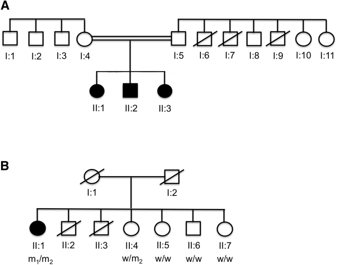
Reports of primary isolated dystonia inherited in an autosomal-recessive (AR) manner, often lumped together as "DYT2 dystonia," have appeared in the scientific literature for several decades, but no genetic cause has been identified to date. Using a combination of homozygosity mapping and whole-exome sequencing in a consanguineous kindred affected by AR isolated dystonia, we identified homozygous mutations in HPCA, a gene encoding a neuronal calcium sensor protein found almost exclusively in the brain and at particularly high levels in the striatum, as the cause of disease in this family. Subsequently, compound-heterozygous mutations in HPCA were also identified in a second independent kindred affected by AR isolated dystonia. Functional studies suggest that hippocalcin might play a role in regulating voltage-dependent calcium channels. The identification of mutations in HPCA as a cause of AR primary isolated dystonia paves the way for further studies to assess whether "DYT2 dystonia" is a genetically homogeneous condition or not.


Figures

Figure 1
Figure 1
Genetic Pedigrees for Families Affected by Mutations in HPCA Abbreviated genetic pedigrees are shown for the core members of (A) the index family and (B) the second family identified to be affected by compound-heterozygous mutations in HPCA. For the family in (B), the results of the segregation analysis are shown under each individual: WT, wild-type allele; M1, c.212C>A (p.Thr71Asn) mutation; M2, c.568G>C (Ala190Thr) mutation. The results are consistent with AR inheritance of dystonia due to biallelic mutations in HPCA. Individual II:6 did not report any symptoms suggestive of dystonia; however, this could not be confirmed by examination because he did not live in the UK.
Figure 2
Figure 2
Expression Data for LAPTM5 and HPCA (A and B) Publically available expressed sequence tag (EST) data for (A) LAPTM5 and (B) HPCA demonstrate that both genes show relatively tissue-specific expression patterns. LAPTM5 is predominantly expressed in hemopoietic tissues, whereas HPCA is almost exclusively expressed in the brain. (C) Boxplot of mRNA expression levels for HPCA in ten CNS regions. Data are based on in-house exon array experiments and plotted on a log2 scale (y axis). A full description of the samples used, the methods of RNA isolation and processing, and data-analysis steps can be found in Trabzuni et al. This plot shows significant variation in HPCA transcript expression across the ten CNS regions analyzed: putamen (PUTM, n = 129), hippocampus (HIPP, n = 122), temporal cortex (TCTX, n = 119), frontal cortex (FCTX, n = 127), occipital cortex (OCTX, n = 129), thalamus (THAL, n = 124), cerebellar cortex (CRBL, n = 130), substantia nigra (SNIG, n = 101), intralobular white matter (WHMT, n = 131), and medulla (specifically inferior olivary nucleus, MEDU, n = 109). HPCA mRNA expression is highest in the putamen, followed closely by the hippocampus. Expression is also high in the cortex. Whiskers extend from the box to 1.5× the inter-quartile range.
Figure 3
Figure 3
Summary of the Functional Studies in Hpca-Knockdown Neuronal-Astrocytic Co-cultures Astrocytes and neurons from primary cortical co-culture were loaded for 30 min at room temperature with 5 μM fura-2 AM and 0.005% pluronic acid in a HEPES-buffered salt solution composed of 156 mM NaCl, 3 mM KCl, 2 mM MgSO4, 1.25 mM KH2PO4, 2 mM CaCl2, 10 mM glucose, and 10 mM HEPES (pH was adjusted to 7.35 with NaOH). Fluorescence measurements were obtained on an epifluorescence inverted microscope equipped with a 20× fluorite objective. [Ca2+]c was monitored in single cells with excitation light provided by a Xenon arc lamp, and the beam passed through a monochromator at 340 and 380 nm (Cairn Research). Emitted fluorescence light was reflected through a 515-nm longpass filter to a charge-coupled-device camera (Retiga, QImaging) and digitized to a 12-bit resolution. All imaging data were collected and analyzed with software from Andor IQ. The fura-2 data were not calibrated in terms of [Ca2+]c because of the uncertainty arising from the use of different calibration techniques. Areas for the analysis were chosen depending on the GFP fluorescence intensity, and four independent experiments were performed for each condition. The figure shows representative traces of [Ca2+]c response to physiological stimuli as measured by changes in fura-2 fluorescence intensity. Compared to neurons from the scrambled or empty control (A, black triangle trace), Hpca-knockdown neurons showed no rise in [Ca2+]c in response to depolarization of the plasma membrane with 50 mM KCl (A, dark-gray trace; B, dark-gray bars). In addition, the amplitude of the response to physiological concentration of glutamate (5 μM) was lower in the Hpca-knockdown neurons than in control cells (B, black bars), although this decrease was not statistically significant. Hpca knockdown also diminished the amplitude of the [Ca2+]c response of astrocytes to an ATP stimulus (100 μM) (C and D, light-gray trace and bars). Error bars represent the SEM, and asterisks represents statistical significance (∗∗∗p < 0.0001, p < 0.05).

Comment in

References

    1. Fahn S. Concept and classification of dystonia. Adv. Neurol. 1988;50:1–8. - PubMed
    1. Ozelius L.J., Hewett J.W., Page C.E., Bressman S.B., Kramer P.L., Shalish C., de Leon D., Brin M.F., Raymond D., Corey D.P. The early-onset torsion dystonia gene (DYT1) encodes an ATP-binding protein. Nat. Genet. 1997;17:40–48. - PubMed
    1. Fuchs T., Gavarini S., Saunders-Pullman R., Raymond D., Ehrlich M.E., Bressman S.B., Ozelius L.J. Mutations in the THAP1 gene are responsible for DYT6 primary torsion dystonia. Nat. Genet. 2009;41:286–288. - PubMed
    1. Xiao J., Uitti R.J., Zhao Y., Vemula S.R., Perlmutter J.S., Wszolek Z.K., Maraganore D.M., Auburger G., Leube B., Lehnhoff K., LeDoux M.S. Mutations in CIZ1 cause adult onset primary cervical dystonia. Ann. Neurol. 2012;71:458–469. - PMC - PubMed
    1. Charlesworth G., Plagnol V., Holmström K.M., Bras J., Sheerin U.M., Preza E., Rubio-Agusti I., Ryten M., Schneider S.A., Stamelou M. Mutations in ANO3 cause dominant craniocervical dystonia: ion channel implicated in pathogenesis. Am. J. Hum. Genet. 2012;91:1041–1050. - PMC - PubMed

Publication types