. 2015 Mar 23;11(3):e1004735.
doi: 10.1371/journal.ppat.1004735. eCollection 2015 Mar.

An 18 kDa scaffold protein is critical for Staphylococcus epidermidis biofilm formation

Affiliations

An 18 kDa scaffold protein is critical for Staphylococcus epidermidis biofilm formation

Rahel Decker et al. PLoS Pathog. .

Abstract

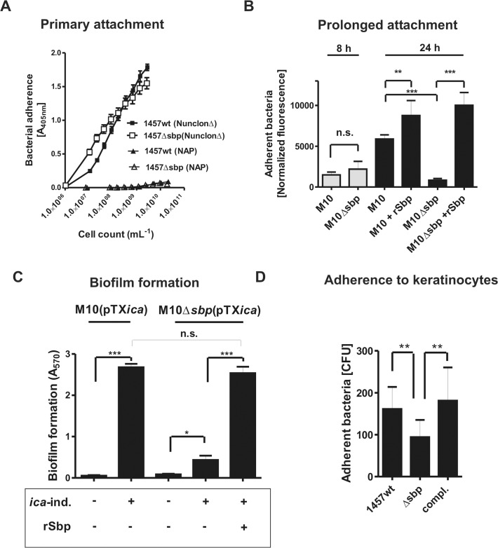
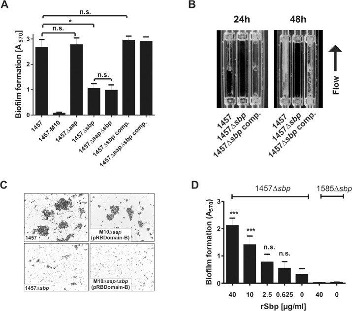
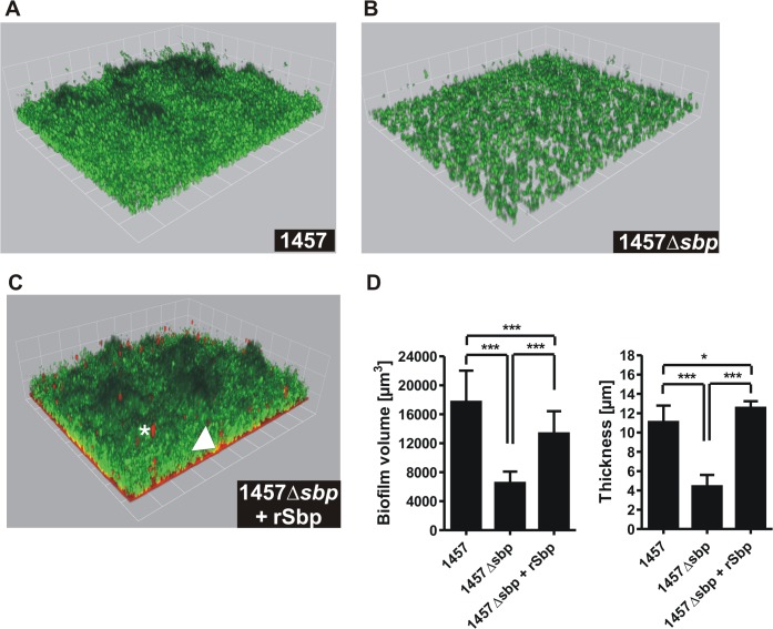
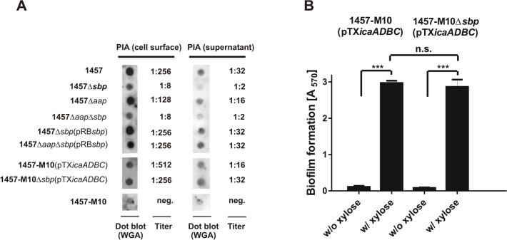
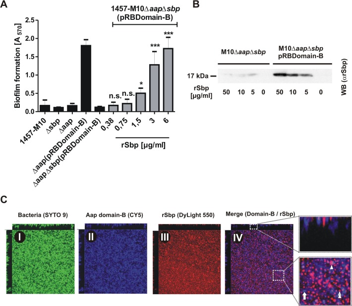
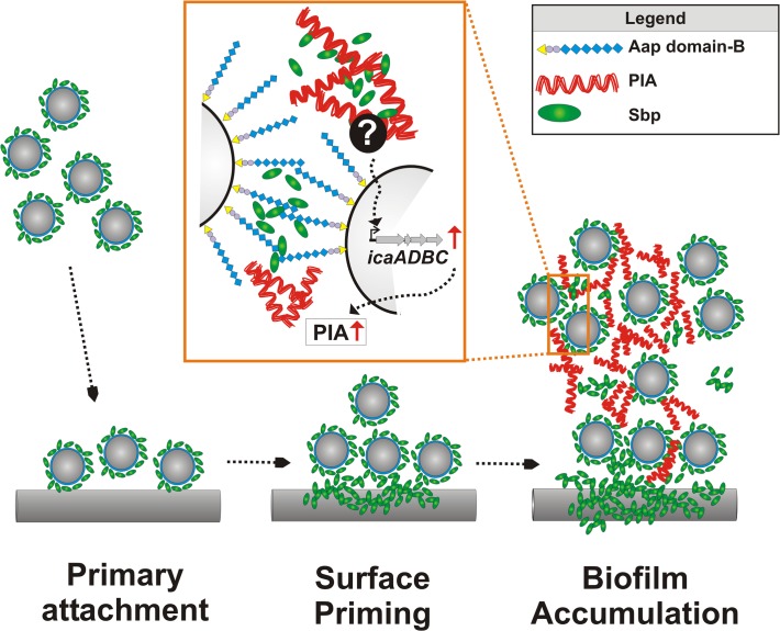
Virulence of the nosocomial pathogen Staphylococcus epidermidis is crucially linked to formation of adherent biofilms on artificial surfaces. Biofilm assembly is significantly fostered by production of a bacteria derived extracellular matrix. However, the matrix composition, spatial organization, and relevance of specific molecular interactions for integration of bacterial cells into the multilayered biofilm community are not fully understood. Here we report on the function of novel 18 kDa Small basic protein (Sbp) that was isolated from S. epidermidis biofilm matrix preparations by an affinity chromatographic approach. Sbp accumulates within the biofilm matrix, being preferentially deposited at the biofilm-substratum interface. Analysis of Sbp-negative S. epidermidis mutants demonstrated the importance of Sbp for sustained colonization of abiotic surfaces, but also epithelial cells. In addition, Sbp promotes assembly of S. epidermidis cell aggregates and establishment of multilayered biofilms by influencing polysaccharide intercellular-adhesin (PIA) and accumulation associated protein (Aap) mediated intercellular aggregation. While inactivation of Sbp indirectly resulted in reduced PIA-synthesis and biofilm formation, Sbp serves as an essential ligand during Aap domain-B mediated biofilm accumulation. Our data support the conclusion that Sbp serves as an S. epidermidis biofilm scaffold protein that significantly contributes to key steps of surface colonization. Sbp-negative S. epidermidis mutants showed no attenuated virulence in a mouse catheter infection model. Nevertheless, the high prevalence of sbp in commensal and invasive S. epidermidis populations suggests that Sbp plays a significant role as a co-factor during both multi-factorial commensal colonization and infection of artificial surfaces.


Conflict of interest statement

The authors have declared that no competing interests exist.

Figures

Fig 1
Fig 1. Affinity purification of Aap domain-B interaction partners from crude biofilm matrix preparations.
(A) SDS-PAGE analysis of fractions collected from affinity chromatography in which recombinant Aap domain-B was coupled to an NHS-activated sepharose column (GE Life Sciences, Freiburg, Germany). Lane 1 shows the input protein preparation that was loaded onto the column. Lanes 2–8: fractions collected during washing with PBS (pH 7.4). Lanes 9–11: fractions collected during elution with Tris-HCl. Proteins were made visible by silver staining (Pierce silver stain kit). Mass spectrometry identified proteins from lanes 9–11 as AtlE (arrows) and hypothetical protein YP_187866 (star). (B) Ligand binding assay showing binding of rDomain-B to proteins eluted during affinity purification. Fractions 9–11 were separated by SDS-PAGE and blotted onto a PVDF membrane. After blocking the membrane was incubated with biotinylated rDomain-B (5 μg/ml) and bound ligand was detected by peroxidase-coupled streptavidin and chemiluminescence.
Fig 2
Fig 2. General features of Small basic protein (Sbp) and Biochemical analysis of rDomain-B—Sbp interactions.
(A) Schematic representation of Sbp. The protein with an anticipated MW of 18 kDa is Encoded by a 513 nt ORF. Bioinformatic analysis identified an N-terminal export signal (E; aa 1–20) but no additional conserved domains. (B) Binding of rSbp to rDomain-B immobilized on wells of a 96-well microtiter plate. Increasing amounts of rSbp were allowed to bind to immobilized rDomain-B and bound rSbp was detected by rabbit anti-rSbp antiserum and a alkaline phosphatase coupled goat ant-rabbit IgG. Absorbance at 405 nM is taken as a function of rSbp binding. The untreated (native) polystyrene surface served as a control. Each data point represents mean of 4 values obtained in two independent experiments. Error bars indicate standard deviation. (C) Competitive inhibition of rSbp binding to immobilized rDomain-B. rSbp (150 μg/ml) was incubated with increasing amounts of rDomain-B as indicated. After one hour, binding of rSbp to immobilized rDomain-B was tested as described above. Inhibition of rSbp binding was estimated using the formula (1-A405 rSbp w/ rDomain-B / A405 rSbp w/o rDomain-B) x 100. Each data point represent mean of four values obtained in two independent experiments. Error bars indicate standard deviation. (D) Effect of divalent cations on rSbp–rDomain-B interactions. rSbp was allowed to bind to immobilized rDomain-B in the absence or presence of ZnCl2 and MgCl2, respectively. The unmodified polystyrene surface served as a control (Cntrl). Bound rSbp was detected as described above. Columns represent mean of four values obtained in two independent experiments. Error bars indicate standard deviation. Significant binding differences in the presence of ZnCl2 or MgCl2 as compared to the control without additional salts (p<0.05, one-way ANOVA with Dunnett’s correction for multiple testing) are indicated (***, p<0.001). n.s., not significant.
Fig 3
Fig 3. Spatial distribution of Sbp in S. epidermidis cultures.
(A) Preparations of cell wall associated proteins and 10-fold concentrated supernatants from S. epidermidis 1457 (biofilm-positive) and 1585 (biofilm-negative) after static overnight growth were separated by SDS PAGE and blotted onto PVDF-membranes. Sbp was detected after incubation with rabbit anti-rSbp antiserum and anti-rabbit IgG coupled to peroxidase by chemiluminescence. (B) Growth phase dependent regulation of Sbp. Cell wall associated proteins were prepared from S. epidermidis 1457, 1457ΔsarA, and 1457ΔrnaIII at different time points during adherent growth in TSB. At each time point cell numbers were adjusted to an identical A600 before cell surface associated proteins were isolated by boiling in LDS buffer. After separation of surface associated proteins by SDS-PAGE and blotting onto a PVDF membrane Sbp was detected by chemiluminescence using a rabbit anti-rSbp antiserum and a peroxidase-coupled anti-rabbit IgG. SDS-PAGE analysis proved loading of gels with similar total protein amounts (S2 Fig.).
Fig 4
Fig 4. Distribution of Sbp in living biofilms.
Confocal laser scanning microscopy images of S. epidermidis 1457 biofilms grown overnight under static conditions. Bacteria were stained using DAPI (blue), while PIA and Sbp were detected by WGA coupled to Alexa 568 (red) and rabbit anti-rSbp / anti-rabbit IgG-Alexa488 (green), respectively. (A, B) Confocal images of DAPI-labelled S. epidermidis 1457 (blue), PIA (red) and Sbp (green). Two dimensional projections along the XY / XZ-axes (A) and a three dimensional reconstruction (B) clearly show a preferential localization of Sbp at the biofilm—substrate interface (arrowheads) but also within higher parts of the biofilm (asterisk) where the protein partially co-localizes with PIA. Scale bar = 2 μm (A), grid unit = 1.76 μm (B). (C) Quantitative analyses of the image by planimetry shows the distribution of the labelled components along the z-axis in a 109x109μm2 field of view. The majority of Sbp localizes near the coverslip, whereas PIA is found throughout all layers of the biofilm. 10 images were analyzed and the measured area was plotted as mean +/- SEM.
Fig 5
Fig 5. Functional role of Sbp in primary attachment.
(A) Quantification of surface bound bacteria on pro-adherent polystyrene after one hour (rapid adherence). Binding of 1457 and 1457Δsbp to cell culture treated polystyrene (NunclonΔ, Roskilde, Denmark) and non-adhesive polystyrene (NAP; Greiner, Frickenhausen, Germany). Each data point represents the mean A405 value derived from 18 replicate measurements obtained in two independent experiments. Error bars indicate standard deviation. (B) Prolonged S. epidermidis adherence to unmodified and rSbp-coated non-adhesive polystyrene (NAP). Gfp-expressing 1457-M10(pGFP) and 1457-M10Δsbp(pGFP) were grown in TSB under static conditions for 8 and 24 hours. After washing adherent cells were quantified by determining fluorescence intensity (excitation 485 nm, emission 535 nm). Columns represent normalized fluorescence values (i.e. fluorescence intensities normalized against respective bacterial cell densities). Error bars indicate standard deviation. Significant differences (p<0.05; one-way ANOVA with Bonferroni’s correction for multiple testing) are indicated by stars (**, p<0.01; ***, p<0.001). n.s., not significant. (C) Biofilm accumulation on non-adhesive polystyrene (NAP). 1457-M10(pTXicaADBC) and 1457-M10Δsbp(pTXicaADBC) were grown under static conditions for 24 hours in the presence or absence of 3% [wt/vol] xylose. After washing, adherent cells were stained with Gentiana violet and biofilm formation was quantified at 570 nm. Biofilm formation of 1457-M10Δsbp(pTXicaADBC) under inducing was also tested after coating of NAP surfaces with rSbp. Columns represent means of 6 values obtained in 3 independent experiments. 1457-M10 and 1457-M10Δsbp complemented with plasmid pTXicaADBC (allowing for inducible PIA production) was used here to avoid confounding effects of sbp inactivation on PIA production (as demonstrated in Fig. 8A). Error bars indicate standard deviation. Significant differences (p<0.05; one-way ANOVA with Bonferroni’s correction for multiple testing) are indicated by stars (*, p<0.05; ***, p<0.001). n.s., not significant. (D) Adhesion of S. epidermidis strain 1457, 1457Δsbp and the complemented mutant to HaCaT keratinocytes. Columns represent mean of 10 values obtained in 5 independent experiments, error bars depict standard deviation. Differences between 1457 and 1457Δsbp as well as 1457Δsbp and the complemented mutant were significant different (p<0.01, Wilcoxon rank sum test).
Fig 6
Fig 6. Functional role of Sbp in S. epidermidis 1457 biofilm formation.
(A) Photometric quantification of biofilm formation after overnight growth of 1457, 1457Δsbp, 1457Δaap, 1457ΔaapΔsbp, 1457Δsbp complemented with pRBsbp and 1457ΔaapΔsbp complemented with pRBsbp. 1457-M10 (PIA-negative) served as a control. Adherent cells were stained with gentiana violet before absorption at 570 nm was assessed. Columns represent means of twelve values obtained in three independent experiments. Error bars indicate standard deviations. Significant differences (Kruskall-Wallis one-way ANOVA with Dunn’s multiple comparison test) are indicated (*, p<0.05). n.s. not significant. (B) Analysis of biofilm formation under flow conditions. S. epidermidis 1457, 1457Δsbp and the complemented mutant were grown in TSB + 0.5% glucose for 48 h at a flow rate of 1 ml/min. (C) Microscopic analysis of S. epidermidis 1457, 1457Δsbp, 1457-M10Δaap(pRBDomain-B), and 1457-M10ΔaapΔsbp(pRBDomain-B). Cells were scraped from cell culture plates after static over night growth and appropriate dilutions were allowed to dry on glass cover slips. Bacteria were Gram-stained. Images were taken at 1000 x magification. (D) Induction of biofilm formation by exogenous recombinant rSbp. Biofilm formation of 1457Δsbp and 1585Δsbp was quantitatively assessed in the presence of varying amounts of purified rSbp. After overnight growth, adherent cells were stained using gentiana violet, and biofilms were quantified spectrophotometrically at 570 nm. Columns represent means of 6 values obtained in three independent experiments. Error bars indicate standard deviation. Significant differences compared to the control (no exogenous rSbp; One-way ANOVA with Dunnett’s correction for multiple testing) are indicated (***, p<0.001). n.s., not significant.
Fig 7
Fig 7. Microscopic analysis of 1457Δsbp.
(A—C) Three dimensional reconstruction of biofilms from Gfp-expressing S. epidermidis 1457 (A), 1457Δsbp (B) and 1457Δsbp grown in the presence of fluorescence-labelled rSbp-DyLight550 (1.5 μg/ml; red) (C). The arrowhead indicates localization of rSbp at the biofilm—substrate interface, the asterisk highlights localization of rSbp within the biofilm matrix. A detailed demonstration of rSbp-DyLight550 distribution is shown in S6A Fig.. Fluorescence labelling did not alter the biofilm-inducing properties of rSbp (S6B Fig.). Grid unit = 11.62 μm (D) Quantification of mean biofilm volume and thickness. 18 randomly chosen biofilm CLSM images obtained in three independent experiments were analyzed for each strain. Analysis was carried out using the Volocity software package. Error bars depict standard deviations. Significant differences (p<0.05; one-way ANOVA with Bonferroni’s correction for multiple testing) are indicated (*. p<0.05; ***, p<0.001). Differences indicate between wild type 1457 and 1457Δsbp complemented with rSbp indicate the potential importance of the Sbp source for S. epidermidis biofilm structure. rSbp-DyLight550 fully reconstituted biofilm formation in 1457Δsbp in standard micro titer plate biofilm assays (S6B Fig.).
Fig 8
Fig 8. Interconnection between Sbp production and PIA synthesis.
(A) Quantification of PIA production in S. epidermidis 1457, 1457Δsbp 1457Δaap and 1457ΔaapΔsbp by dot blot analysis. Serial dilutions of cell wall extracts were spotted onto PVDF membranes which were then incubated with WGA coupled to peroxidase. Bound WGA was then visualized by chemiluminescence. PIA titers were defined as the highest dilution giving a signal above the background (as determined by parallel analysis of 1457-M10). Dot blots show results obtained at 1:8 (cell wall preparations) and 1:2 (supernatants) dilution. 1457-M10(pTXicaADBC) and 1457-M10Δsbp(pTXicaADBC) were grown in the presence of 3% [wt/vol] xylose for induction of icaADBC expression. (B) Analysis of biofilm formation by 1457-M10 and 1457-M10Δsbp complemented with pTXicaADBC allowing for xylose-inducible in trans expression of icaADBC. Bacteria were grown overnight in TSB or TSB supplemented with xylose (3% [w/v]), respectively. Following washing procedures adherent bacteria were stained with gentiana violet. Columns represent means of 12 values obtained in three independent experiments. Error bars indicate standard deviation. Significant differences (p<0.05; one-way ANOVA with Bonferroni’s correction for multiple testing) are indicated by stars (***, p<0.001). n.s., not significant.
Fig 9
Fig 9. Functional role of Sbp in Aap-mediated S. epidermidis biofilm formation.
(A) Biofilm phenotypes of 1457-M10, 1457-M10Δaap, 1457-M10Δsbp, 1457-M10ΔaapΔsbp, 1457-M10Δaap(pRBDomain-B) and 1457-M10ΔaapΔsbp(pRBDomain-B) were tested in static biofilm assays in the absence (black columns) or presence of varying rSbp concentrations (grey columns). All columns represent mean of 12 values obtained in 3 independent experiments. Error bars represent standard deviations. Significant differences (p< 0.05; one-way ANOVA with Dunnett’s correction for multiple testing) are indicated (*, p<0.05; ***, p<0.001). n.s., not significant. (B) Recruitment of rSbp to the surface of 1457-M10ΔaapΔsbp in the presence of absence of in trans expressed Aap domain-B. Western blot of cell surface protein extracts from identical numbers of bacteria suspended in PBS containing 50, 10, or 5 μg/ml rSbp. PBS without rSbp served as a negative control. rSbp was detected by bioluminescence using a polyclonal rabbit anti-rSbp antiserum and anti-rabbit IgG coupled to peroxidase. (C) Distribution of Aap domain-B and rSbp in living biofilms. 1457-M10ΔaapΔsbp(pRBDomain-B) was grown in the presence of 1.5 μg/ml rSbp-DyLight550. Bacteria were stained with SYTO 9, Aap domain-B was detected using a polyclonal anti-rDomain-B antiserum and a Cy5-labelled anti-rabbit IgG antibody. Panels I–III represent images of each fluorescence channel, image IV is a merge depicting Aap domain-B and rSbp (IV). A zoom-in shows a representative area with Aap domain-B–rSbp co-localizations (purple; double arrow head). Arrow, Aap domain-B expressing cells without Sbp-recruitment (blue); arrow head; Sbp deposition independent from Aap domain-B (red).
Fig 10
Fig 10. Relevance of Sbp in a mouse catheter infection-model.
Implanted catheter segments were injected with (A) 1457-M10, 1457-M10Δaap, 1457-M10Δsbp, and 1457-M10ΔaapΔsbp (n = 10 mice per strain) or (B) 1585 (n = 9 mice) and 1585Δsbp (n = 10 mice). Animals were sacrificed after 7 days and numbers of catheter adherent cells [CFU/ml] and bacteria from the peri-catheter tissue [CFU/g] were enumerated. n.s., not significant compared to the Sbp producing parent strain (1457-M10 or 1585, respectively).
Fig 11
Fig 11. Integrated model of Sbp functions in S. epidermidis biofilm formation.
Free-floating S. epidermidis decorated with cell surface bound Sbp adhere to artificial surfaces. The fast primary attachment phase is apparently independent from Sbp. While S. epidermidis adheres to the surface, Sbp localizes to the bacterial–substrate interface. Sbp deposition is a surface priming process necessary for stable S. epidermidis–foreign material interactions and sustained adherence during biofilm accumulation. Most likely, priming and accumulation are processes running in parallel. Sbp is part of the extracellular biofilm matrix, partly co-localizing with PIA (Zoom in). PIA-dependent biofilm formation indirectly depends on the presence of Sbp that, via so far unknown mechanisms modulates icaADBC transcription and subsequent PIA synthesis. In addition, Sbp serves as a necessary factor during Aap domain-B mediated bacterial aggregation, potentially through direct molecular interactions. Here, the involvement of additional (protein) factors cannot be excluded.

References

    1. Foster CB, Sabella C. Health care—associated infections in children. JAMA 2011. Apr 13;305(14):1480–1. 10.1001/jama.2011.449 - DOI - PubMed
    1. Wisplinghoff H, Seifert H, Tallent SM, Bischoff T, Wenzel RP, Edmond MB. Nosocomial bloodstream infections in pediatric patients in United States hospitals: epidemiology, clinical features and susceptibilities. Pediatr Infect Dis J 2003. Aug;22(8):686–91. - PubMed
    1. Wisplinghoff H, Seifert H, Wenzel RP, Edmond MB. Current trends in the epidemiology of nosocomial bloodstream infections in patients with hematological malignancies and solid neoplasms in hospitals in the United States. Clin Infect Dis 2003. May 1;36(9):1103–10. - PubMed
    1. Ammerlaan HS, Harbarth S, Buiting AG, Crook DW, Fitzpatrick F, Hanberger H, et al. Secular trends in nosocomial bloodstream infections: antibiotic-resistant bacteria increase the total burden of infection. Clin Infect Dis 2013. Mar;56(6):798–805. 10.1093/cid/cis1006 - DOI - PubMed
    1. Darouiche RO. Treatment of infections associated with surgical implants. N Engl J Med 2004. Apr 1;350(14):1422–9. - PubMed

Publication types

MeSH terms

Substances

LinkOut - more resources