. 2015 Mar 23;10(3):e0120593.
doi: 10.1371/journal.pone.0120593. eCollection 2015.

The effects of secretion factors from umbilical cord derived mesenchymal stem cells on osteogenic differentiation of mesenchymal stem cells

Affiliations

The effects of secretion factors from umbilical cord derived mesenchymal stem cells on osteogenic differentiation of mesenchymal stem cells

Kui-Xing Wang et al. PLoS One. .

Abstract

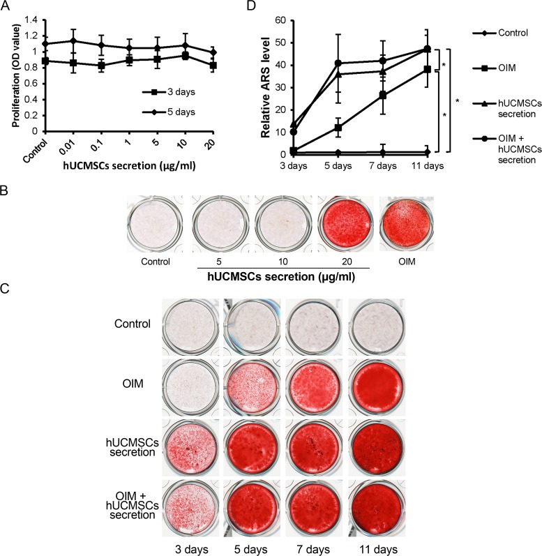
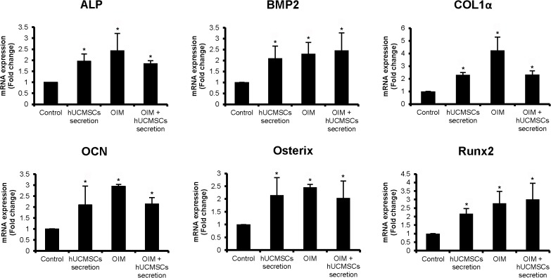
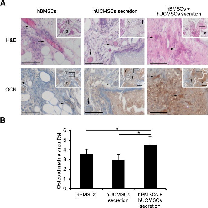
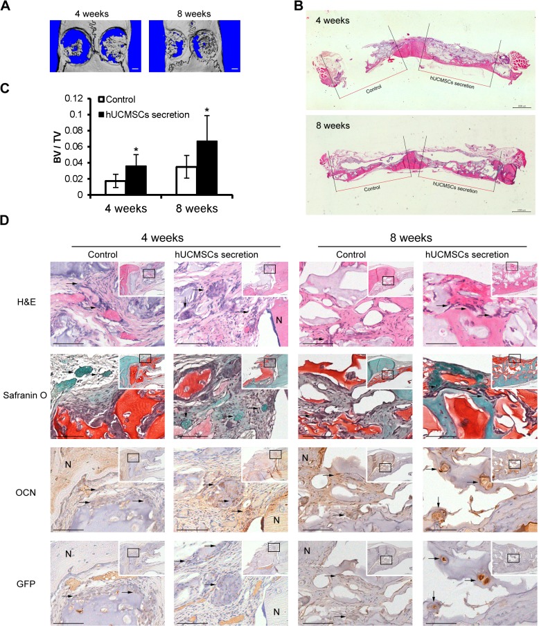
Factors synthesized by mesenchymal stem cells (MSCs) contain various growth factors, cytokines, exosomes and microRNAs, which may affect the differentiation abilities of MSCs. In the present study, we investigated the effects of secretion factors of human umbilical cord derived mesenchymal stem cells (hUCMSCs) on osteogenesis of human bone marrow derived MSCs (hBMSCs). The results showed that 20 μg/ml hUCMSCs secretion factors could initiate osteogenic differentiation of hBMSCs without osteogenic induction medium (OIM), and the amount of calcium deposit (stained by Alizarin Red) was significantly increased after the hUCMSCs secretion factors treatment. Real time quantitative reverse transcription-polymerase chain reaction (real time qRT-PCR) demonstrated that the expression of osteogenesis-related genes including ALP, BMP2, OCN, Osterix, Col1α and Runx2 were significantly up-regulated following hUCMSCs secretion factors treatment. In addition, we found that 10 μg hUCMSCs secretion factors together with 2×10(5) hBMSCs in the HA/TCP scaffolds promoted ectopic bone formation in nude mice. Local application of 10 μg hUCMSCs secretion factors with 50 μl 2% hyaluronic acid hydrogel and 1×10(5) rat bone marrow derived MSCs (rBMSCs) also significantly enhanced the bone repair of rat calvarial bone critical defect model at both 4 weeks and 8 weeks. Moreover, the group that received the hUCMSCs secretion factors treatment had more cartilage and bone regeneration in the defect areas than those in the control group. Taken together, these findings suggested that hUCMSCs secretion factors can initiate osteogenesis of bone marrow MSCs and promote bone repair. Our study indicates that hUCMSCs secretion factors may be potential sources for promoting bone regeneration.


Conflict of interest statement

Competing Interests: The authors have declared that no competing interests exist.

Figures

Fig 1
Fig 1. hUCMSCs secretion factors promote mineralization in osteogenic differentiation of hBMSCs.
(A) hBMSCs were treated with hUCMSCs secretion factors (from 0.01 μg/ml to 20 μg/ml) for 3 days or 5 days. MTT assay was performed to test the cell proliferation rate. DMSO served as control. The hUCMSCs secretion factors have no significant effect on cell proliferation. (B) The dose-dependent assay of hUCMSCs secretion factors (5, 10 and 20 μg/ml) on osteogenic differentiation of hBMSCs that were treated for 11 days in 24-well plates. Calcium deposits formation was rare in the 5 μg/ml or 10 μg/ml group, whereas significant calcium nodules were formed in the 20 μg/ml group. (C) Time-dependent assay of hUCMSCs secretion factors at a dose of 20 μg/ml on osteogenic differentiation of hBMSCs that were treated for 3, 5, 7 and 11 days in 24-well plates. Alizarin Red staining showed that the amount of calcium deposits in both hUCMSCs secretion factors and OIM + hUCMSCs secretion factors groups were higher than those of the OIM group at all the time points. (D) Alizarin Red was dissolved in 10% (w/v) cetylpyridinium chloride, and the amount was determined by absorbance measurement at 570 nm. The amount of Alizarin Red in both hUCMSCs secretion factors and OIM + hUCMSCs secretion factors groups was significantly higher than that in the control group at day 3, 5, 7 and 11. (OIM: osteogenic induction medium. hUCMSCs secretion concentration: 20 μg/ml * p<0.05, compared to control).
Fig 2
Fig 2. hUCMSCs secretion factors up-regulated expression of osteogenic marker genes of hBMSCs.
hBMSCs were incubated with 20 μg/ml hUCMSCs secretion, osteogenic induction medium and osteogenic induction medium with 20 μg/ml hUCMSCs secretion for 11 days before RNA extraction and real time qRT-PCR. The expression of ALP, BMP2, Col1α, OCN, Osterix and Runx2 was significantly increased in the hUCMSCs secretion factors group, OIM group and OIM + hUCMSCs secretion factors group compared to that of the control group. Cells treated with α-MEM medium served as a control. (* p<0.05, compared to control)
Fig 3
Fig 3. hUCMSCs secretion factors together with hBMSCs promoted ectopic bone formation in nude mice.
(A) The sections were stained with H&E and OCN immunostaining. Arrows indicate the areas of osteoid matrices(H&E staining) or OCN positive cells (OCN immunostaining). OCN positive cells distributed in the areas of osteoid, which was consistent with that in H&E staining. The data showed that there was more OCN positive staining in the hBMSCs + hUCMSCs secretion factors group than in either of the other two groups. (B) Quantification of osteoid matrices areas showed significantly more osteoid matrices in the MSCs + hUCMSCs secretion factors group compared to the other two groups. Box areas of inset indicate the region shown in higher magnification. Scale bars = 100 μm (A), 200 μm (inset of A). S: HA/TCP scaffolds. T: regenerated tissue. *p<0.05.
Fig 4
Fig 4. hUCMSCs secretion factors with rBMSCs enhanced rat calvarial bone repair.
(A) 3D reconstruction of calvarial bone samples of 4 weeks and 8 weeks following treatment. The left side: PBS +GFP-rBMSCs; the right side: 10 μg hUCMSCs secretion + GFP-rBMSCs. The image showed increased bone formation in the hUCMSCs secretion factors group (the right side) than in the control group (the left side) at 4 weeks and 8 weeks. (B) H&E staining of cranial bone (transverse plane) showed increased regenerated tissue in hUCMSCs secretion factor group (the right side). (C) microCT analysis showed the new bone volume in the hUCMSCs secretion factors group was significantly increased compared to that of the control group at both time points. (D) The sections were stained with H&E, Safranin O, and immunostaining with OCN and GFP. OCN positive immunostaining illustrated the bone tissues. GFP positive cells were derived from allogenic rBMSCs in new tissue. More OCN and GFP positive cells were seen in the hUCMSCs secretion factors group than in the control group. Arrows indicate the newly formed bone tissues (in H&E and Safranin O staining), or positive cells (in OCN and GFP immunostaining). Box areas of inset indicate the region shown in higher magnification. Scale bars = 1000 μm (A,B), 100μm (D), 200 μm (inset of D). N: native bone. *p<0.05.

References

    1. Pittenger MF, Mackay AM, Beck SC, Jaiswal RK, Douglas R, Mosca JD, et al. Multilineage potential of adult human mesenchymal stem cells. Science. 1999;284: 143–147. - PubMed
    1. Jiang Y, Jahagirdar BN, Reinhardt RL, Schwartz RE, Keene CD, Ortiz-Gonzalez XR, et al. Pluripotency of mesenchymal stem cells derived from adult marrow. Nature. 2002;418: 41–49. - PubMed
    1. Bruder SP, Fink DJ, Caplan AI. Mesenchymal stem cells in bone development, bone repair, and skeletal regeneration therapy. J Cell Biochem. 1994;56: 283–294. - PMC - PubMed
    1. Krampera M, Pizzolo G, Aprili G, Franchini M. Mesenchymal stem cells for bone, cartilage, tendon and skeletal muscle repair. Bone. 2006;39: 678–683. - PubMed
    1. Wang HS, Hung SC, Peng ST, Huang CC, Wei HM, Guo YJ, et al. Mesenchymal stem cells in the Wharton's jelly of the human umbilical cord. Stem Cells. 2004;22: 1330–1337. - PubMed

Publication types