. 2015 Mar 23;10(3):e0119661.
doi: 10.1371/journal.pone.0119661. eCollection 2015.

Low-dose irradiation affects expression of inflammatory markers in the heart of ApoE -/- mice

Affiliations

Low-dose irradiation affects expression of inflammatory markers in the heart of ApoE -/- mice

Daniel Mathias et al. PLoS One. .

Erratum in

Abstract

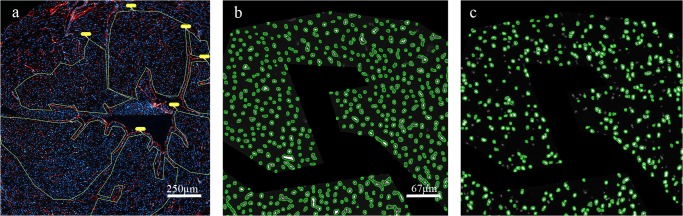
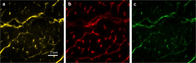
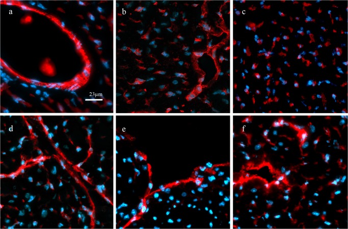
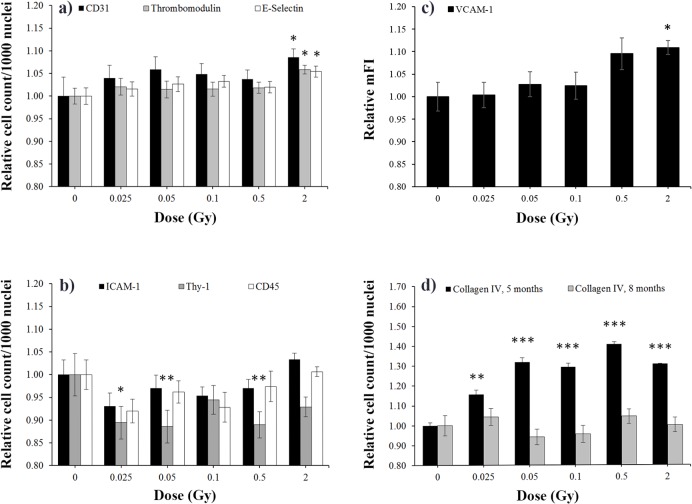
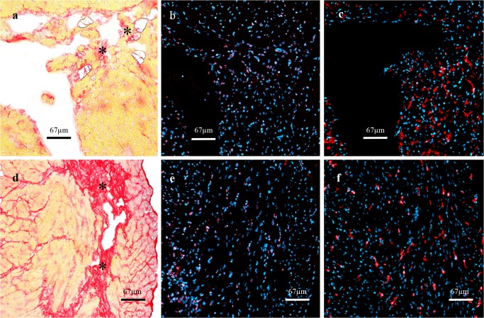
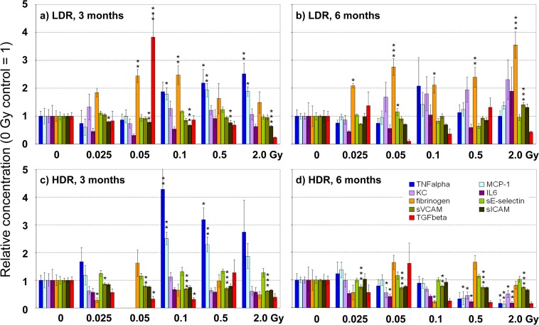
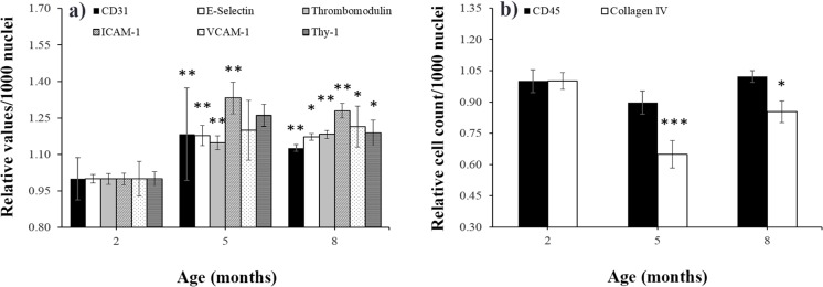
Epidemiological studies indicate long-term risks of ionizing radiation on the heart, even at moderate doses. In this study, we investigated the inflammatory, thrombotic and fibrotic late responses of the heart after low-dose irradiation (IR) with specific emphasize on the dose rate. Hypercholesterolemic ApoE-deficient mice were sacrificed 3 and 6 months after total body irradiation (TBI) with 0.025, 0.05, 0.1, 0.5 or 2 Gy at low (1 mGy/min) or high dose rate (150 mGy/min). The expression of inflammatory and thrombotic markers was quantified in frozen heart sections (CD31, E-selectin, thrombomodulin, ICAM-1, VCAM-1, collagen IV, Thy-1, and CD45) and in plasma samples (IL6, KC, MCP-1, TNFα, INFγ, IL-1β, TGFβ, INFγ, IL-10, sICAM-1, sE-selectin, sVCAM-1 and fibrinogen) by fluorescence analysis and ELISA. We found that even very low irradiation doses induced adaptive late responses, such as increases of capillary density and changes in collagen IV and Thy-1 levels indicating compensatory regulation. Slight decreases of ICAM-1 levels and reduction of Thy 1 expression at 0.025-0.5 Gy indicate anti-inflammatory effects, whereas at the highest dose (2 Gy) increased VCAM-1 levels on the endocardium may represent a switch to a pro-inflammatory response. Plasma samples partially confirmed this pattern, showing a decrease of proinflammatory markers (sVCAM, sICAM) at 0.025-2.0 Gy. In contrast, an enhancement of MCP-1, TNFα and fibrinogen at 0.05-2.0 Gy indicated a proinflammatory and prothrombotic systemic response. Multivariate analysis also revealed significant age-dependent increases (KC, MCP-1, fibrinogen) and decreases (sICAM, sVCAM, sE-selectin) of plasma markers. This paper represents local and systemic effects of low-dose irradiation, including also age- and dose rate-dependent responses in the ApoE-/- mouse model. These insights in the multiple inflammatory/thrombotic effects caused by low-dose irradiation might facilitate an individual evaluation and intervention of radiation related, long-term side effects but also give important implications for low dose anti-inflammatory radiotherapy.


Conflict of interest statement

Competing Interests: The authors have declared that no competing interests exist.

Figures

Fig 1
Fig 1. Quantitative analysis of staining by Tissue Fax/Quest.
a) Regions of interest (ROI) with cross-sectioned capillaries only, were manually marked and fields of view (FOV) were scanned. Blue = nuclei stained with DAPI, red = capillaries stained with CD31, yellow = ROI, b) DAPI-stained nuclei are counted as reference parameter and represent the whole cell count within the FOV, c) CD31-stained events are counted in corresponding areas.
Fig 2
Fig 2. Detection of CD31+ve and Lectin+ve vessels in serial sections.
a) Lectin (yellow), b) serial section (7 μm apart) stained for CD31 (red), c) Matching areas are indicated in green (artificial colours).
Fig 3
Fig 3. Immunofluorescence staining of endothelial markers.
Specific expression of endothelial proteins is shown in red (artificial colour) and nuclei counterstaining in blue (DAPI). CD31 a), E-selectin b), thrombomodulin c), ICAM-1 d), VCAM-1 e) and, Thy-1 f) were expressed on capillaries, small vessels (arterioles, venules) and endocard. Staining was quantitatively assessed only on capillaries, except for VCAM, which was evaluated on the endocard only.
Fig 4
Fig 4. Dose-specific effects on tissue markers.
Quantification of immunofluorescence stainings is presented in panel a) for CD31, thrombomodulin, E-selectin; b) ICAM-1, Thy-1, CD45; c) for VCAM, and d) for collagen IV. The number of specifically stained cells per 1000 nuclei around the left ventricle of the heart was normalized on 0 Gy values (= 1). Only for VCAM-1 c), relative values of mean fluorescence intensity (mFI) from endocard stainings are presented. Except for collagen IV d), where age-specific data are shown, data are given as mean of all subgroups (HDR and LDR, at 5 and 8 months, corresponding to 3 and 6 months after irradiation, base and apex) ± SEM, n = 16. Asterisks indicate p-values ≤ 0.05: *, p ≤ 0.01: **, p ≤ 0.001: ***.
Fig 5
Fig 5. Histological findings.
Upper row: one animal of the 5-month group (0.5 Gy, LDR) showed disseminated collageneous stripes (*) after sirius red staining a), corresponding accumulations of b) CD45+ve and c) collagen IV+ve cells. Lower row: in four animals of the 8-month group (2 Gy, LDR) similar findings were found: broad collagenous scars d) going along with CD45+ve e) and collagen IV+ve f) immunoreactivity.
Fig 6
Fig 6. Dose-dependent effects on plasma markers.
Concentrations of plasma markers were measured 3 (a, c) and 6 (b, d) months after irradiation at the indicated doses at low dose rate (LDR, a,b) and high dose rate (HDR, c,d). Values are presented as mean ± SEM, relative to sham-irradiated control (0 Gy = 1). Missing bars in Fig. 6C are due to analytical failure. Asterisks indicate p-values ≤ 0.05: *, p ≤ 0.01: **, p ≤ 0.001: *** compared to control.
Fig 7
Fig 7. Age-specific effects on tissue markers in nonirradiated mice.
The expression of a) CD31, E-selectin, thrombomodulin, ICAM-1, VCAM-1, Thy-1 and of b) CD45 and collagen IV+ve cells is presented in sham-irradiated 0 Gy control mice at 2, 5, and 8 months of age. The relative number of specifically stained cells per 1000 nuclei around the left ventricle of the heart is given (mice at 2 months = 1, mean ± SEM, n = 8). Asterisks indicate p-values ≤ 0.05: *, p ≤ 0.01: **, p ≤ 0.001: *** compared to the 2-month group.

References

    1. Takahashi I, Abbott RD, Ohshita T, Takahashi T, Ozasa K, Akahoshi M, et al. A prospective follow-up study of the association of radiation exposure with fatal and non-fatal stroke among atomic bomb survivors in Hiroshima and Nagasaki (1980–2003). BMJ Open 2012;2: e000654 10.1136/bmjopen-2011-000654 - DOI - PMC - PubMed
    1. Baker JE, Moulder JE, Hopewell JW. Radiation as a risk factor for cardiovascular disease. Antioxid Redox Signal. 2011;15: 1945–1956. 10.1089/ars.2010.3742 - DOI - PMC - PubMed
    1. Sakata R, Grant EJ, Ozasa K. Long-term follow-up of atomic bomb survivors. Maturitas. 2012;72: 99–103. 10.1016/j.maturitas.2012.02.009 - DOI - PubMed
    1. Jurcut R, Savu O, Giusca S, Deleanu D, Ciudin R, Ginghina C. Between Scylla and Charybdis: long-term cardiovascular complications after radiotherapy for Hodgkin's lymphoma. Hellenic J Cardiol. 2009;50: 538–543. - PubMed
    1. Galper SL, Yu JB, Mauch PM, Strasser JF, Silver B, Lacasce A, et al. Clinically significant cardiac disease in patients with Hodgkin lymphoma treated with mediastinal irradiation. Blood. 2011;117: 412–418. 10.1182/blood-2010-06-291328 - DOI - PubMed

Publication types

MeSH terms