. 2015 Mar 23;10(3):e0121305.
doi: 10.1371/journal.pone.0121305. eCollection 2015.

Clostridium perfringens type E virulence traits involved in gut colonization

Affiliations

Clostridium perfringens type E virulence traits involved in gut colonization

Leandro M Redondo et al. PLoS One. .

Abstract

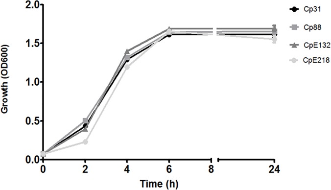
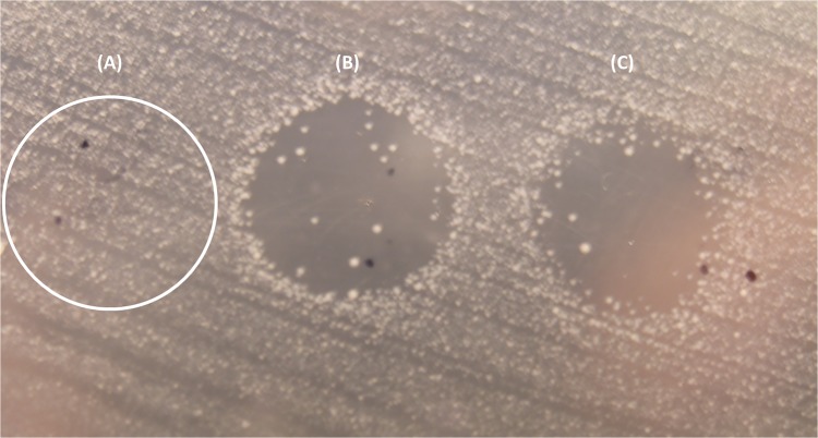
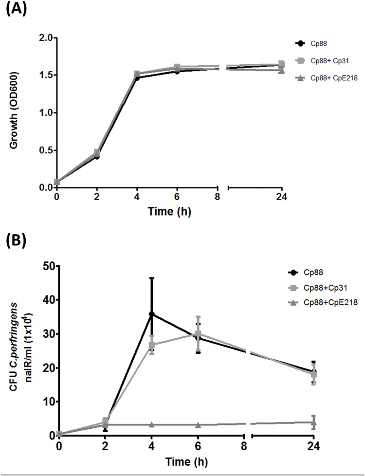
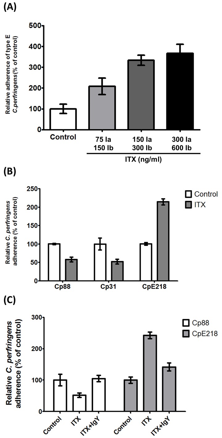
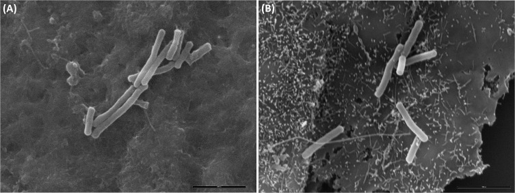
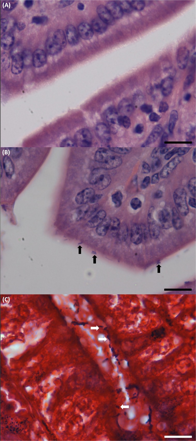
Clostridium perfringens type E disease in ruminants has been characterized by hemorrhagic enteritis or sudden death. Although type E isolates are defined by the production of alpha and iota toxin, little is known about the pathogenesis of C. perfringens type E infections. Thus far, the role of iota toxin as a virulence factor is unknown. In this report, iota toxin showed positive effects on adherence and colonization of C. perfringens type E while having negative effect on the adherence of type A cells. In-vitro and in-vivo models suggest that toxinotype E would be particularly adapted to exploit the changes induced by iota toxin in the surface of epithelial cells. In addition, type E strains produce metabolites that affected the growth of potential intra-specific competitors. These results suggest that the alteration of the enterocyte morphology induced by iota toxin concomitantly with the specific increase of type E cell adhesion and the strong intra-specific growth inhibition of other strains could be competitive traits inherent to type E isolates that improve its fitness within the bovine gut environment.


Conflict of interest statement

Competing Interests: The authors have declared that no competing interests exist.

Figures

Fig 1
Fig 1. Growth curves of selected C. perfringens strains.
The growth of two type E (CpE218 and CpE132) and two type A (Cp88 and Cp31) strains was measured by OD600 over 24 h. The patterns of growth were similar for all the strains. Results shown represent the average of three independent experiments; error bars indicate the standard error of the mean (SEM).
Fig 2
Fig 2. Result of the agar spot lawn diffusion assay.
(A) A drop of C. perfringens type A strain (Cp31) supernatant was stabbed on a lawn of an indicator type A strain (Cp88) and partially through the agar beneath, no inhibition was observed. When type E supernatant was used, CpE218 and CpE132 (B and C respectively) a clear inhibition zone was observed.
Fig 3
Fig 3. C. perfringens type E (CpE218) strain inhibits the growth of type A strain (Cp88).
Both strains were co-cultured under anaerobic conditions. (A) Optical density at 600 nm shows no differences (P>0.05) in total clostridia cell density between monocultures (Cp88) and co-cultures with inhibitor type E strain (CP88 + CpE218) and non-inhibitor strain (Cp88 + Cp31). (B) Samples were analyzed for nalidixic acid resistant (nalR) CFU/ml every 2 h of culture. Compared to the expected number of nalidixic acid resistant Cp88 grown in monoculture, there was a significant reduction in the number of nalidixic acid resistant CFU counts since 4 h of culture onwards. No significant differences were observed when cultivated with a nalidixic acid sensitive non-inhibitor type A strain (Cp31). Results are the mean CFU counts obtained from three independent experiments; error bars indicate the standard deviation (SD).
Fig 4
Fig 4. Type E clostridia reduce type A cell adhesion.
(A) C. perfringens type E (CpE218) strain reduces adherence of type A strain (Cp88) to Caco-2 cells. Caco-2 cells were incubated with DMEM medium containing bacteria. Cp88: Nalidixic acid resistant type A strain alone; Cp88+Cp31: Mix of nalidixic acid resistant type A strain with a non-inhibitor type A strain but sensitive to nalidixic acid; Cp88+CpE218: Mix of nalidixic resistant type A strain with a type E strain. As mentioned, both strains were added simultaneously. Adhesion of Cp88 alone was considered as a control for the adhesion assay. (B) Type E cells displace type A cells attached to Caco-2 cells only at higher type A/type E ratio. The number below each column represents the ratio between attached type A and free competing type E cells. Results are the mean nalidixic acid resistant (nalR) CFU counts from three independent experiments; error bars indicate the standard deviation (SD).
Fig 5
Fig 5. Iota toxin increases type E adherence while decreases type A adherence.
(A) ITX treatment increases the adherence of C. perfringens type E (CpE218). Cells were incubated with the indicated toxin concentrations. 75ng/ml Ia +150ng/ml Ib, 150ng/ml Ia +300ng/ml Ib and 300ng/ml Ia +600ng/ml Ib. After ITX treatment, cells monolayers were incubated with type E bacteria. Cells were lysed and total clostridia were plated onto neomycin blood agar plates for counting. (B) ITX treatment increases the adherence of type E bacteria (CpE218), and reduce adherence of type A bacteria (Cp88—Cp31). Cells were treated with 150 ng/ml Ia and 300 ng/ml Ib (ITX), and washed 3 times with PBS. After ITX treatment, cells monolayers were incubated with the indicated strains. (C) ITX induced changes in bacterial adherence were neutralized by addition of anti-ITX IgY. Cells were treated with 150 ng/ml Ia and 300 ng/ml Ib (ITX) with or without neutralizing IgY. After treatment, cells monolayers were incubated with the indicated strains. Attachment levels were quantified as percent of bacterial adherence relative to untreated cells. Attachment levels were quantified as percent of bacterial adherence relative to untreated cells. Results shown represent the average of three independent experiments; error bars indicate the standard error of the mean (SEM).
Fig 6
Fig 6. Scanning electron microscopy of C. perfringens interacting with epithelial cells.
Caco-2 cells were incubated for 2 h with PBS buffer (A) or ITX (B). No superficial protrusions were observed in PBS treated cells, although bacteria were attached to the cells surface after 2 h of anaerobic incubation. ITX treatment induced the formation of membrane protrusions in Caco-2 cells and clostridia cells were adhered to these cellular deformations.
Fig 7
Fig 7. Histological analysis of sample sections of ileum of the C. perfringens challenged mice.
Mice were intragastrically inoculated with the indicated strains and then sacrificed 48 h post-infection. (A) Hematoxylin and eosin staining of sample sections of ileum of the mice challenged with type A strain (Cp88), no C. perfringens compatible attached bacteria were observed. Scale bar, 20 μm. (B) In type E challenged mice many rod shaped bacteria compatible with C. perfringens (black arrows) were observed intimately attached to the intestinal mucosa. Scale bar, 20 μm. (C) Gram staining of a sample section of ileum of one of the type E challenged mice: Gram positive rod shaped bacteria compatible with C. perfringens attached to the gut mucosa (white arrows). Scale bar, 20 μm.
Fig 8
Fig 8. Immunohistochemical analysis of sample sections of ileum of the C. perfringens challenged mice.
Mice were intragastrically inoculated with the indicated strains and then sacrificed 48 h post-infection. (A) Sample sections of ileum of the mice challenged with type A strain (Cp88), no attached C. perfringens were observed. Scale bar, 20 μm. (B) In type E challenged mice, many red bacilli corresponding with C. perfringens (black arrows) were observed intimately attached to the intestinal mucosa. Scale bar, 20 μm.

References

    1. McClane BA, Uzal FA, Fernandez Miyakawa ME, Lyerly DM, Wilkins TD. The enterotoxic clostridia In Dworkin M., Falkow S., Rosenberg E., Schleifer K.-H., & Stackebrandt E., Editors. The Prokaryotes: Springer US; 2006; pp. 698–752. 10.1007/0-387-30744-3 - DOI
    1. Songer JG. Clostridial enteric diseases of domestic animals. Clinical Microbiology Reviews. 1996; 9: 216–34. - PMC - PubMed
    1. Li J, Miyamoto K, McClane BA. Comparison of virulence plasmids among Clostridium perfringens type E isolates. Infection and Immunity. 2007; 75: 1811–9. 10.1128/IAI.01981-06 - DOI - PMC - PubMed
    1. Baskerville M, Wood M, Seamer JH. Clostridium perfringens type E enterotoxaemia in rabbits. Veterinary Record. 1980; 107: 18–19. - PubMed
    1. Hart B, Hooper PT. Enterotoxamemia of calves due to Clostridium welchii type E. Australian Veterinary Journal. 1967; 43: 360–363. 10.1111/j.1751-0813.1967.tb04882.x - DOI - PubMed

Publication types

Substances

LinkOut - more resources