. 2015 May 28;521(7553):537-540.
doi: 10.1038/nature14216. Epub 2015 Mar 23.

MAD2L2 controls DNA repair at telomeres and DNA breaks by inhibiting 5' end resection

Affiliations

MAD2L2 controls DNA repair at telomeres and DNA breaks by inhibiting 5' end resection

Vera Boersma et al. Nature. .

Abstract

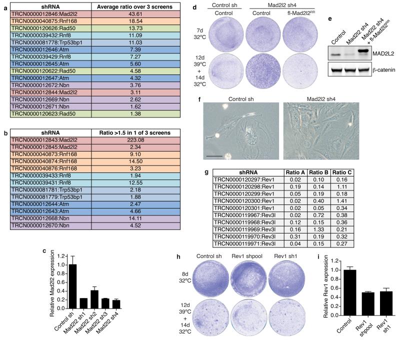
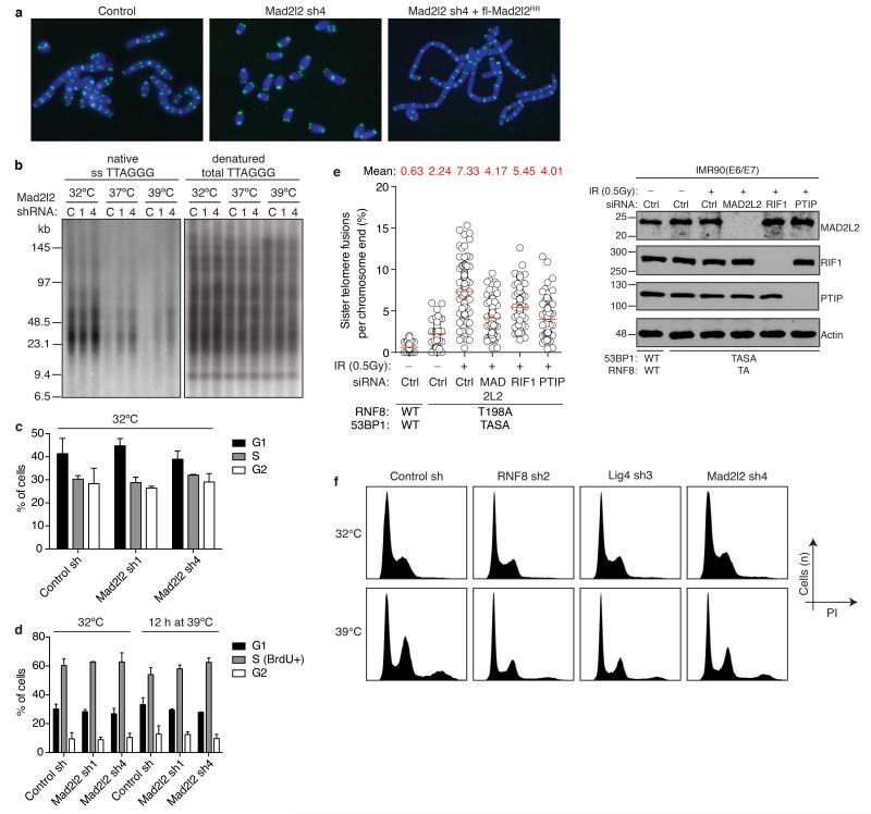
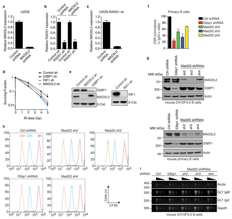
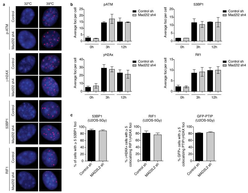
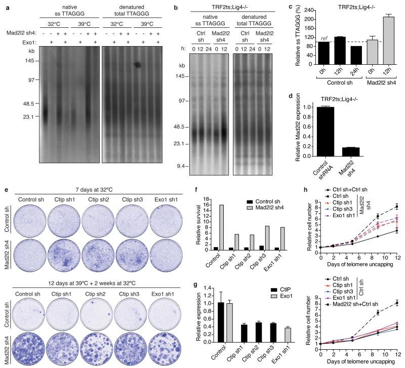
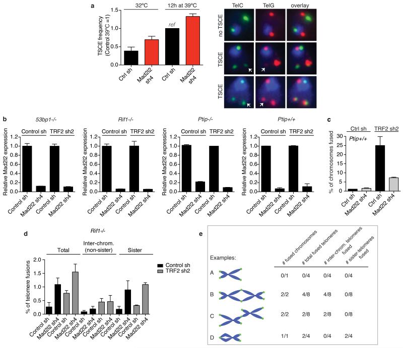
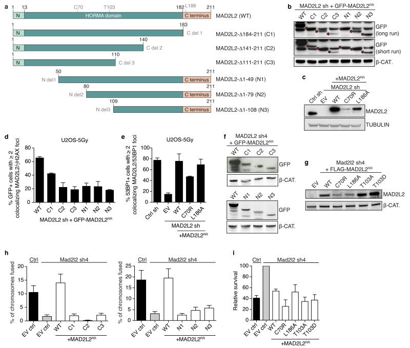
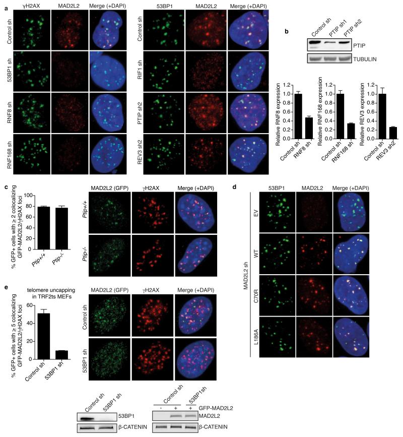
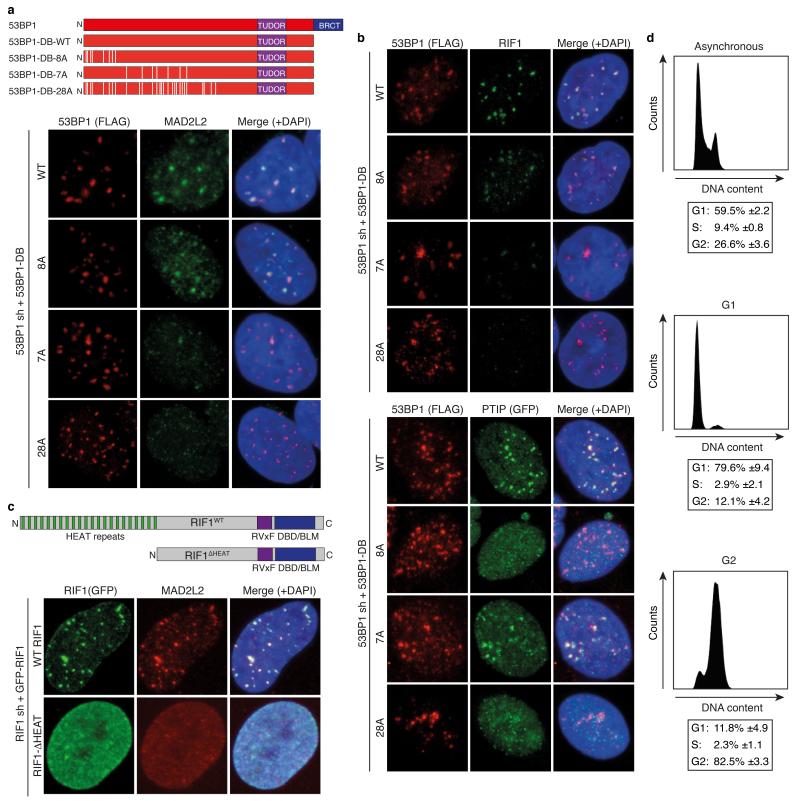
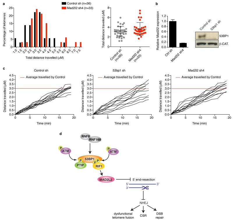
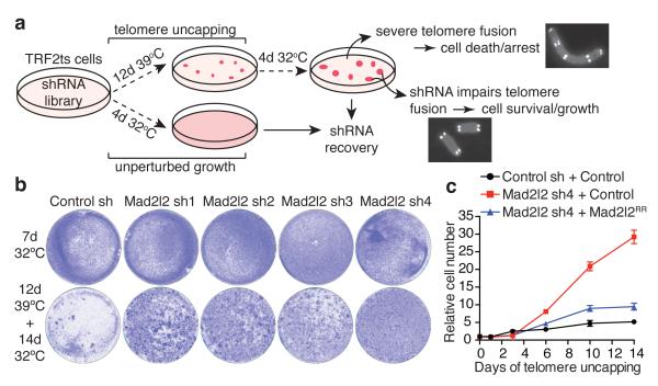
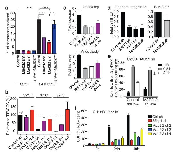
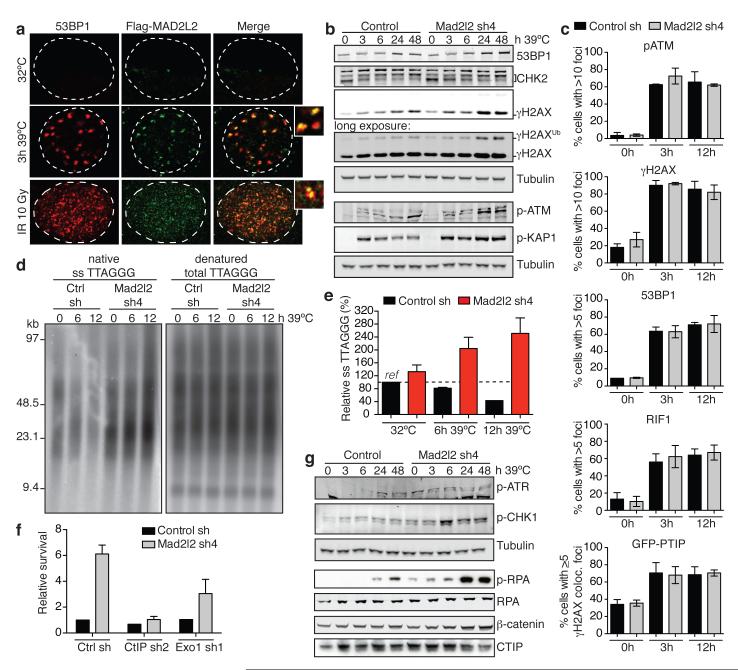
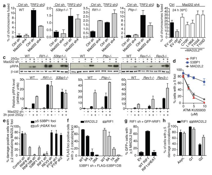
Appropriate repair of DNA lesions and the inhibition of DNA repair activities at telomeres are crucial to prevent genomic instability. By fuelling the generation of genetic alterations and by compromising cell viability, genomic instability is a driving force in cancer and ageing. Here we identify MAD2L2 (also known as MAD2B or REV7) through functional genetic screening as a novel factor controlling DNA repair activities at mammalian telomeres. We show that MAD2L2 accumulates at uncapped telomeres and promotes non-homologous end-joining (NHEJ)-mediated fusion of deprotected chromosome ends and genomic instability. MAD2L2 depletion causes elongated 3' telomeric overhangs, indicating that MAD2L2 inhibits 5' end resection. End resection blocks NHEJ while committing to homology-directed repair, and is under the control of 53BP1, RIF1 and PTIP. Consistent with MAD2L2 promoting NHEJ-mediated telomere fusion by inhibiting 5' end resection, knockdown of the nucleases CTIP or EXO1 partially restores telomere-driven genomic instability in MAD2L2-depleted cells. Control of DNA repair by MAD2L2 is not limited to telomeres. MAD2L2 also accumulates and inhibits end resection at irradiation-induced DNA double-strand breaks and promotes end-joining of DNA double-strand breaks in several settings, including during immunoglobulin class switch recombination. These activities of MAD2L2 depend on ATM kinase activity, RNF8, RNF168, 53BP1 and RIF1, but not on PTIP, REV1 and REV3, the latter two acting with MAD2L2 in translesion synthesis. Together, our data establish MAD2L2 as a crucial contributor to the control of DNA repair activity by 53BP1 that promotes NHEJ by inhibiting 5' end resection downstream of RIF1.


Figures

Extended Data Figure 1
Related to Figure 1 a, A functional genetic screen for telomere-induced genomic instability regulators (TIGIRs) identifies independent shRNAs against Mad2l2 and the previously identified regulators of NHEJ-mediated telomere fusion Atm, Nbs1 (a.k.a. Nbn), Rad50, 53bp1 (a.k.a. Trp53bp1) and Rnf8. Listed are the independent shRNAs enriched >1.5 fold in at least 2 out of 3 TIGIR-screens, with their average ratio of enrichment over all 3 screens. Ratios reflect shRNA abundance after 12 days of telomere uncapping at 39°C followed by 4 days recovery at 32°C versus shRNA abundance after growth for 4 days at 32°C. b, Additional shRNAs targeting Mad2l2 or known TIGIRs that were enriched >1.5 fold in 1 of 3 screens, with their ratio. Not shown are additional shRNAs against factors not previously implicated in control of telomere fusion, that were significantly enriched in these TIGIR-screens but await validation. c, qRT-PCR analysis of Mad2l2 expression levels in TRF2ts MEFs transduced with 4 independent shRNAs targeting Mad2l2 and used in Fig. 1b (Error bars: s.d.). d, Survival assay of TRF2ts cells infected with control or Mad2l2 sh4 shRNAs, complemented with empty control or RNAi-resistant Flag-Mad2l2RR and grown as indicated. e, Western blot showing expression of endogenous MAD2L2 and exogenous Flag-MAD2L2 in TRF2ts cells used in d and in Fig. 1c. f, Photograph of shRNA-transduced TRF2ts cells grown for 12 days at 39°C. Scale bar, 100 μm. g, None of 10 shRNAs targeting Rev1 or Rev3 were significantly enriched in any of 3 independent DDR TIGIR-screens. h, shRNA-mediated Rev1 knockdown does not increase survival upon prolonged telomere uncapping in survival assays of TRF2ts MEFs. Of note, Rev3 knockdown compromised viability and was therefore not informative in these assays. i, qRT-PCR analysis of mouse Rev1 expression levels of cells shown in h (Error bars: s.d.).
Extended Data Figure 2
Related to Figure 2 a, Representative metaphase spreads of TRF2ts MEFs transduced as indicated, harvested after 24 h at 39°C for telomere fluorescence in situ hybridization (FISH). b, Representative telomeric single-strand G-overhang (ss TTAGGG) and total telomere (total TTAGGG) analysis in TRF2ts MEFs at 32°C and after 48 h at 37°C or 39°C. c-d, Mad2l2 knockdown does not affect cell cycle parameters in TRF2ts MEFs. Cell cycle phase analysis was based on PI staining of asynchronously growing cells (c), as well as on 1 h incubation with BrdU, followed by detection of BrdU incorporation and PI staining for DNA content (d) (n=3, ± s.d.). e, MAD2L2 is required for sister-telomere fusion upon activation of DNA repair in mitosis. Sister-telomere fusions were quantified in IMR90 cells expressing exogenous WT 53BP1 and RNF8, or 53BP1-T1608A/S1618A (TASA) and RNF8-T198A (TA) mutant alleles, and depleted for endogenous RNF8 and 53BP1, as well as depleted for MAD2L2, RIF1 or PTIP (n=4). f, Examples of DNA content profiles of PI stained TRF2ts cells transduced with the indicated shRNAs and grown at 32°C or for 48 h at 39°C. Analysis of the fraction of cells with 8N (tetraploid) or > 4N (aneuploid) DNA content was done on corresponding dotplots of which the results are shown in Fig. 2c.
Extended Data Figure 3
Related to Figure 2 a, qRT-PCR analysis of MAD2L2 expression levels in U2OS cells infected with control or MAD2L2 shRNAs, and used in the repair assays shown in Fig. 2d (Error bars: s.d.). b, c, qRT-PCR analysis of RAD51 (b) and MAD2L2 (c) expression levels in RAD51-depleted, E6E7-expressing U2OS cells used in the assays shown in Fig. 2e (Error bars: s.d.). d, Clonogenic survival assays of U2OS cells transduced with non-targeting control or 53BP1, RIF1 or MAD2L2 shRNAs and treated with the indicated doses of IR (n=3-4, ± s.e.m.). e, Western blot analysis of 53BP1, MAD2L2 and RIF1 in U2OS cells transduced with the indicated shRNAs. f, CSR in shRNA-transduced primary B cells (n=2, ± s.d.). g, Western blot analysis of MAD2L2 and 53BP1 in CH12F3-2 B cells and mouse primary B cells transduced with the indicated shRNAs. h, MAD2L2 depletion does not affect cellular proliferation in murine B cells. CH12F3-2 cells transduced with control, 53bp1 or Mad2l2 shRNAs were loaded with CFSE and analysed at 0, 24 and 48 h post-stimulation by flow-cytometry. Profiles from all time points are plotted in the same histogram. i, MAD2L2 depletion does not affect the transcription of critical genes implicated in CSR. RT-PCR analysis of AID (Aicda) mRNA, IgM (GLT IgM) and IgA (GLT IgA) germline transcript levels using 2-fold serial dilutions of cDNA made from activated CH12F3-2 B cells transduced with the indicated shRNAs. Gapdh was used as a control for transcript expression.
Extended Data Figure 4
Related to Figure 3 a, Representative images of IF detection of p-ATM(S1981), γ-H2AX, 53BP1 and RIF1 in TRF2ts MEFs transduced with control or Mad2l2 shRNAs and grown at 32°C or for 12 h at 39°C to induce telomere uncapping. DNA was stained with DAPI. b, Quantification of the number of p-ATM, γ-H2AX, 53BP1 and RIF1 foci per cell in TRF2ts MEFs transduced with control or Mad2l2 shRNAs and grown at 32°C or for 3 and 12 h at 39°C (n=2, ± s.d.). c, Quantifications of 53BP1, RIF1 and PTIP foci in U2OS cells transduced with control or MAD2L2 shRNAs, 3 h after 5Gy (n=2, ± s.d.).
Extended Data Figure 5
Related to Figure 3 a, Telomeric single-strand G-overhang assay of TRF2ts MEFs transduced with control or Mad2l2 sh4 shRNAs, showing that the increase in overhang signal upon Mad2l2 knockdown is due to 3′ terminal sequences because the signal is removed by treatment with Escherichia coli 3′ exonuclease EXO1. b, Mad2l2 knockdown causes increased ss telomeric G-overhang signals in TRF2ts;Lig4−/−;TRF2ts MEFs. c, Quantification of relative telomeric G-overhang signals in TRF2ts;Lig4−/− MEFs transduced with control or Mad2l2 shRNAs and grown at 32°C or for 12 or 24 h at 39°C (n=2, ± s.e.m.). d, qRT-PCR analysis of Mad2l2 expression levels in TRF2ts;Lig4−/− MEFs infected with control or Mad2l2 shRNA lentivirus (Error bars: s.d.). e, Survival assays of TRF2ts MEFs transduced with control or Mad2l2 shRNAs and subsequently with control, Ctip or Exo1 shRNAs. f, Quantification of the survival assays shown in e. g, qRT-PCR analysis of Ctip and EXO1 expression levels of cells shown in e, f, h and in Fig. 3f (Error bars: s.d.). h, Growth curves at 39°C of TRF2ts MEFs transduced with non-targeting control or Mad2l2 shRNAs and subsequently with control, Ctip or Exo1 shRNAs (Error bars: s.e.m.).
Extended Data Figure 6
Related to Figures 3 and 4 a, TSCE analysis in shRNA-transduced TRF2ts MEFs grown at 32°C or for 12 h at 39°C to uncap telomeres (n=2, ± s.d., counting >1,000 chromosomes per condition, per experiment). TSCE frequency in control cells is set at 1 (corresponding to an average of 6.9% of chromosomes with a TSCE event). Shown on the right are examples of chromosomes without and with TSCE in cells quantified on the left. b, qRT-PCR analysis of Mad2l2 expression levels in 53bp1−/−, Rif1−/−, Ptip−/− and Ptip+/+ MEFs used in the chromosome fusion analysis shown in Fig. 4a and in c (Error bars: s.d.). c, Percentage of chromosomes fused upon TRF2 inhibition in the Ptip+/+ MEFs matching with the Ptip−/− MEFs shown in Fig. 4a (n=2, ± s.e.m.). d, Analysis of different types of telomere fusions in Rif1−/− MEFs. Depletion of MAD2L2 in Rif1−/− MEFs does not reduce inter-chromosomal telomere fusions induced by TRF2 inhibition, indicating epistasis. However, irrespective of TRF2 inhibition, MAD2L2 depletion in Rif1−/− MEFs induces association between sister-telomeres, causing an increase in total fusions scored for MAD2L2-depleted Rif1−/− MEFs, as also visible in Fig. 4a (n=2, ± s.e.m., >1,300-2,000 chromosomes were analysed per condition, per independent experiment). e, Explanation of scoring different types of telomere fusions shown in d.
Extended Data Figure 7
Related to Figure 4 a, Schematic overview of C-terminal and N-terminal deletion mutants of MAD2L2. b, c, Expression analysis in U2OS cells of GFP-tagged WT MAD2L2 and C- and N-terminal MAD2L2 deletion mutants (b) and of FLAG-tagged WT MAD2L2 and MAD2L2-C70R and −L186A (c). d, Analysis of DDR foci formation of GFP-tagged WT MAD2L2 and C- and N-terminal MAD2L2 deletion mutants by IF detection of GFP and γH2AX in U2OS at 3 h after IR (n=2 for N-terminals, n=3 for C-terminals, ± s.e.m.). e, Analysis of WT, C70R and L186A MAD2L2 accumulation into DDR foci by IF detection of MAD2L2 and 53BP1 in U2OS at 3 h post IR (n=2, ± s.e.m.). f, g, Expression analysis in TRF2ts MEFs of GFP-tagged MAD2L2 and C- and N-terminal MAD2L2 deletion mutants (f) and of FLAG-tagged WT MAD2L2 and MAD2L2-C70R, −L186A, −T103A and −T103D (g). h, Quantification of chromosome fusions after 24 h of telomere deprotection at 39°C in TRF2ts MEFs transduced with control or Mad2l2 shRNAs and complemented with empty vector control or RNAi-resistant GFP-tagged WT MAD2L2 and C- or N-terminal MAD2L2 deletion mutants (Error bars: s.e.m.). i, Quantification of survival assays of TRF2ts MEFs transduced with control or Mad2l2 shRNAs and subsequently with empty vector control, WT MAD2L2, MAD2L2 C70R, L186A, T103A or T103D retroviruses (n=2, ± s.e.m.).
Extended Data Figure 8
Related to Figure 4 a, Representative images of IF detection of endogenous MAD2L2 and γH2AX or 53BP1 in U2OS cells transduced with control, 53BP1, RNF8, RNF168, RIF1, PTIP or REV3 shRNA lentiviruses, irradiated with 5Gy and processed for IF after 3h (quantifications are shown in Fig. 4e). b, Western blot or qRT-PCR analysis of PTIP, RNF8, RNF168 and REV3 levels in shRNA-transduced U2OS cells (Error bars: s.d.). c, Quantification and representative images of IF detection of γH2AX and GFPMAD2L2 in Ptip+/+ or Ptip−/− MEFs, 3h after 5Gy (n=2, ± s.d.). d, Representative images of IF for 53BP1 and exogenous Flag-MAD2L2 WT, C70R or L186A in U2OS cells depleted for endogenous MAD2L2 with lentiviral shRNA, processed for IF 3h after 5Gy (quantifications are shown in Extended Data Fig. 7e). e, Quantification and representative IF images of GFP-MAD2L2 localisation to uncapped telomeres in TRF2ts MEFs transduced with control or 53BP1 shRNAs (n=2, ± s.d.).
Extended Data Figure 9
Related to Figure 4 a, Schematic representation of the 53BP1 alleles with wild-type and substituted S/TQ sites used to address MAD2L2 IRIF localisation dependence on 53BP1. Representative IF images are displayed that show colocalisation of endogenous MAD2L2 with 53BP1-DB-WT and −8A, but not with 53BP1-DB-7A or −28A. Quantifications are provided in Fig. 4f. b, Top: Representative IF images showing colocalisation of endogenous RIF1 with 53BP1-DB-WT and −8A, but not 53BP1-DB-7A or −28A. Quantifications are presented in Fig. 4f. Bottom: Representative IF images showing colocalisation of GFP-PTIP with 53BP1-DB-WT and −7A, but impaired colocalisation of GFP-PTIP with 53BP1-DB-8A or −28A. c, Schematic representation of GFP-tagged wild-type RIF1 (WT) and GFP-tagged RIF1 lacking the N-terminal HEAT repeats (ΔHEAT), used to address MAD2L2 IRIF localisation dependence on the HEAT repeats of RIF1. Representative IF images are shown. Quantifications are presented in Fig. 4g. d, Cell cycle phase distributions of the RPE cells used in Fig. 4h to address cell cycle dependence of endogenous RIF1 and MAD2L2 localisation to IRIFs (n=3, ± s.d.).
Extended Data Figure 10
Related to Figure 4 a, Frequency distribution (left) and scatter plot (right) of total distances travelled by uncapped telomeres in control or Mad2l2 knockdown cells. b, qRT-PCR analysis of Mad2l2 expression levels and Western blot analysis of 53BP1 proteins levels in TRF2ts cells used in the experiments shown in a and c (Error bars: ± s.d.). c, Distance travelled by 10 representative uncapped telomeres for each condition. While multiple uncapped telomeres in 53BP1-depleted cells have reduced mobility, this is not seen for uncapped telomeres in MAD2L2-depleted cells. d, Model of the role of MAD2L2 in promoting NHEJ.
Figure 1
Figure 1. A functional genetic screen identifies Mad2l2 as a critical factor in telomere-driven genomic instability
a, Outline of TIGIR-screen to identify factors controlling telomere-driven genomic instability. b, Survival assays of TRF2ts MEFs infected with control or Mad2l2 shRNAs, stained after growth as indicated. c, Growth curves at 39°C of TRF2ts cells transduced with control or Mad2l2 sh4 shRNAs and complemented with empty control or RNAi-resistant Flag-Mad2l2RR (quadruplicate ± s.d.).
Figure 2
Figure 2. Mad2l2 facilitates telomeric G-overhang degradation, NHEJ-mediated telomere fusion, repair and CSR
a, Chromosome fusion in shRNA-transduced TRF2ts MEFs (n=2-4, ± s.e.m., ****p< 0.0001, ***p=0.0002, Kruskal Wallis test, ANOVA). b, Telomeric single-strand G-overhang quantification in TRF2ts MEFs at 32°C and after 48 h at 37°C or 39°C. (n=4 except Mad2l2 sh4 at 37°C: n=1, ± s.e.m.). c, Tetraploidy and aneuploidy upon telomere uncapping in TRF2ts MEFs (n=2, ± s.d.). d, NHEJ-mediated repair in U2OS cells analysed by random plasmid integration (left, n=3) or an EJ5-GFP reporter (right, n=6). Error bars: s.d.. e, MAD2L2 depletion impairs resolution of DDR foci after IR (2Gy) in HR-deficient U2OS cells, indicating defective NHEJ (n=2, ± s.e.m.). f, CSR in shRNA-transduced CH12F3-2 cells (n=2, ± s.d.).
Figure 3
Figure 3. Mad2l2 inhibits end-resection and promotes telomere-induced genomic instability in a CTIP and EXO1 dependent manner
a, Flag-MAD2L2 and 53BP1 localisation in TRF2ts MEFs at 32°C and 39°C, and in p53−/− MEFs after IR. b, Immunoblotting for DDR factors in TRF2ts MEFs upon telomere uncapping at 39°C. c, p-ATM, γ-H2AX, 53BP1, RIF1 and PTIP-GFP foci in TRF2ts MEFs grown for 0, 3 or 12 h at 39°C (n=2, ± s.d.). d-e, Representative example (d) and quantification (e) of telomeric single-strand G-overhang analysis in TRF2ts MEFs grown as specified (n=3, ± s.e.m.). f, Quantification of survival assays of TRF2ts MEFs shRNA-transduced as indicated (n=2, ± s.e.m.). g, Immunoblotting for p-ATR(S428), p-CHK1(S345), p-RPA32(S4/S8), RPA34 and CTIP in TRF2ts MEFs upon telomere uncapping at 39°C.
Figure 4
Figure 4. Mad2l2 localizes to DSBs, inhibits end-resection and promotes telomere-NHEJ in a 53BP1 and RIF1 dependent manner
a, Chromosome fusion upon TRF2 inhibition in MEFs (n=2, ± s.e.m.). WT: controls to 53bp1−/− and Rif1−/− MEFs. See Extended Data Fig. 6c for Ptip+/+ MEFs. b, Chromosome fusion in TRF2ts MEFs (Error bars: s.e.m.). c, pRPA levels in MEFs (n=2, ± s.e.m.). d, IR-induced MAD2L2, RIF1 and 53BP1 foci in ATM inhibitor-treated U2OS cells (n=3, ± s.e.m.). e, MAD2L2 foci in irradiated shRNA-transduced U2OS cells (n=2, ± s.d.). f, MAD2L2 and RIF1 foci in irradiated U2OS cells expressing 53BP1-DB alleles with wild-type or substituted S/TQ sites (schematic in Extended Data Fig. 9a; n=2, ± s.d.). g, MAD2L2 foci in irradiated U2OS cells expressing wild-type RIF1 or RIF1 lacking heat repeats (n=2, ± s.d.). h, MAD2L2 and RIF1 foci in irradiated asynchronous, G1 or G2 synchronized RPE cells (n=3, ± s.d.).

Comment in

Similar articles

Cited by

References

    1. Hanahan D, Weinberg RA. Hallmarks of cancer: the next generation. Cell. 2011;144:646–74. - PubMed
    1. Lopez-Otin C, Blasco MA, Partridge L, Serrano M, Kroemer G. The hallmarks of aging. Cell. 2013;153:1194–217. - PMC - PubMed
    1. Zimmermann M, de Lange T. 53BP1: pro choice in DNA repair. Trends Cell Biol. 2014;24:108–17. - PMC - PubMed
    1. Waters LS, et al. Eukaryotic translesion polymerases and their roles and regulation in DNA damage tolerance. Microbiol Mol Biol Rev. 2009;73:134–54. - PMC - PubMed
    1. Konishi A, de Lange T. Cell cycle control of telomere protection and NHEJ revealed by a ts mutation in the DNA-binding domain of TRF2. Genes Dev. 2008;22:1221–30. - PMC - PubMed

Additional references to Full Methods

    1. Bennardo N, Gunn A, Cheng A, Hasty P, Stark JM. Limiting the persistence of a chromosome break diminishes its mutagenic potential. PLoS Genet. 2009;5:e1000683. - PMC - PubMed
    1. Kato H, et al. Involvement of RBP-J in biological functions of mouse Notch1 and its derivatives. Development. 1997;124:4133–41. - PubMed
    1. Celli GB, Denchi EL, de Lange T. Ku70 stimulates fusion of dysfunctional telomeres yet protects chromosome ends from homologous recombination. Nat Cell Biol. 2006;8:885–90. - PubMed

Publication types

MeSH terms

Substances