

Review
doi: 10.1007/s00281-015-0475-7.
Epub 2015 Mar 24.
CD8+ T cells in cutaneous leishmaniasis: the good, the bad, and the ugly
Affiliations
- PMID: 25800274
- PMCID: PMC4439344
- DOI: 10.1007/s00281-015-0475-7
Item in Clipboard
Review
CD8+ T cells in cutaneous leishmaniasis: the good, the bad, and the ugly
Semin Immunopathol.
2015 May.
Abstract
CD8(+) T lymphocytes are components of the adaptive immune response and play an important role in protection against many viral and bacterial infections. However, their role in parasitic infections is less well understood. In leishmaniasis, a disease caused by intracellular protozoan parasites of the genus Leishmania, CD8(+) T cells have been shown to be protective. However, increasing evidence indicates that CD8(+) T cells may also exacerbate disease. In this review, we will describe the situations where CD8(+) T cells are either good or bad for the outcome of the infection and attempt to reconcile the dual role played by CD8(+) T cells in cutaneous leishmaniasis.
Figures
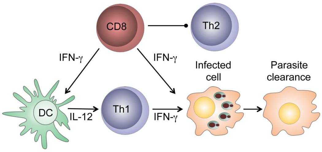
CD8+ T cells producing IFN-γ activate macrophages, leading to parasite clearance. In addition, the IFN-γ produced by CD8+ T cells promotes increased production of IL-12, which amplifies the development of protective CD4+ Th1 cells. With low doses of parasites, CD8+ T cells are essential for blocking CD4 Th2 cells development.
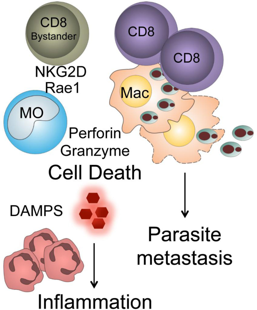
Leishmania-specific CD8+ T cells migrate to leishmanial lesions and lyse infected cells, leading to the release of proinflammatory molecules, including molecules with damage-associated molecular patterns (DAMPS). Lysis of infected cells leads to release of parasites, which may promote increased metastasis of the parasites. Memory CD8+ T cells generated from prior non-leishmanial infections are also recruited to leishmanial lesions. Leishmania infection leads to upregulation of ligands for NKG2D, such as Rae1γ, and thus if the recruited CD8+ T cells express NKG2D they lyse target cells, also leading to cell death and increased inflammation.
Similar articles
-
Phenotyping of circulating CD8⁺ T cell subsets in human cutaneous leishmaniasis.Microbes Infect. 2012 Aug;14(9):702-11. doi: 10.1016/j.micinf.2012.02.006. Epub 2012 Feb 28. Microbes Infect. 2012. PMID: 22421108
-
CD8 cytotoxic T cells in cutaneous leishmaniasis.Parasite Immunol. 2007 Dec;29(12):671-8. doi: 10.1111/j.1365-3024.2007.00991.x. Parasite Immunol. 2007. PMID: 18042173 Review.
-
CD8 alpha- and Langerin-negative dendritic cells, but not Langerhans cells, act as principal antigen-presenting cells in leishmaniasis.Eur J Immunol. 2004 Jun;34(6):1542-50. doi: 10.1002/eji.200324586. Eur J Immunol. 2004. PMID: 15162423
-
Immunomodulation mediated through Leishmania donovani protein disulfide isomerase by eliciting CD8+ T-cell in cured visceral leishmaniasis subjects and identification of its possible HLA class-1 restricted T-cell epitopes.J Biomol Struct Dyn. 2017 Jan;35(1):128-140. doi: 10.1080/07391102.2015.1134349. Epub 2016 Apr 12. J Biomol Struct Dyn. 2017. PMID: 26727289
-
CD8+ T cell immunity in an encephalitis model of Toxoplasma gondii infection.Semin Immunopathol. 2015 May;37(3):271-9. doi: 10.1007/s00281-015-0483-7. Epub 2015 May 6. Semin Immunopathol. 2015. PMID: 25944514 Free PMC article. Review.
Cited by
-
In situ cellular immune response in non-ulcerated skin lesions due to Leishmania (L.) infantum chagasi infection.J Venom Anim Toxins Incl Trop Dis. 2021 Feb 26;27:e20200149. doi: 10.1590/1678-9199-JVATITD-2020-0149. eCollection 2021. J Venom Anim Toxins Incl Trop Dis. 2021. PMID: 33708246 Free PMC article.
-
A third generation vaccine for human visceral leishmaniasis and post kala azar dermal leishmaniasis: First-in-human trial of ChAd63-KH.PLoS Negl Trop Dis. 2017 May 12;11(5):e0005527. doi: 10.1371/journal.pntd.0005527. eCollection 2017 May. PLoS Negl Trop Dis. 2017. PMID: 28498840 Free PMC article. Clinical Trial.
-
Evaluation of the Ability of Miltefosine Associated with Topical GM-CSF in Modulating the Immune Response of Patients with Cutaneous Leishmaniasis.J Immunol Res. 2020 Aug 6;2020:2789859. doi: 10.1155/2020/2789859. eCollection 2020. J Immunol Res. 2020. PMID: 32851099 Free PMC article.
-
Therapeutic control of leishmaniasis by inhibitors of the mammalian target of rapamycin.PLoS Negl Trop Dis. 2018 Aug 22;12(8):e0006701. doi: 10.1371/journal.pntd.0006701. eCollection 2018 Aug. PLoS Negl Trop Dis. 2018. PMID: 30133440 Free PMC article.
-
In-situ immune profile of polymorphic vs. macular Indian Post Kala-azar dermal leishmaniasis.Int J Parasitol Drugs Drug Resist. 2019 Dec;11:166-176. doi: 10.1016/j.ijpddr.2019.08.005. Epub 2019 Aug 22. Int J Parasitol Drugs Drug Resist. 2019. PMID: 31542359 Free PMC article.
References
-
- Turetz ML, Machado PR, Ko AI, Alves F, Bittencourt A, Almeida RP, Mobashery N, Johnson WD, Jr, Carvalho EM. Disseminated leishmaniasis: a new and emerging form of leishmaniasis observed in northeastern Brazil. J Infect Dis. 2002;186:1829–1834. - PubMed
-
- Thiery J, Lieberman J. Perforin: a key pore-forming protein for immune control of viruses and cancer. Subcell Biochem. 2014;80:197–220. - PubMed
Publication types
MeSH terms
Substances
Grants and funding
LinkOut - more resources
Full Text Sources
Other Literature Sources
Research Materials
