. 2015 Aug;23(8):1254-66.
doi: 10.1016/j.joca.2015.02.778. Epub 2015 Mar 20.

Translational development of an ADAMTS-5 antibody for osteoarthritis disease modification

Affiliations

Translational development of an ADAMTS-5 antibody for osteoarthritis disease modification

J Larkin et al. Osteoarthritis Cartilage. 2015 Aug.

Abstract

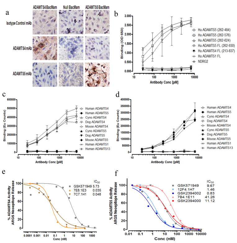
Objective/method: Aggrecanase activity, most notably ADAMTS-5, is implicated in pathogenic cartilage degradation. Selective monoclonal antibodies (mAbs) to both ADAMTS-5 and ADAMTS-4 were generated and in vitro, ex vivo and in vivo systems were utilized to assess target engagement, aggrecanase inhibition and modulation of disease-related endpoints with the intent of selecting a candidate for clinical development in osteoarthritis (OA).

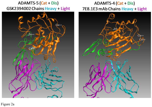
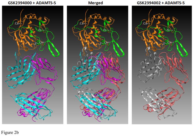
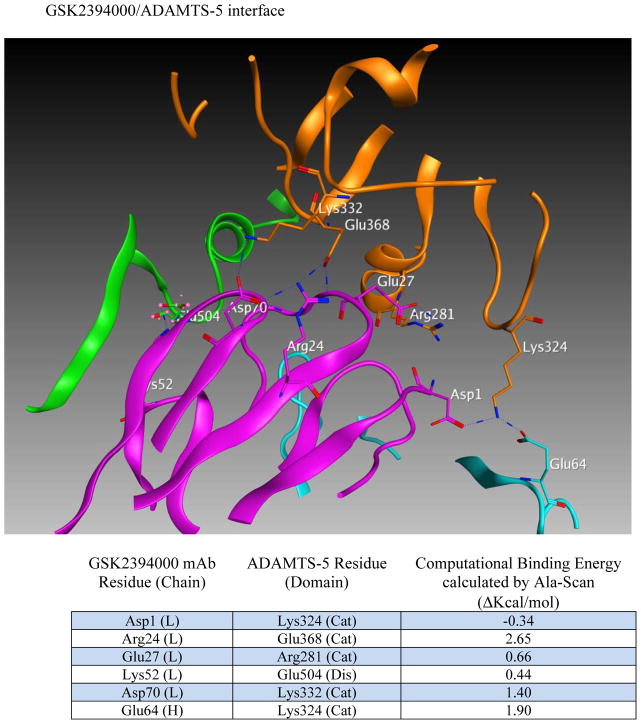
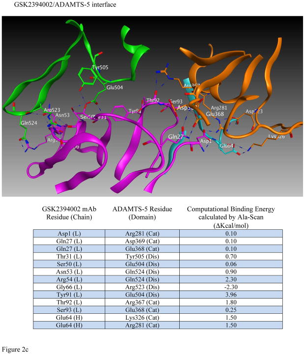
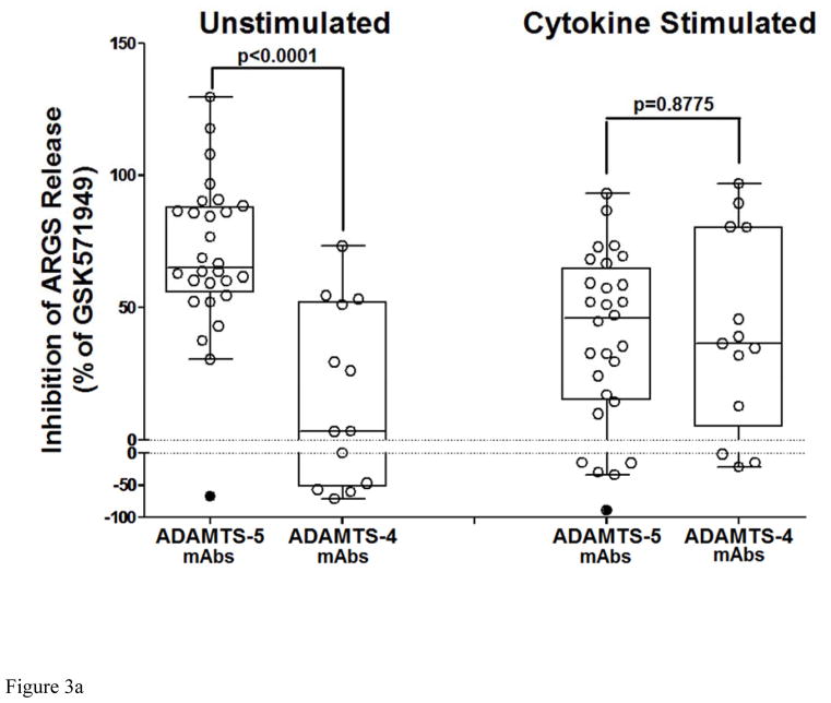
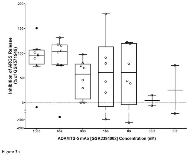
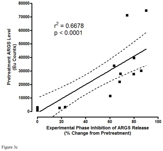
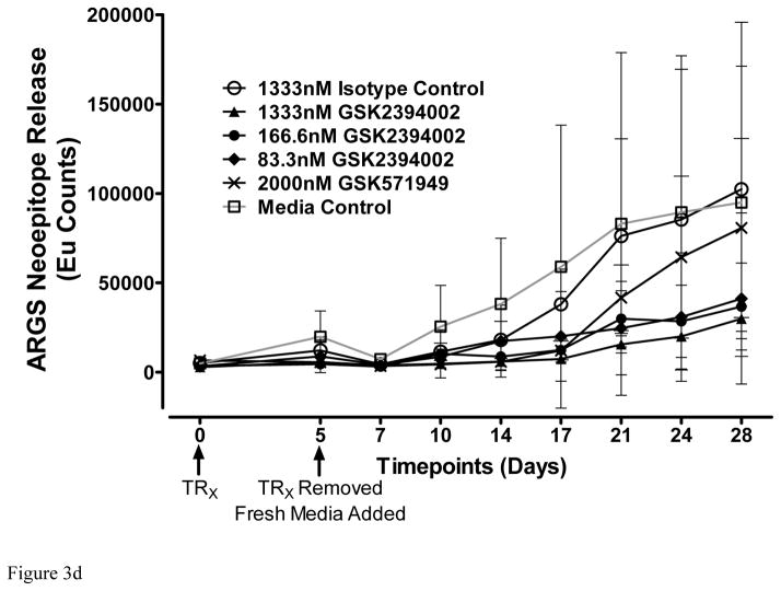
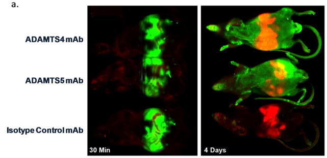
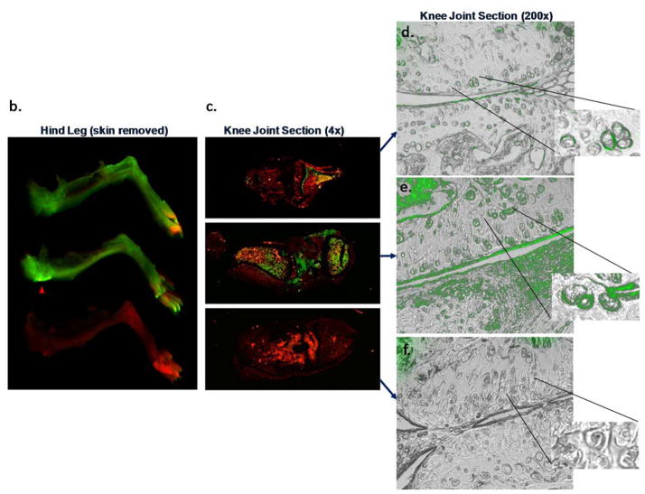
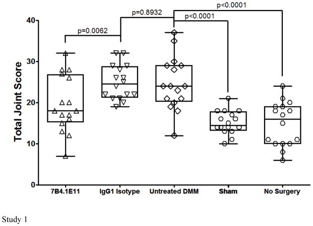
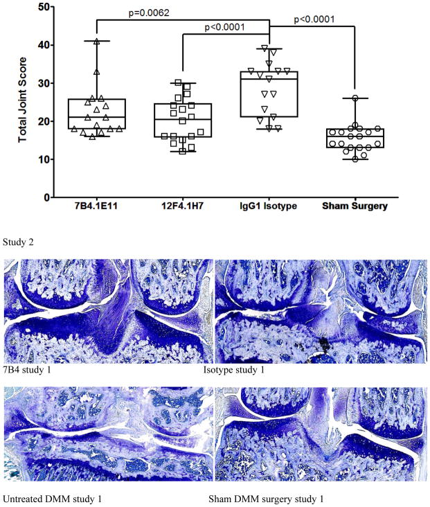
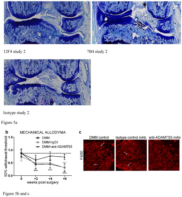
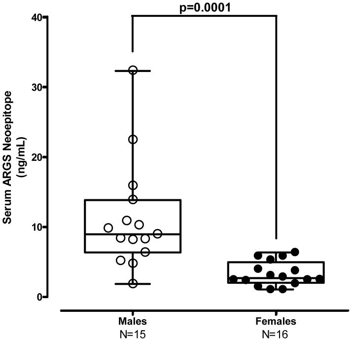
Results: Structural mapping predicts the most potent mAbs employ a unique mode of inhibition by cross-linking the catalytic and disintegrin domains. In a surgical mouse model of OA, both ADAMTS-5 and ADAMTS-4-specific mAbs penetrate cartilage following systemic administration, demonstrating access to the anticipated site of action. Structural disease modification and associated alleviation of pain-related behavior were observed with ADAMTS-5 mAb treatment. Treatment of human OA cartilage demonstrated a preferential role for ADAMTS-5 inhibition over ADAMTS-4, as measured by ARGS neoepitope release in explant cultures. ADAMTS-5 mAb activity was most evident in a subset of patient-derived tissues and suppression of ARGS neoepitope release was sustained for weeks after a single treatment in human explants and in cynomolgus monkeys, consistent with high affinity target engagement and slow ADAMTS-5 turnover.

Conclusion: This data supports a hypothesis set forth from knockout mouse studies that ADAMTS-5 is the major aggrecanase involved in cartilage degradation and provides a link between a biological pathway and pharmacology which translates to human tissues, non-human primate models and points to a target OA patient population. Therefore, a humanized ADAMTS-5-selective monoclonal antibody (GSK2394002) was progressed as a potential OA disease modifying therapeutic.

Keywords: ADAMTS-4; ADAMTS-5; Cartilage; Disease-modifying osteoarthritis drugs (DMOADs); Monoclonal antibody; Osteoarthritis.


Conflict of interest statement

Competing interests

JL, TAL, LE, JS, JLS, RM, YX, FL, CG, CCM and CJM are current or former employees of, and are shareholders in, GlaxoSmithKline. REM, PBT and AMM declare no competing interests.

Figures

Figure 1
Figure 1
ADAMTS-4 and ADAMTS-5 monoclonal antibodies specifically bind to and potently inhibit aggrecanase activity. (a) Selective binding of mAbs for ADAMTS-4 (7E8.1E3) and ADAMTS-5 (7B4.1E11) is demonstrated by immunocytochemistry using ADAMTS-4, ADAMTS-5 and Null BacMam transduced CHO cells. ADAMTS-5 specific domain mapping and selective binding to purified recombinant proteins is profiled by ELISA (b; 12F4.1H7) and DELFIA (c; 7E8.1E3 and d; 12F4.1H7) (Results shown are n=2 independent experiments with 2 dependent replicates for each concentration ± 95% CI). Functional potency of ADAMTS-4 (e) and ADAMTS-5 (f) mAbs and non-selective small molecule (GSK579149) is quantified vs isotype controls (not shown) by measuring percent inhibition of selective aggrecanase-mediated ARGS neoepitope formation from purified bovine aggrecan substrate. IC50 values for each mAb and GSK571949 are shown (inset, e and f) in relation to isotype control mAbs and DMSO, respectively, which did not demonstrate any detectible inhibitory activity. Consistent with binding specificity, no inhibition of aggrecanase activity was observed in this assay for ADAMTS-4 with ADAMTS-5 mAbs and ADAMTS-5 with ADAMTS-4 mAbs (not shown).
Figure 2
Figure 2
in silico structural modeling of antigen/antibody interactions suggest a conformational binding epitope for select ADAMTS-4 (7E8.1E3) and ADAMTS-5 (GSK2394002) mAbs which spans the catalytic and disintegrin domains of each protease and predicts an ‘allosteric lock’ mechanism of inhibition (a). In all panels the top ranked docking orientation is shown; cyan/light blue is the light chain and magenta/purple the heavy chain of the monoclonal antibodies; orange is the catalytic domain and green the disintegrin domain of ADAMTS-4 or -5. Individual and merged overlay structural models of the inhibitory ADAMTS-5 mAbs (b) and amino acid residues spanning the catalytic and disintegrin domains of ADAMTS-5 predicted to form hydrogen bond interactions (GSK2394000, six bonds and GSK2394002, thirteen bonds) are shown with bond energy predictions from an Alanine substitution scan of the predicted protease/mAb interface (c).
Figure 2
Figure 2
in silico structural modeling of antigen/antibody interactions suggest a conformational binding epitope for select ADAMTS-4 (7E8.1E3) and ADAMTS-5 (GSK2394002) mAbs which spans the catalytic and disintegrin domains of each protease and predicts an ‘allosteric lock’ mechanism of inhibition (a). In all panels the top ranked docking orientation is shown; cyan/light blue is the light chain and magenta/purple the heavy chain of the monoclonal antibodies; orange is the catalytic domain and green the disintegrin domain of ADAMTS-4 or -5. Individual and merged overlay structural models of the inhibitory ADAMTS-5 mAbs (b) and amino acid residues spanning the catalytic and disintegrin domains of ADAMTS-5 predicted to form hydrogen bond interactions (GSK2394000, six bonds and GSK2394002, thirteen bonds) are shown with bond energy predictions from an Alanine substitution scan of the predicted protease/mAb interface (c).
Figure 3
Figure 3
Potent and sustained inhibition of aggrecanolysis from human OA cartilage explants by ADAMTS-5 mAb treatment. ADAMTS-5 mAb treatment inhibits ARGS neoepitope release from sequentially unstimulated and IL-1b/OSM stimulated human OA cartilage explants (a), whereas only modest inhibition is observed with ADAMTS-4 mAb treatment, preferentially under cytokine-stimulated conditions. Mean percent inhibition of mAbs is shown relative to isotype control mAb (0% inhibition, dashed lines in a and b) and GSK571949 (100% inhibition) treatment. Experiments were conducted with 7 replicates for each treatment condition from 8 timepoints across multiple 28-day experiments consisting of a 21 day unstimulated phase followed by a 7-day cytokine-stimulated phase. A total of 13–28 independent OA patient experiments were conducted for each treatment condition. ADAMTS-4 (7E8.1E3 or 7C71.H1) and ADAMTS-5 (12F4.1H7 or 7B4.1E11) mAb treatments are shown as compiled data associated with each mAb specificity. All mAb treatments were held constant at 670nM throughout the experiment and GSK571949 was similarly held at 2 μM. (b) Dose-dependent inhibition of ARGS neoepitope release is evident with GSK2394002 treatment in the absence of exogenous cytokine stimulation using cartilage from individual OA donors with elevated pretreatment ARGS neoepitope levels (n=2–8 independent donor experiments per treatment concentration and each point represents mean % inhibition from 21 day unstimulated phase, as described in (a)). Plots (a and b) are presented in standard box-and-whisker format with Tukey outliers noted (>1.5 times IQR, filled symbols). (c) Linear regression analysis suggests the response to GSK2394002 treatment correlates with pre-treatment ARGS neoepitope levels. Sustained suppression of ARGS neoepitope release is observed with pulse-chase GSK2394002 treatment in relation to small molecule (GSK571949) in individual donor experiments using cultures with elevated pretreatment ARGS neoepitope levels (n=4; each timepoint and ±95% CI error bar represents mean of 4 independent donor experiments where 7 biological replicates for each donor/timepoint was used to calculate a within donor mean for each timepoint) (d).
Figure 3
Figure 3
Potent and sustained inhibition of aggrecanolysis from human OA cartilage explants by ADAMTS-5 mAb treatment. ADAMTS-5 mAb treatment inhibits ARGS neoepitope release from sequentially unstimulated and IL-1b/OSM stimulated human OA cartilage explants (a), whereas only modest inhibition is observed with ADAMTS-4 mAb treatment, preferentially under cytokine-stimulated conditions. Mean percent inhibition of mAbs is shown relative to isotype control mAb (0% inhibition, dashed lines in a and b) and GSK571949 (100% inhibition) treatment. Experiments were conducted with 7 replicates for each treatment condition from 8 timepoints across multiple 28-day experiments consisting of a 21 day unstimulated phase followed by a 7-day cytokine-stimulated phase. A total of 13–28 independent OA patient experiments were conducted for each treatment condition. ADAMTS-4 (7E8.1E3 or 7C71.H1) and ADAMTS-5 (12F4.1H7 or 7B4.1E11) mAb treatments are shown as compiled data associated with each mAb specificity. All mAb treatments were held constant at 670nM throughout the experiment and GSK571949 was similarly held at 2 μM. (b) Dose-dependent inhibition of ARGS neoepitope release is evident with GSK2394002 treatment in the absence of exogenous cytokine stimulation using cartilage from individual OA donors with elevated pretreatment ARGS neoepitope levels (n=2–8 independent donor experiments per treatment concentration and each point represents mean % inhibition from 21 day unstimulated phase, as described in (a)). Plots (a and b) are presented in standard box-and-whisker format with Tukey outliers noted (>1.5 times IQR, filled symbols). (c) Linear regression analysis suggests the response to GSK2394002 treatment correlates with pre-treatment ARGS neoepitope levels. Sustained suppression of ARGS neoepitope release is observed with pulse-chase GSK2394002 treatment in relation to small molecule (GSK571949) in individual donor experiments using cultures with elevated pretreatment ARGS neoepitope levels (n=4; each timepoint and ±95% CI error bar represents mean of 4 independent donor experiments where 7 biological replicates for each donor/timepoint was used to calculate a within donor mean for each timepoint) (d).
Figure 4
Figure 4
ADAMTS-4 and ADAMTS-5 mAbs engage their targets within the cartilage following systemic administration in a mouse OA model. IR800-labeled ADAMTS-4 (7E8.1E3), ADAMTS-5 (7B4.1E11) or Isotype control mAbs were administered ([16mg/kg] IP) to mice (n=1/group) six weeks after surgical destabilization of the medial meniscus and periodically imaged to monitor systemic biodistribution and tissue penetration. Within 30 minutes of dosing a strong IR800 signal (green) can be observed in the abdomen of all animals (a, left) indicative of accurate administration. The red abdominal signal is related to the inherent infrared signature of the animal diet traversing the digestive system. Systemic IR800 signal is observed in mice receiving ADAMTS-4 or ADAMTS-5 mAb (and to a much lesser extent the isotype control treated animal) 4 days following administration (a, right) when the animals were sacrificed to assess tissue distribution. The right hind leg of each animal is shown (b) with skin removed showing a strong signal intensity in the joint region of the ADAMTS-5 mAb treated mouse (red arrowhead) compared to the other treatments. Similar qualitative differences between treatment groups can be observed in low (c; 4X magnification) and high power (d–f; 200X magnification) images of the knee joint, as well as evidence for cartilage penetration and deposition in the cartilage for the ADAMTS-4 and ADAMTS-5 mAbs in the superficial cartilage zone and pericellular region of the articular chondrocytes (d–e, inset). Merged microscopic bright-field/IR800 images are shown. In the ADAMTS-5 mAb panel (e) staining in the meniscus can be observed (bottom half of image). No signal was observed following administration of a labeled isotype control mAb (f) or following administration of a non-reactive dye control, which was undetectable within 24 hours after dosing (not shown).
Figure 4
Figure 4
ADAMTS-4 and ADAMTS-5 mAbs engage their targets within the cartilage following systemic administration in a mouse OA model. IR800-labeled ADAMTS-4 (7E8.1E3), ADAMTS-5 (7B4.1E11) or Isotype control mAbs were administered ([16mg/kg] IP) to mice (n=1/group) six weeks after surgical destabilization of the medial meniscus and periodically imaged to monitor systemic biodistribution and tissue penetration. Within 30 minutes of dosing a strong IR800 signal (green) can be observed in the abdomen of all animals (a, left) indicative of accurate administration. The red abdominal signal is related to the inherent infrared signature of the animal diet traversing the digestive system. Systemic IR800 signal is observed in mice receiving ADAMTS-4 or ADAMTS-5 mAb (and to a much lesser extent the isotype control treated animal) 4 days following administration (a, right) when the animals were sacrificed to assess tissue distribution. The right hind leg of each animal is shown (b) with skin removed showing a strong signal intensity in the joint region of the ADAMTS-5 mAb treated mouse (red arrowhead) compared to the other treatments. Similar qualitative differences between treatment groups can be observed in low (c; 4X magnification) and high power (d–f; 200X magnification) images of the knee joint, as well as evidence for cartilage penetration and deposition in the cartilage for the ADAMTS-4 and ADAMTS-5 mAbs in the superficial cartilage zone and pericellular region of the articular chondrocytes (d–e, inset). Merged microscopic bright-field/IR800 images are shown. In the ADAMTS-5 mAb panel (e) staining in the meniscus can be observed (bottom half of image). No signal was observed following administration of a labeled isotype control mAb (f) or following administration of a non-reactive dye control, which was undetectable within 24 hours after dosing (not shown).
Figure 5
Figure 5
ADAMTS-5 mAb treatment suppresses joint disease severity in a mouse model of osteoarthritis. Mice treated prophylactically with anti-ADAMTS-5 (1x/week, i.p. 10–16 mg/kg) were protected from developing (a) histologic cartilage degeneration (two independent studies shown, n=8–10 animals/group each with two DMM knees/animal, although not all knees in study 2 could be scored due to isolated tissue processing errors and in one group loss of a single mouse between surgery and treatment commencement, as demonstrated by individual knee symbols in each group ranging from 15–19/group). Representative toluidine blue stained knee joint sections are shown for each study group. (b) Mice treated prophylactically with anti-ADAMTS5 mAb (1x/week, i.p. 10 mg/kg) were protected from developing mechanical allodynia through 8 weeks post DMM, *** p<0.001 vs time 0, n=12/group. Mean ±95% CI. (c) Prophylactic treatment with anti-ADAMTS5 mAb also protected mice from macrophage infiltration (F4/80 marker) into the DRG at 8 weeks post DMM. Arrows indicate examples of infiltrating macrophages in the DMM control and Isotype control mAb (IgG1) conditions.
Figure 5
Figure 5
ADAMTS-5 mAb treatment suppresses joint disease severity in a mouse model of osteoarthritis. Mice treated prophylactically with anti-ADAMTS-5 (1x/week, i.p. 10–16 mg/kg) were protected from developing (a) histologic cartilage degeneration (two independent studies shown, n=8–10 animals/group each with two DMM knees/animal, although not all knees in study 2 could be scored due to isolated tissue processing errors and in one group loss of a single mouse between surgery and treatment commencement, as demonstrated by individual knee symbols in each group ranging from 15–19/group). Representative toluidine blue stained knee joint sections are shown for each study group. (b) Mice treated prophylactically with anti-ADAMTS5 mAb (1x/week, i.p. 10 mg/kg) were protected from developing mechanical allodynia through 8 weeks post DMM, *** p<0.001 vs time 0, n=12/group. Mean ±95% CI. (c) Prophylactic treatment with anti-ADAMTS5 mAb also protected mice from macrophage infiltration (F4/80 marker) into the DRG at 8 weeks post DMM. Arrows indicate examples of infiltrating macrophages in the DMM control and Isotype control mAb (IgG1) conditions.
Figure 6
Figure 6
Figure 6a – Male cynomolgus monkeys exhibit generally higher endogenous circulating ARGS neoepitope concentrations as compared to females. Figure 6b – Serum ARGS neoepitope levels rapidly and dose-dependently decline in response to ADAMTS-5 mAb (GSK2394002) treatment in non-human primates. Monkeys (n/group at each timepoint is shown from two independent studies, both males and females included at each timepoint except days 14–28 which is only females) were dosed every two weeks starting immediately after a pre-dose blood sample at day 0. Serum and plasma was drawn on the 3rd day after dosing, immediately prior to the next dose and periodically throughout the study as shown to monitor circulating PK and PD endpoints. Mid study samples were not taken for the 100mg/kg group and some animals in the other groups, as shown. Data represents mean percent change from pre-dose ± 95% CI with statistical significance (∞p=0.023, %p=0.0046, #p=0.0027, ∇p=0.0001 *p<0.0001) observed for all GSK2394002 dose groupings and at all post-treatment timepoints relative to the vehicle control treated group.
Figure 6
Figure 6
Figure 6a – Male cynomolgus monkeys exhibit generally higher endogenous circulating ARGS neoepitope concentrations as compared to females. Figure 6b – Serum ARGS neoepitope levels rapidly and dose-dependently decline in response to ADAMTS-5 mAb (GSK2394002) treatment in non-human primates. Monkeys (n/group at each timepoint is shown from two independent studies, both males and females included at each timepoint except days 14–28 which is only females) were dosed every two weeks starting immediately after a pre-dose blood sample at day 0. Serum and plasma was drawn on the 3rd day after dosing, immediately prior to the next dose and periodically throughout the study as shown to monitor circulating PK and PD endpoints. Mid study samples were not taken for the 100mg/kg group and some animals in the other groups, as shown. Data represents mean percent change from pre-dose ± 95% CI with statistical significance (∞p=0.023, %p=0.0046, #p=0.0027, ∇p=0.0001 *p<0.0001) observed for all GSK2394002 dose groupings and at all post-treatment timepoints relative to the vehicle control treated group.

Comment in

References

    1. Loeser RF. Age-Related Changes in the Musculoskeletal System and the Development of Osteoarthritis. Clin Geriatr Med. 2010;26:371–386. - PMC - PubMed
    1. Woolfe AD, Pfleger B. Burden of major musculoskeletal conditions. Bull World Health Organ. 2003;81:646–656. - PMC - PubMed
    1. Troeberg L, Nagase H. Proteases involved in cartilage matrix degradation in osteoarthritis. Biochim Biophys Acta. 2012;1824:133–145. - PMC - PubMed
    1. Glasson SS, Askew R, Sheppard B, Carito B, Blanchet T, Ma HL, et al. Deletion of active ADAMTS5 prevents cartilage degradation in a murine model of osteoarthritis. Nature. 2005;434:644–648. - PubMed
    1. Stanton H, Rogerson FM, East CJ, Golub SB, Lawlor KE, Meeker CT, et al. ADAMTS5 is the major aggrecanase in mouse cartilage in vivo and in vitro. Nature. 2005;434:648–652. - PubMed

Publication types