. 2015 Apr 8;17(4):526-35.
doi: 10.1016/j.chom.2015.02.011. Epub 2015 Mar 19.

Plasmodium vivax liver stage development and hypnozoite persistence in human liver-chimeric mice

Affiliations

Plasmodium vivax liver stage development and hypnozoite persistence in human liver-chimeric mice

Sebastian A Mikolajczak et al. Cell Host Microbe. .

Abstract

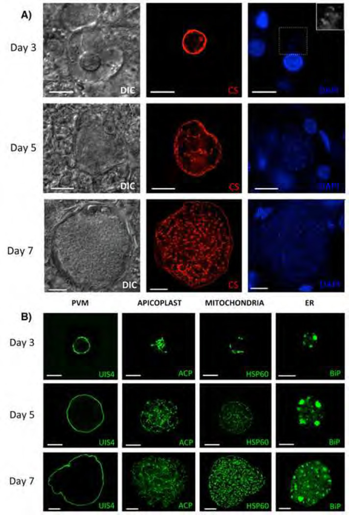
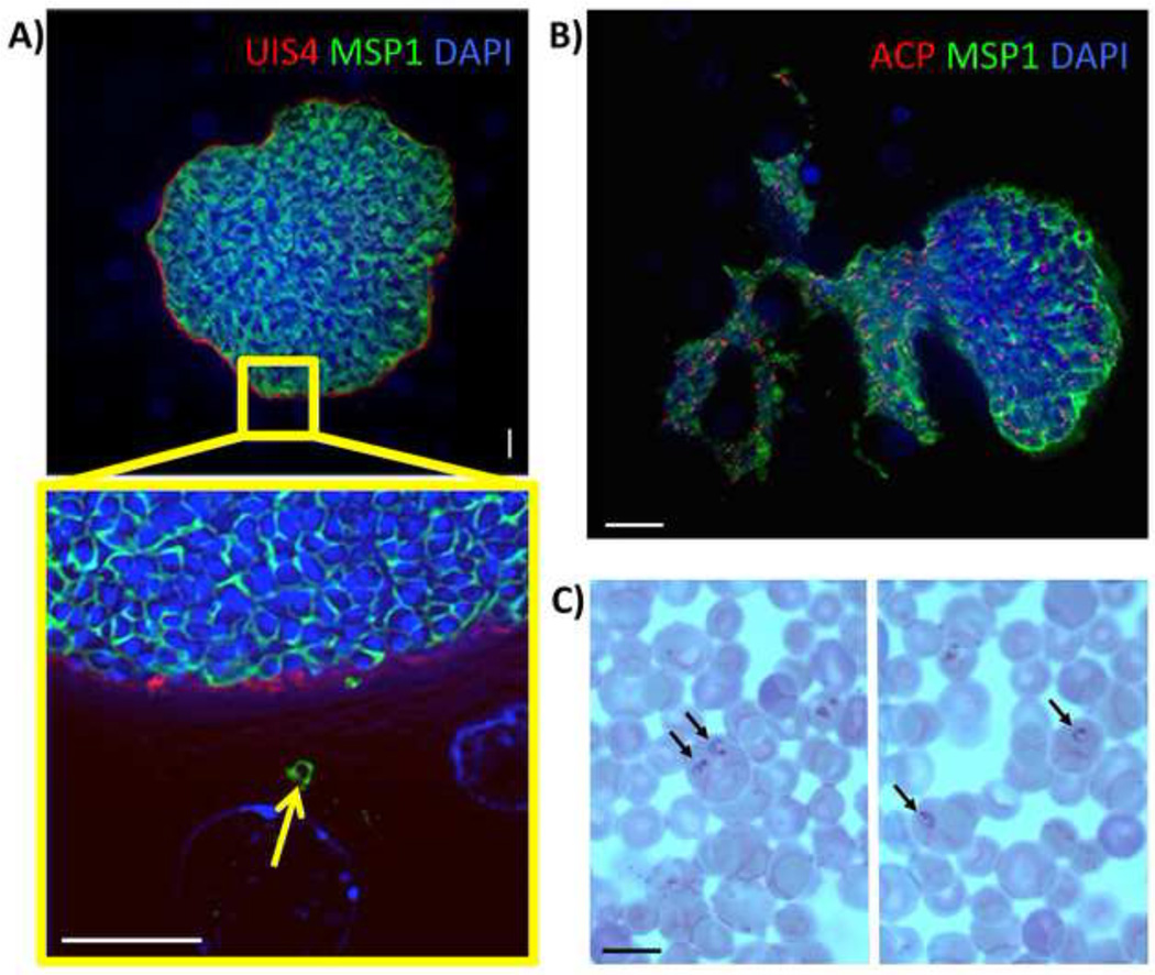
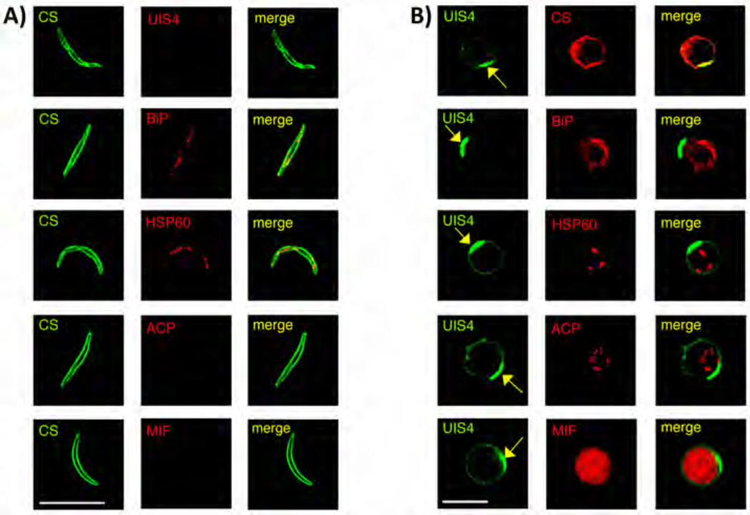
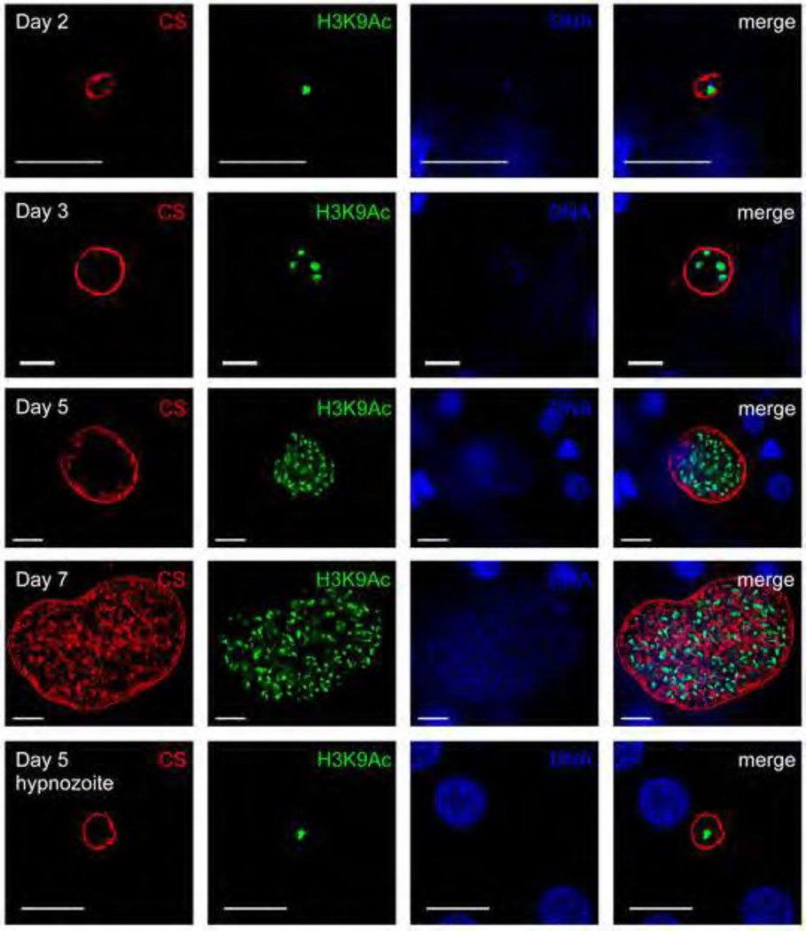
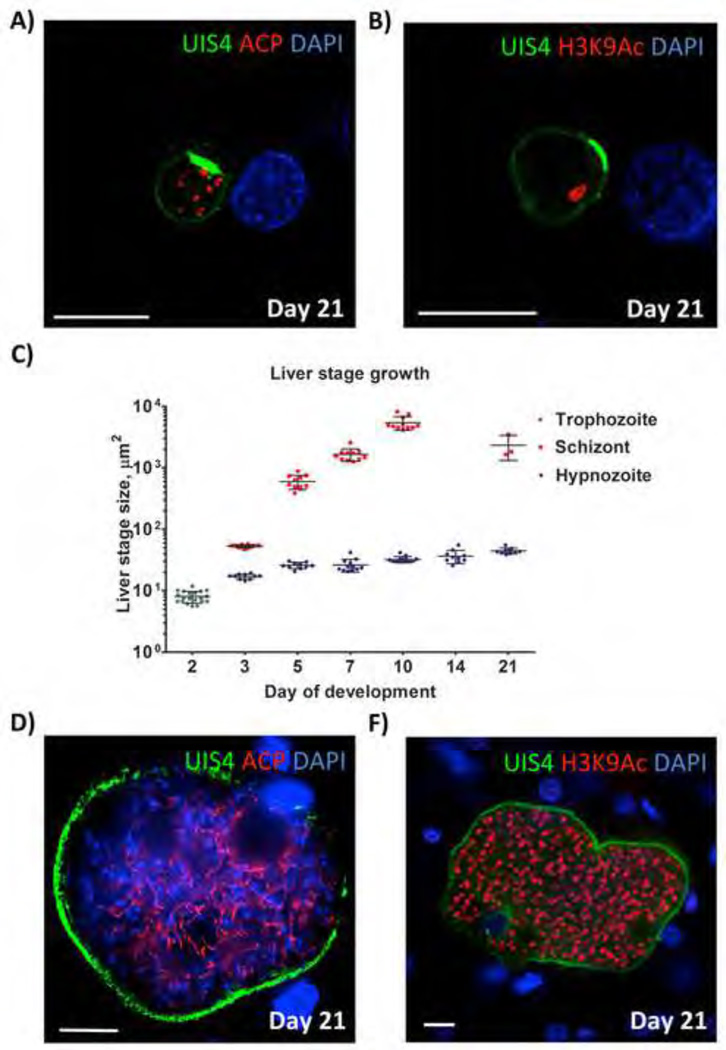
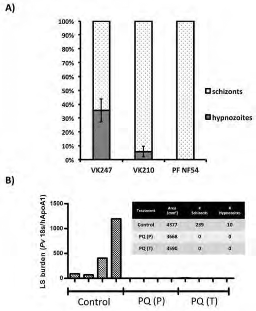
Plasmodium vivax malaria is characterized by periodic relapses of symptomatic blood stage parasite infections likely initiated by activation of dormant liver stage parasites-hypnozoites. The lack of tractable P. vivax animal models constitutes an obstacle in examining P. vivax liver stage infection and drug efficacy. To overcome this obstacle, we have used human liver-chimeric (huHep) FRG KO mice as a model for P. vivax infection. FRG KO huHep mice support P. vivax sporozoite infection, liver stage development, and hypnozoite formation. We show complete P. vivax liver stage development, including maturation into infectious exo-erythrocytic merozoites as well as the formation and persistence of hypnozoites. Prophylaxis or treatment with the antimalarial primaquine can prevent and eliminate liver stage infection, respectively. Thus, P. vivax-infected FRG KO huHep mice are a model to investigate liver stage development and dormancy and may facilitate the discovery of drugs targeting relapsing malaria.


Conflict of interest statement

The authors declare no conflicts of interest.

Figures

Figure 1
Figure 1. Development of P. vivax liver stages in the FRG KO huHep mouse
(A) Liver stage parasites in infected liver sections three-, five- and seven days after sporozoite infection were visualized with differential interference contrast (DIC) imaging (left panel), with a monoclonal antibody specific for P. vivax circumsporozoite protein (CS; VK247) (central panel), and DAPI for DNA content (right panel). To visualize the DNA content in the small three-day old parasite the area inside the dotted box is magnified in the top right corner. (B) Liver stages at three-, five- and seven days after sporozoite infection were analyzed with monoclonal mouse antibodies to P. vivax Upregulated in Infectious Sporozoites protein 4 (UIS4, localizes to the PVM), acyl carrier protein (ACP, localizes the apicoplast), heat shock protein 60 (HSP60, localizes to the mitochondria) and binding immunoglobulin protein (BiP, localizes to the ER). Scale bar: 10 µm.
Figure 2
Figure 2. Complete maturation of P. vivax liver stages and persistence of hypnozoites in the FRG KO huHep mouse
(A) Indirect immunofluorescence assay of P. vivax liver stages at ten days after sporozoite infection using Pv merozoite surface protein-1 (MSP-1) polyclonal rabbit antibody (green), PvUIS4 mouse monoclonal antibody (red) and DAPI (blue, DNA). MSP-1 expression reveals the presence of differentiated exo-erythrocytic merozoites within the liver stage schizont. The magnification of the area in the yellow box shows a MSP-1- positive exo-erythrocytic merozoite (arrow) outside the confines of the mature liver schizont. Scale bar: 10 µm. (B) IFA of a nine-day old P. vivax liver stage stained with PvMSP-1 polyclonal rabbit antibody (green) to visualize the merozoite surfaces, ACP monoclonal mouse antibody (red) to visualize the apicoplast and DAPI (DNA stain, blue). The mature liver stage is in the process of releasing exo-erythrocytic merozoites into the surrounding liver tissue. Scale bar: 20 µm. (C) Human red blood cells enriched for reticulocytes were injected into FRG KO huHep mice nine days post sporozoite infection. Four hours later, the blood was removed and microscopically analyzed by Giemsa-stained thin blood smear. Black arrows point to P. vivax ring stage parasites within reticulocytes. Scale bar: 10 µm.
Figure 3
Figure 3. Characterization of P. vivax hypnozoites in FRG KO huHep mouse and comparison to sporozoites
(A) IFA of P. vivax sporozoites stained with mouse monoclonal antibody to PvCS (green) and rabbit polyclonal antibodies to PvUIS4, BiP, PvHSP60, ACP and Pv macrophage inhibitory factor (MIF, red). (B) Hypnozoites at day seven post sporozoite infection were localized with antibodies to PvUIS4 (rabbit polyclonal and mouse monoclonal) (green), monoclonal mouse antibody to PvCS (red) and rabbit polyclonal antibodies to BiP, PvHSP60, ACP and PvMIF (red). The yellow arrow points to a PvUIS4-postive PVM prominence that is notable in the PVM of all hypnozoites. Scale bar: 10 µm.
Figure 4
Figure 4. P. vivax liver stage genome replication
Antibodies to acetylated lysine 9 of histone H3 (H3K9Ac) (green) was used to analyze genome replication of liver stage parasites in infected FRG KO huHep mice. Schizonts and hypnozoites were also stained with antibody to CS (VK247) (red). DNA was visualized with DAPI (blue). A single H3K9Ac-positive structure was detected within liver stage trophozoites two days after sporozoite infection. Hypnozoites (shown here five days after sporozoite infection) also contained a single H3K9Ac-positive structure. Multiple H3K9Ac-positive structures were observed for replicating liver stage schizonts at day three-, five- and seven after sporozoite infection. Scale bar: 10 µm.
Figure 5
Figure 5. P. vivax hypnozoite persistence and hypnozoite activation in FRG KO huHep mice
(A, B) Persistent hypnozoites at day 21 after sporozoite infection. Parasites were visualized with antibodies to PvUIS4 (green), antibodies to acetylated lysine 9 of histone H3 (H3K9Ac) (red) and host hepatocyte nuclei were visualized with DAPI. The UIS4-postive PVM prominence is maintained and notable in the PVM of all persistent hypnozoites. Hypnozoites contained a single H3K9Ac-positive structure. (C) Size comparison of liver stage trophozoites, liver stage schizonts and hypnozoites at different time points of infection. The size (liver stage area at the greatest circumference of the parasite) was calculated. Measurements were taken for at least 10 liver stages at each time point. The average size +/− SEM is shown on the dot plots. Note that hypnozoites show some growth over time but remain smaller than the three-day old schizonts. No schizonts were detected in infections at 14 days after sporozoite infection biut were again detected at day 21. (D, E) show examples replicating liver stages at day 21 post sporozoite infection, suggesting that they originated from hypnozoites that activated and entered schizogony. Antibodies used for IFA were mouse monoclonal antibody to PvUIS4 (green) rabbit polyclonal antibodies to ACP in the left panel and H3K9Ac (red) in the right panel. DNA was stained with DAPI (blue). Scale bar: 10 µm.
Figure 6
Figure 6. Thai P. vivax hypnozoite frequencies and outcomes of experimental drug treatments in FRG KO huHep mice
(A) A graphical representation of hypnozoite and liver stage schizont abundance in infections. Liver stages are denoted as schizonts or hypnozoites. For quantitative assessment, liver stages observed on ten, non-serial liver sections were totaled for each infected mouse. Results for two Thai P. vivax isolates are shown (CS VK247 genotype (two independent experiments with different patient isolates) and CS VK210 genotype (four independent experiments with different patient isolates). Error bars: standard deviation. (B) Four mice were treated with primaquine (30 mg/kg) at days one- through day three post sporozoite infection (PQ prophylaxis (P)) or days three through seven post sporozoite infection (PQ treatment (T)). Parasites of the CS VK210 genotype were used for the experiments. Four untreated mice served as controls (Control). Eight days after sporozoite infection, all mice were sacrificed and cDNA was produced from three separate liver tissue samples for each mouse. Liver stage burden using qRT-PCR and was used to normalize P. vivax 18S rRNA transcription to human ApoAI transcription. The graph insert shows microscopic quantitation of liver stage schizonts (LS) and hypnozoites observed by IFA. Area of tissue refers to cumulative area of tissue analyzed for each treatment where similar number of sections from each technical replicate was processed.

Similar articles

Cited by

References

    1. Anstey NM, Douglas NM, Poespoprodjo JR, Price RN. Plasmodium vivax: Clinical Spectrum, Risk Factors and Pathogenesis. Advances in parasitology. 2012;80:151–201. - PubMed
    1. Azuma H, Paulk N, Ranade A, Dorrell C, Al-Dhalimy M, Ellis E, Strom S, Kay MA, Finegold M, Grompe M. Robust expansion of human hepatocytes in Fah−/−/Rag2−/−/Il2rg−/− mice. Nat Biotechnol. 2007;25:903–910. - PMC - PubMed
    1. Bissig KD, Le TT, Woods NB, Verma IM. Repopulation of adult and neonatal mice with human hepatocytes: a chimeric animal model. Proc Natl Acad Sci U S A. 2007;104:20507–20511. - PMC - PubMed
    1. Carlton JM, Sina BJ, Adams JH. Why is Plasmodium vivax a neglected tropical disease? PLoS neglected tropical diseases. 2011;5:e1160. - PMC - PubMed
    1. Collins WE, Contacos PG, Jumper JR, Smith CS, Skinner JC. Studies on human malaria in aotus monkeys. 3. Exoerythrocytic stages of the Salvador II strain of Plasmodium vivax. The Journal of parasitology. 1973;59:859–866. - PubMed

Publication types