. 2015 Apr 8;17(4):452-65.
doi: 10.1016/j.chom.2015.02.009. Epub 2015 Mar 19.

Calnexin induces expansion of antigen-specific CD4(+) T cells that confer immunity to fungal ascomycetes via conserved epitopes

Affiliations

Calnexin induces expansion of antigen-specific CD4(+) T cells that confer immunity to fungal ascomycetes via conserved epitopes

Marcel Wüthrich et al. Cell Host Microbe. .

Abstract

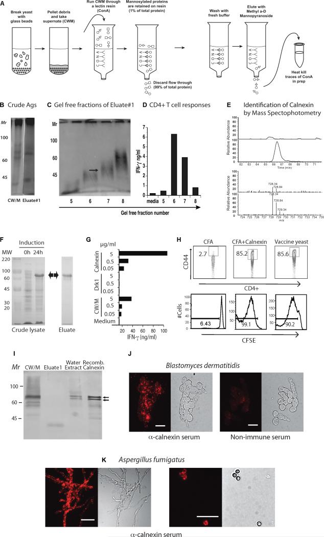
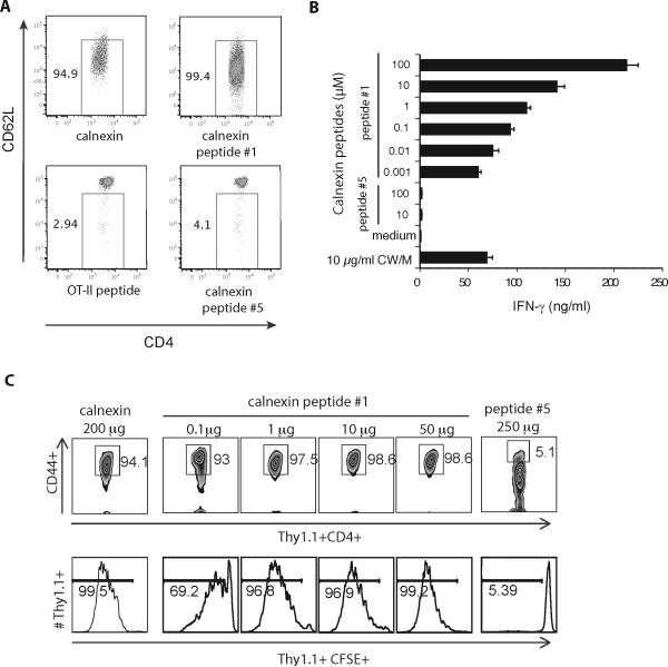
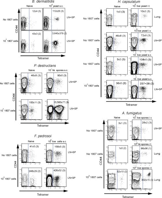
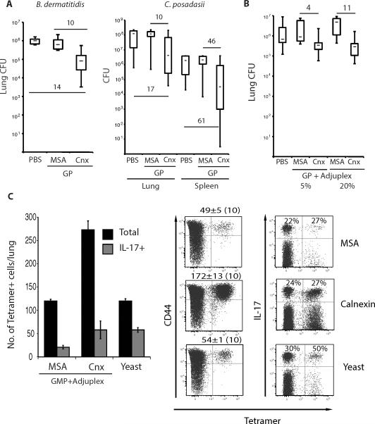
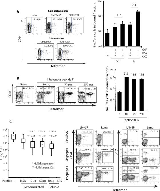
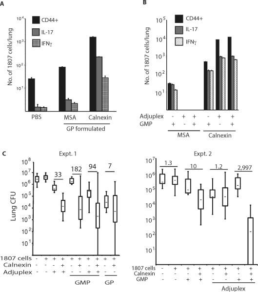
Fungal infections remain a threat due to the lack of broad-spectrum fungal vaccines and protective antigens. Recent studies showed that attenuated Blastomyces dermatitidis confers protection via T cell recognition of an unknown but conserved antigen. Using transgenic CD4(+) T cells recognizing this antigen, we identify an amino acid determinant within the chaperone calnexin that is conserved across diverse fungal ascomycetes. Calnexin, typically an ER protein, also localizes to the surface of yeast, hyphae, and spores. T cell epitope mapping unveiled a 13-residue sequence conserved across Ascomycota. Infection with divergent ascomycetes, including dimorphic fungi, opportunistic molds, and the agent causing white nose syndrome in bats, induces expansion of calnexin-specific CD4(+) T cells. Vaccine delivery of calnexin in glucan particles induces fungal antigen-specific CD4(+) T cell expansion and resistance to lethal challenge with multiple fungal pathogens. Thus, the immunogenicity and conservation of calnexin make this fungal protein a promising vaccine target.


Figures

Fig. 1
Fig. 1. Identity of shared fungal Ag
A. Generation of eluate #1. B. Silver stain of PAGE of B. dermatitidis Ags. C. Gel free separation of Eluate #1 into fractions. D. Stimulation of 1807 TCR Tg cells in vitro by fractions from panel C, as measured by IFN-γ response. Arrow in fraction 7 denotes material analyzed by MS/MS. E. Identification of calnexin by MS/MS. The panel shows data collected for one calnexin-derived peptide, as an example. The top set of paired traces is a comparison of the HPLC separation of the non-stimulatory control fraction (upper) and the stimulatory fraction #7 (lower). The peak in fraction #7 is not present in the control. MS of this peak (bottom traces) identified the peptide: LQNSLNCGGAYMK [728.34Da; +2H] and this mass is better represented in stimulatory fraction #7 (lower) vs. non-stimulatory control (upper). Adjacent peaks are representative of isotopic variants. E. Induction of E. coli produced r-calnexin (63kD). F. r-calnexin stimulates 1807 T cells to produce IFN-γ in vitro. G. r-calnexin activates (CD44) and induces proliferation (CFSE) of transferred 1807 cells in vivo. H. Western of crude Ags. I&J. Surface stain of B. dermatitidis yeast (strain#55) and A. fumigatus hyphae (left) and spores (right) with anti-calnexin oligospecific antibody. Bar = 10 microns.
Fig. 2
Fig. 2. Identification of calnexin's T cell epitope recognized by 1807 cells
A. In vitro activation of 1807 T cells by calnexin peptide #1. 105 BMDC were loaded with calnexin (50μg/ml) or peptide (10μM) and co-cultured with 3 × 10⁵ CD4+ purified 1807 T cells. 3d later T-cells were analyzed for activation by flow cytometry. B. Naïve 1807 T cells were co-cultured as in Panel A, and culture supernatants analyzed for IFN-γ. C. Mice received 106 naïve, CFSE-labeled 1807 cells prior to s.c. vaccination with 200 μg rcalnexin, a dilution series of peptide #1 and 250 μg peptide #5 (as a negative control). 7d later the skin draining lymph nodes were harvested and CFSE profiles and CD44-expression of 1807 cells analyzed.
Fig. 3
Fig. 3. Tetramer enrichment of endogenous, fungal-specific T cells ex vivo
Mice received naïve 1807 T cells or not and were infected by doses and routes shown for B. dermatitidis yeast, F. pedrosoi spores, A. fumigatus spores, H. capsulatum yeast and P. destructans spores. 7d post-infection, the skin draining lymph nodes (LN), spleen (SP) or lungs were collected. The number of calnexin peptide #1-specific CD4+ T cells were analyzed and quantified after tetramer enrichment as detailed in the Methods. Tetramer positive cells are shown to the right of the gate in each dot plot. The number represents the geometric mean ± SEM of tetramer-positive cells, with number of mice studied in parenthesis.
Fig. 4
Fig. 4. Vaccine-induced resistance mediated by calnexin
A. Mice were vaccinated s.c. thrice, 2 wks apart with 108 glucan particles (GP) loaded with 10μg r-calnexin (Cnx) or mouse serum albumin (MSA) as a control. 2wk after the last boost, mice were challenged with 2×10³ B. dermatitidis 26199 yeast or 86 spores of C. posadasii strain C735. Lung and spleen (latter for C. posadasii infection) CFU were assessed 2wk post-infection. Numbers indicate the fold difference in lung CFUs vs. controls. B. Mice were vaccinated s.c. with 25 μg r-calnexin or MSA mixed with 5 or 20% Adjuplex. 2 wk after the last boost, mice were challenged with 2×10³ B. dermatitidis and lung CFU measured as in A. Numbers are the fold difference in lung CFUs vs. controls. C. IL-17 reporter mice were vaccinated thrice with 25μg of calnexin encapsulated in GMP and mixed with 5% Adjuplex. The histogram shows the mean number of tetramer-positive cells from the bound and unbound fractions combined. Dot plots show the mean ± SEM number of tetramer-positive and percent of IL-17+ (eYFP+) CD4+ T cells among tetramer-positive and -negative cells from the bound fraction, enumerated by FACS. Dot plots represent an overlay of 10 samples/group.
Fig. 5
Fig. 5. Intravenous delivery of calnexin peptide, expansion of endogenous, tetramer-specific T cells, and resistance to infection
A. Wild type C57BL6 mice were vaccinated s.c. or i.v. with 108 glucan mannan particles (GMP) loaded with 10 μg of r-calnexin (Cnx) or MSA as a negative control. B. Mice were vaccinated i.v. with 10-250 μg soluble calnexin peptide #1 and 5 μg LPS. 7d after vaccination in panels A and B, the skin draining lymph nodes and spleen were harvested and the number and activation (CD44) of tetramer-positive T cells assessed. The dot plots represent concatenated samples for 3 - 4 mice (noted in parenthesis) per group. The numbers of tetramer+ CD4+ T cells per concatenated sample is indicated inside the dot plots. The mean ± SEM of tetramer+ CD4+ T cells per mouse is indicated in the histogram (right). The number over a bar denotes the fold change of tetramer+ T cells vs. indicated control mice. C. To assess resistance after i.v. delivery of calnexin peptide, mice were vaccinated thrice with 10 μg soluble peptide #1 plus 5 μg LPS or GP loaded with 10 or 50 μg peptide #1 or MSA as a control. 2wk after the last boost, mice were challenged with 2×10³ B. dermatitidis 26199 yeast. Lung CFU was assayed 2wk post-infection. * and **, denote fold change vs. the GMP/MSA or naïve control groups, respectively. Dot plots show the mean ± SEM number of tetramer+, activated (CD44+) and IL-17 differentiated cells (as determined by eYFP fluorescence with IL-17A fate-reporter mice) in the draining lymph nodes and spleen at the time of challenge, and recalled to the lung 4d post-infection, concatenated for 5 mice/group.
Fig. 6
Fig. 6. Naïve T cell precursor frequency and adjuvant formulation impact the pool size of calnexin primed T cells and resistance to infection
A. Mice received 106 naïve 1807 cells prior to vaccination s.c. with 108 glucan particles (GP) loaded with 10μg r-calnexin or MSA as a negative control. 2wk after the last boost, mice were challenged with 2×10³ B. dermatitidis 26199 yeast and the number of activated (CD44+) and cytokine-producing 1807 cells determined by FACS. B. Mice received 106 naïve 1807 cells before vaccination s.c. with 50 μg calnexin or MSA formulated in GMP or Adjuplex or in GMP and Adjuplex together. At d4 post-challenge, the number of CD44+, IL-17 and IFN-γ producing 1807 cells were determined by FACS. C. Mice received 106 naïve 1807 cells and were vaccinated as in B. 2wk after the last boost, mice were challenged with B. dermatitidis and lung CFU assayed 2wk post-infection when unvaccinated controls were moribund. Numbers in bold are the fold-change vs. MSA vaccinated controls.

Comment in

References

    1. Acosta-Rodriguez EV, Rivino L, Geginat J, Jarrossay D, Gattorno M, Lanzavecchia A, Sallusto F, Napolitani G. Surface phenotype and antigenic specificity of human interleukin 17-producing T helper memory cells. Nature immunology. 2007;8:639–646. - PubMed
    1. Beck O, Topp MS, Koehl U, Roilides E, Simitsopoulou M, Hanisch M, Sarfati J, Latge JP, Klingebiel T, Einsele H, et al. Generation of highly purified and functionally active human TH1 cells against Aspergillus fumigatus. Blood. 2006;107:2562–2569. - PubMed
    1. Blyth E, Clancy L, Simms R, Ma CK, Burgess J, Deo S, Byth K, Dubosq MC, Shaw PJ, Micklethwaite KP, et al. Donor-derived CMV-specific T cells reduce the requirement for CMV-directed pharmacotherapy after allogeneic stem cell transplantation. Blood. 2013;121:3745–3758. - PubMed
    1. Brandhorst TT, Wüthrich M, Warner T, Klein B. Targeted gene disruption reveals an adhesin indispensable for pathogenicity of Blastomyces dermatitidis. The Journal of experimental medicine. 1999;189:1207–1216. - PMC - PubMed
    1. Casadevall A, Nosanchuk JD, Williamson P, Rodrigues ML. Vesicular transport across the fungal cell wall. Trends in microbiology. 2009;17:158–162. - PMC - PubMed

Publication types