[Density and SUV ratios from PET/CT in the detection of mediastinal lymph node metastasis in non-small cell lung cancer]
- PMID: 25800571
- PMCID: PMC6000007
- DOI: 10.3779/j.issn.1009-3419.2015.03.05
[Density and SUV ratios from PET/CT in the detection of mediastinal lymph node metastasis in non-small cell lung cancer]
Abstract
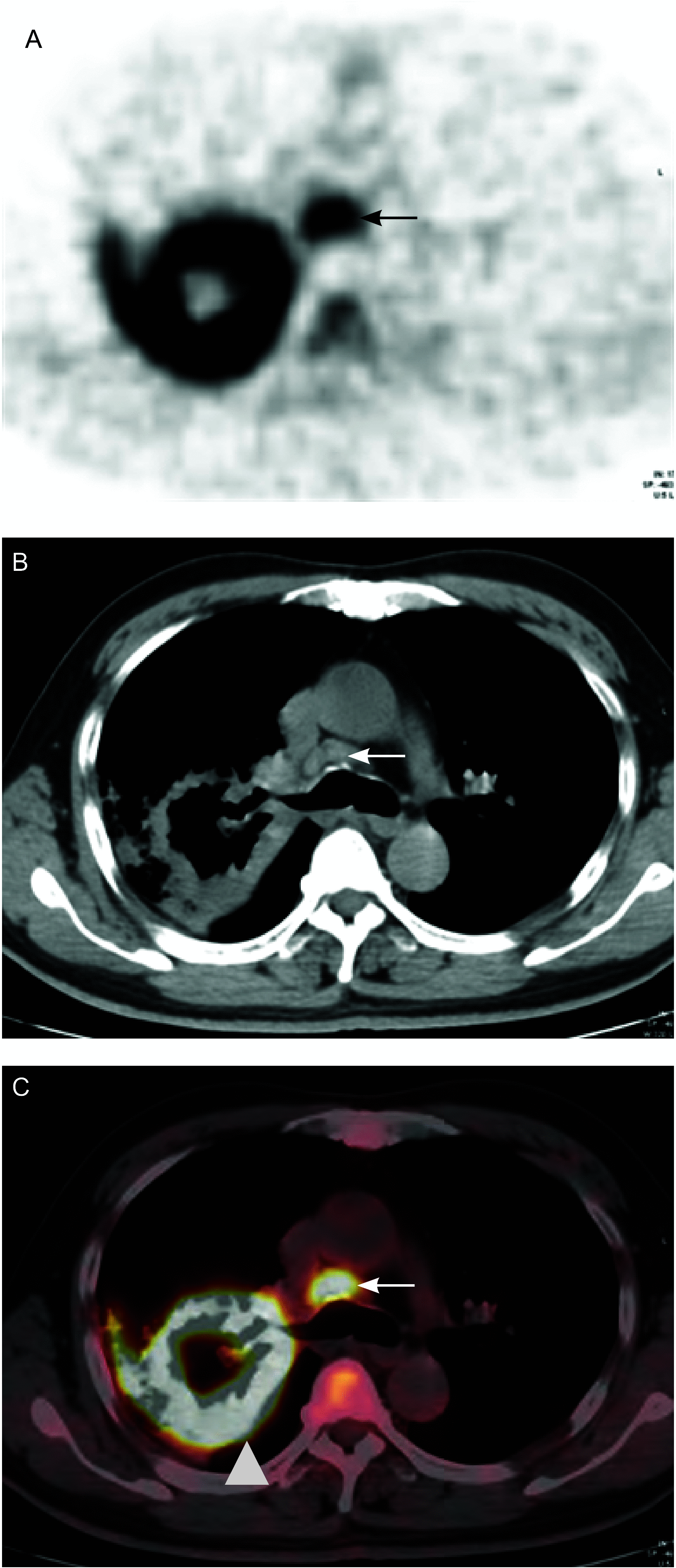
Background and objective: Mediastinal involvement in lung cancer is a highly significant prognostic factor for survival, and accurate staging of the mediastinum will correctly identify patients who will benefit the most from surgery. Positron emission tomography/computed tomography (PET/CT) has become the standard imaging modality for the staging of patients with lung cancer. The aim of this study is to investigate 18-fluoro-2-deoxy-glucose (18F-FDG) PET/CT imaging in the detection of mediastinal disease in lung cancer.
Methods: A total of 72 patients newly diagnosed with non-small cell lung cancer (NSCLC) who underwent preoperative whole-body 18F-FDG PET/CT were retrospectively included. All patients underwent radical surgery and mediastinal lymph node dissection. Mediastinal disease was histologically confirmed in 45 of 413 lymph nodes. PET/CT doctors analyzed patients' visual images and evaluated lymph node's short axis, lymph node's maximum standardized uptake value (SUVmax), node/aorta density ratio, node/aorta SUV ratio, and other parameters using the histopathological results as the reference standard. The optimal cutoff value for each ratio was determined by receiver operator characteristic curve analysis.
Results: Using a threshold of 0.9 for density ratio and 1.2 for SUV ratio yielded high accuracy for the detection of mediastinal disease. The lymph node's short axis, lymph node's SUVmax, density ratio, and SUV ratio of integrated PET/CT for the accuracy of diagnosing mediastinal lymph node was 95.2%. The diagnostic accuracy of mediastinal lymph node with conventional PET/CT was 89.8%, whereas that of PET/CT comprehensive analysis was 90.8%.
Conclusions: Node/aorta density ratio and SUV ratio may be complimentary to conventional visual interpretation and SUVmax measurement. The use of lymph node's short axis, lymph node's SUVmax, and both ratios in combination is better than either conventional PET/CT analysis or PET/CT comprehensive analysis in the assessment of mediastinal disease in NSCLC patients. .
背景与目的 肺癌纵隔淋巴结转移是非常重要的生存预后因素,准确的纵隔分期可以使患者最大程度地受益于手术,正电子发射体层显像/计算机体层成像(positron emission tomography/computed tomography, PET/CT )已成为肺癌患者分期的常规手段。本研究旨在探讨18氟-氟代脱氧葡萄糖(18-fluoro-2-deoxy-glucose, 18F-FDG )PET/CT在判断肺癌纵隔淋巴结转移上的价值。方法 回顾性分析72例肺癌患者术前全身PET/CT显像结果。72例患者均行根治性手术及系统纵隔淋巴结清扫,共取出413枚淋巴结,其中转移淋巴结为45枚。以病理结果作为标准,测量淋巴结短径、CT值、标准化摄取值(standardized uptake value, SUV)及纵隔血池的CT值与SUV等参数,计算淋巴结与纵隔血池密度比值以及淋巴结与纵隔血池SUV摄取比值,应用受试者工作特征(receiver operating characteristic, ROC)曲线计算截断点,分析密度比、摄取比与淋巴结良恶性关系,并与常规PET/CT法、PET/CT综合分析法比较诊断纵隔淋巴结的准确性。结果 密度比对淋巴结诊断的截断点为0.9,摄取比的截断点为1.2,当密度比≤0.9、摄取比≥1.2时,PET/CT对纵隔淋巴结诊断的准确率较高,将淋巴结短径、淋巴结最大标准化摄取值(maximum standardized uptake value, SUVmax)、密度比、摄取比综合计算PET/CT对纵隔淋巴结诊断的准确率为95.2%,而常规PET/CT法对纵隔内淋巴结诊断的准确率为89.8%,PET/CT综合分析法诊断的准确率为90.8%。结论 将PET/CT密度比、摄取比与淋巴结短径及SUVmax综合在一起对纵隔淋巴结诊断的准确率较高,优于常规PET/CT法及PET/CT综合分析法。.
Figures


Similar articles
-
Node/aorta and node/liver SUV ratios from (18)F-FDG PET/CT may improve the detection of occult mediastinal lymph node metastases in patients with non-small cell lung carcinoma.Acad Radiol. 2012 Jun;19(6):685-92. doi: 10.1016/j.acra.2012.02.013. Epub 2012 Mar 28. Acad Radiol. 2012. PMID: 22459646
-
Increasing the accuracy of 18F-FDG PET/CT interpretation of "mildly positive" mediastinal nodes in the staging of non-small cell lung cancer.Eur J Radiol. 2014 May;83(5):843-7. doi: 10.1016/j.ejrad.2014.01.016. Epub 2014 Jan 28. Eur J Radiol. 2014. PMID: 24581594
-
18F-FDG PET for mediastinal staging of lung cancer: which SUV threshold makes sense?J Nucl Med. 2007 Nov;48(11):1761-6. doi: 10.2967/jnumed.107.044362. Epub 2007 Oct 17. J Nucl Med. 2007. PMID: 17942814
-
Preoperative intrathoracic lymph node staging in patients with non-small-cell lung cancer: accuracy of integrated positron emission tomography and computed tomography.Eur J Cardiothorac Surg. 2009 Sep;36(3):440-5. doi: 10.1016/j.ejcts.2009.04.003. Epub 2009 May 22. Eur J Cardiothorac Surg. 2009. PMID: 19464906 Review.
-
Test performance of positron emission tomography and computed tomography for mediastinal staging in patients with non-small-cell lung cancer: a meta-analysis.Ann Intern Med. 2003 Dec 2;139(11):879-92. doi: 10.7326/0003-4819-139-11-200311180-00013. Ann Intern Med. 2003. PMID: 14644890 Review.
Cited by
-
Diagnostic value of NSE factor combined with ultrasound hemodynamic indexes in cervical lymph node metastasis of lung cancer.Oncol Lett. 2020 Jul;20(1):699-704. doi: 10.3892/ol.2020.11621. Epub 2020 May 14. Oncol Lett. 2020. PMID: 32565995 Free PMC article.
-
Role of CT Density in PET/CT-Based Assessment of Lymphoma.Mol Imaging Biol. 2018 Aug;20(4):641-649. doi: 10.1007/s11307-017-1155-x. Mol Imaging Biol. 2018. PMID: 29270848
-
Discrimination of mediastinal metastatic lymph nodes in NSCLC based on radiomic features in different phases of CT imaging.BMC Med Imaging. 2020 Feb 5;20(1):12. doi: 10.1186/s12880-020-0416-3. BMC Med Imaging. 2020. PMID: 32024469 Free PMC article.
-
Impact of Computer-Aided CT and PET Analysis on Non-invasive T Staging in Patients with Lung Cancer and Atelectasis.Mol Imaging Biol. 2018 Dec;20(6):1044-1052. doi: 10.1007/s11307-018-1196-9. Mol Imaging Biol. 2018. PMID: 29679299
-
Radiomic Analysis using Density Threshold for FDG-PET/CT-Based N-Staging in Lung Cancer Patients.Mol Imaging Biol. 2017 Apr;19(2):315-322. doi: 10.1007/s11307-016-0996-z. Mol Imaging Biol. 2017. PMID: 27539308
References
-
- Wang SY, Zhang J, Sun GF, et al. The maximum standardized uptake value of 18F-FDG PET/CT combined with the image features on high resolution CT for the diagnosis of lung cancer. http://www.cqvip.com/QK/92974X/201301/44776204.html Zhonghua He Yi Xue Yu Fen Zi Ying Xiang Za Zhi. 2013;33(1):29–33.
- 王 少雁, 张 建, 孙 高峰, et al. 18F-FDG PET/CT最大标准摄取值联合HRCT在肺癌诊断中的价值和影响因素分析. http://www.cqvip.com/QK/92974X/201301/44776204.html 中华核医学与分子影像杂志. 2013;33(1):29–33.
-
- Ding QY, Xu XD, Li TN, et al. Positron emission tomography-CT evaluation of therapeutic effect on lung cancer: a comparative study. Zhonghua Fang She Xue Za Zhi. 2013;47(12):1105–1109. doi: 10.3760/cma.j.issn.1005-1201.2013.12.013. - DOI
- 丁 其勇, 徐 绪党, 李 天女, et al. 正电子发射计算机体层成像-CT评估非小细胞肺癌治疗效果的对照研究. 中华放射学杂志. 2013;47(12):1105–1109. doi: 10.3760/cma.j.issn.1005-1201.2013.12.013. - DOI - PubMed
-
- Darling GE, Maziak DE, Inculet RI, et al. Positron emission tomography/computed tomography compared with invasive mediastinal staging in non-small cell lung cancer: results of mediastinal staging in the early lung positron emission tomography trial. J Thorac Oncol. 2011;6(8):1367–1372. doi: 10.1097/JTO.0b013e318220c912. - DOI - PubMed
Publication types
MeSH terms
Substances
LinkOut - more resources
Full Text Sources
Medical

