

Modulation of conformational changes in helix 69 mutants by pseudouridine modifications
- PMID: 25800680
- PMCID: PMC4414897
- DOI: 10.1016/j.bpc.2015.03.001
Modulation of conformational changes in helix 69 mutants by pseudouridine modifications
Abstract
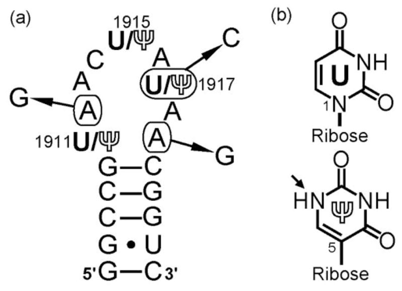
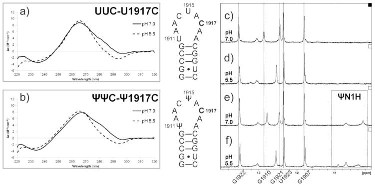
Centrally located at the ribosomal subunit interface and mRNA tunnel, helix 69 (H69) from 23S rRNA participates in key steps of translation. Ribosome activity is influenced by three pseudouridine modifications, which modulate the structure and conformational behavior of H69. To understand how H69 is affected by the presence of pseudouridine in combination with sequence changes, the biophysical properties of wild-type H69 and representative mutants (A1912G, U1917C, and A1919G) were examined. Results from NMR and circular dichroism spectroscopy indicate that pH-dependent structural changes of wild-type H69 and the chosen mutants are modulated by pseudouridine and loop sequence. The effects of the mutations on global stability of H69 are negligible; however, pseudouridine stabilizes H69 at low pH conditions. Alterations to induced conformational changes of H69 likely result in compromised function, as indicated by previous biological studies.
Keywords: CD; NMR; RNA; Ribosome; Thermodynamics.
Copyright © 2015 Elsevier B.V. All rights reserved.
Figures


Similar articles
-
Structure modulation of helix 69 from Escherichia coli 23S ribosomal RNA by pseudouridylations.Nucleic Acids Res. 2014 Apr;42(6):3971-81. doi: 10.1093/nar/gkt1329. Epub 2013 Dec 26. Nucleic Acids Res. 2014. PMID: 24371282 Free PMC article.
-
RluD, a highly conserved pseudouridine synthase, modifies 50S subunits more specifically and efficiently than free 23S rRNA.RNA. 2007 Nov;13(11):1868-76. doi: 10.1261/rna.711207. Epub 2007 Sep 13. RNA. 2007. PMID: 17872507 Free PMC article.
-
Effects of nucleotide substitution and modification on the stability and structure of helix 69 from 28S rRNA.RNA. 2005 Sep;11(9):1420-9. doi: 10.1261/rna.2320605. RNA. 2005. PMID: 16120833 Free PMC article.
-
Pseudouridine in RNA: what, where, how, and why.IUBMB Life. 2000 May;49(5):341-51. doi: 10.1080/152165400410182. IUBMB Life. 2000. PMID: 10902565 Review.
-
Pseudouridine synthases.Chem Biol. 2006 Nov;13(11):1125-35. doi: 10.1016/j.chembiol.2006.09.009. Chem Biol. 2006. PMID: 17113994 Review.
Cited by
-
Advanced reactivity-based sequencing methods for mRNA epitranscriptome profiling.RSC Chem Biol. 2024 Dec 10;6(2):150-169. doi: 10.1039/d4cb00215f. eCollection 2025 Feb 5. RSC Chem Biol. 2024. PMID: 39759443 Free PMC article. Review.
-
Promoted Read-through and Mutation Against Pseudouridine-CMC by an Evolved Reverse Transcriptase.bioRxiv [Preprint]. 2024 Jul 3:2024.07.03.601893. doi: 10.1101/2024.07.03.601893. bioRxiv. 2024. Update in: Commun Biol. 2025 Jan 11;8(1):40. doi: 10.1038/s42003-025-07467-4. PMID: 39005393 Free PMC article. Updated. Preprint.
-
Advancements in pseudouridine modifying enzyme and cancer.Front Cell Dev Biol. 2024 Dec 16;12:1465546. doi: 10.3389/fcell.2024.1465546. eCollection 2024. Front Cell Dev Biol. 2024. PMID: 39737343 Free PMC article. Review.
-
Decoding the 'Fifth' Nucleotide: Impact of RNA Pseudouridylation on Gene Expression and Human Disease.Mol Biotechnol. 2024 Jul;66(7):1581-1598. doi: 10.1007/s12033-023-00792-1. Epub 2023 Jun 21. Mol Biotechnol. 2024. PMID: 37341888 Review.
-
Promoted read-through and mutation against pseudouridine-CMC by an evolved reverse transcriptase.Commun Biol. 2025 Jan 11;8(1):40. doi: 10.1038/s42003-025-07467-4. Commun Biol. 2025. PMID: 39799263 Free PMC article.
References
-
- Nierhaus KH, Wilson DN. Protein Synthesis and Ribosome Structure: Translating the Genome. Weinheim: WILEY-VCH; 2004.
-
- Schuwirth BS, Borovinskaya MA, Hau CW, Zhang W, Vila-Sanjurjo A, Holton JM, Cate JHD. Structures of the bacterial ribosome at 3.5 Å resolution. Science. 2005;310(5749):827–834. http://dx.doi.org/10.1126/science.1117230. - DOI - PubMed
-
- Ben-Shem A, Jenner L, Yusupova G, Yusupov M. Crystal structure of the eukaryotic ribosome. Science. 2010;330(6008):1203–1209. http://dx.doi.org/10.1126/science.1194294. - DOI - PubMed
-
- Jenner LB, Demeshkina N, Yusupova G, Yusupov M. Structural aspects of messenger RNA reading frame maintenance by the ribosome. Nat Struct Mol Biol. 2010;17(5):555–560. http://dx.doi.org/10.1038/nsmb.1790. - DOI - PubMed
-
- Liiv A, O’Connor M. Mutations in the intersubunit bridge regions of 23 S rRNA. J Biol Chem. 2006;281(40):29850–29862. http://dx.doi.org/10.1074/jbc.M603013200. - DOI - PubMed
Publication types
MeSH terms
Substances
Grants and funding
LinkOut - more resources
Full Text Sources
Other Literature Sources
Molecular Biology Databases
