. 2015 Mar 31;10(12):1947-56.
doi: 10.1016/j.celrep.2015.02.053. Epub 2015 Mar 19.

UHRF1 is a sensor for DNA interstrand crosslinks and recruits FANCD2 to initiate the Fanconi anemia pathway

Affiliations

UHRF1 is a sensor for DNA interstrand crosslinks and recruits FANCD2 to initiate the Fanconi anemia pathway

Chih-Chao Liang et al. Cell Rep. .

Abstract

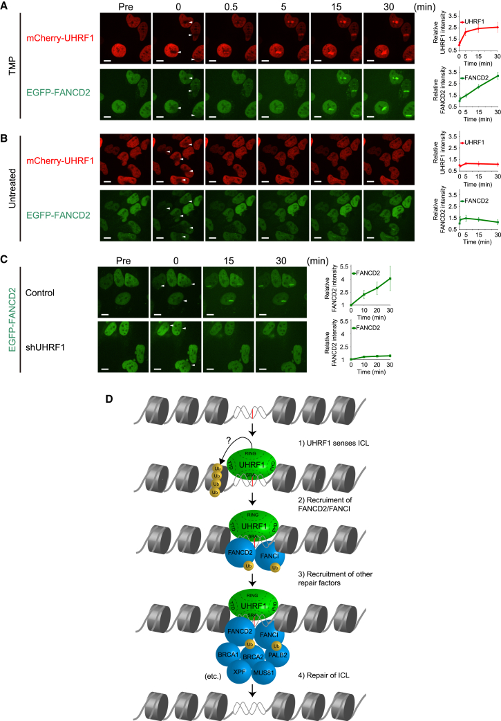
The Fanconi anemia (FA) pathway is critical for the cellular response to toxic DNA interstrand crosslinks (ICLs). Using a biochemical purification strategy, we identified UHRF1 as a protein that specifically interacts with ICLs in vitro and in vivo. Reduction of cellular levels of UHRF1 by RNAi attenuates the FA pathway and sensitizes cells to mitomycin C. Knockdown cells display a drastic reduction in FANCD2 foci formation. Using live-cell imaging, we observe that UHRF1 is rapidly recruited to chromatin in response to DNA crosslinking agents and that this recruitment both precedes and is required for the recruitment of FANCD2 to ICLs. Based on these results, we describe a mechanism of ICL sensing and propose that UHRF1 is a critical factor that binds to ICLs. In turn, this binding is necessary for the subsequent recruitment of FANCD2, which allows the DNA repair process to initiate.


Figures

None
Graphical abstract
Figure 1
Figure 1
Purification of UHRF1 (A) Purification scheme for ICL-interacting proteins. Biotinylated DNA substrate is incubated with HeLa nuclear extract and captured by streptavidin-sepharose beads. The bound proteins are eluted, separated by SDS-PAGE, and analyzed by silver stain. (B) Schematic of the biotinylated ICLs used in the purification. (C) Analysis of the crosslinked ICL1 on an 8 M urea 20% polyacrylamide gel. (D) Proteins purified from HeLa nuclear extract were stained by silver stain. The polypeptide identified by MS is indicated. (E) List of identified peptides that match UHRF1. There were 76 peptides derived from UHRF1. Amino acids contained in the identified peptides are indicated in red.
Figure 2
Figure 2
UHRF1 Interacts Directly with DNA ICLs (A) Coomassie blue stain of recombinant FLAG-HA-tagged UHRF1 and FLAG-HA-tagged FANCL purified from Sf9 cells. (B) Schematic of the biotinylated ICL2 used in the in vitro DNA binding assay in (C). (C) In vitro DNA binding assay showing that recombinant UHRF1 binds more strongly to crosslinked ICL2-XL DNA than to normal ICL2 DNA. (D) Schematic of ICL8 and CPG3 DNA substrates used in (E). (E) EMSA showing that UHRF1 forms stronger protein-DNA complexes with crosslinked ICL8-XL and hemimethylated CPG3-Me DNA substrates than with the corresponding unmodified DNA molecules. Super-shift using antibody against the HA-tag on recombinant UHRF1 confirms that the protein/DNA complex is formed by UHRF1. (F) Coomassie blue stain of recombinant FLAG-HA-tagged UHRF1-ΔSRA purified from Sf9 cells. (G) EMSA using recombinant UHRF1-ΔSRA lacking the SRA domain shows that the SRA domain of UHRF1 is responsible for the interaction with the ICL. (H) Fluorescence anisotropy assay determining the characteristics of UHRF1 binding to either ICL8 or ICL8-XL. Normalized and averaged anisotropy ± SEM.
Figure 3
Figure 3
Knockdown of UHRF1 Sensitizes Cells to MMC (A) A quantitative western blot analysis comparing the serial dilution of the lysate of HeLa cells expressing non-targeting shRNA (HeLa.Scramble), and HeLa cells expressing shRNA targeting UHRF1 (HeLa.shUHRF1), determines the efficiency of UHRF1 knockdown to be ∼95%. (B) A clonogenic survival assay of HeLa.Scramble and HeLa.shUHRF1 cells shows that UHRF1 is required for cell survival after MMC treatment. (C) A clonogenic survival assay of HeLa.Scramble and HeLa.shUHRF1 cells shows that UHRF1 is partly required for cell survival after cisplatin treatment. (D) A clonogenic survival assay of HeLa.Scramble and HeLa.shUHRF1 cells shows that UHRF1 is required for cell survival after TMP/UVA treatment. (E) A clonogenic survival assay of HeLa.Scramble and HeLa.shUHRF1 cells shows that UHRF1 is not required for cell survival after IR treatment. (F) A clonogenic survival assay of HeLa.Scramble and HeLa.shUHRF1 cells shows that UHRF1 is partly required for cell survival after UVC treatment. (G) A clonogenic survival assay of HeLa.Scramble, HeLa.shFANCD2, HeLa.shUHRF1, and HeLa.shFANCD2/shUHRF1 shows that UHRF1 and FANCD2 are epistatic. Error bars in (B)–(G) show SD. See also Figure S1.
Figure 4
Figure 4
UHRF1 Is Required for Normal FANCD2 Foci Formation In Vivo (A) FANCD2 foci accumulate after MMC treatment in control HeLa cells, whereas FANCD2 foci formation is defective in the absence of UHRF1. Scale bar, 10 μm. (B) Quantification of the percentage of cells with more than 20 foci per cell. The error bars are calculated based on two individual experiments and show SD. See also Figure S1.
Figure 5
Figure 5
UHRF1 Is Rapidly Recruited to ICLs In Vivo and Precedes the Recruitment of FANCD2 (A and B) HeLa cells expressing mCherry-tagged UHRF1 and EGFP-tagged FANCD2 were (A) pre-treated with TMP or (B) untreated, and microirradiated at the indicated areas (white arrows). Charts on the right show quantification of mCherry-UHRF1 and EGFP-FANCD2 at the ICL sites. UHRF1 and FANCD2 were recruited to TMP-induced ICLs sites (A), but not to irradiated sites in the absence of TMP (B). Scale bar, 10 μm. (C) HeLa cells expressing EGFP-tagged FANCD2 with or without UHRF1 knockdown were microirradiated at the indicated areas (white arrows). Depletion of UHRF1 abrogates the rapid accumulation of FANCD2 at the ICLs. Scale bar, 10 μm. Charts on the right show quantification of EGFP-FANCD2 at the ICL sites. (D) Model showing how UHRF1 is recruited to the ICL, facilitating the recruitment of FANCD2, which again precedes the recruitment of additional DNA repair factors. See also Figure S1.

References

    1. Arita K., Ariyoshi M., Tochio H., Nakamura Y., Shirakawa M. Recognition of hemi-methylated DNA by the SRA protein UHRF1 by a base-flipping mechanism. Nature. 2008;455:818–821. - PubMed
    1. Bostick M., Kim J.K., Estève P.O., Clark A., Pradhan S., Jacobsen S.E. UHRF1 plays a role in maintaining DNA methylation in mammalian cells. Science. 2007;317:1760–1764. - PubMed
    1. Ciccia A., Ling C., Coulthard R., Yan Z., Xue Y., Meetei A.R., Laghmani H., Joenje H., McDonald N., de Winter J.P. Identification of FAAP24, a Fanconi anemia core complex protein that interacts with FANCM. Mol. Cell. 2007;25:331–343. - PubMed
    1. Cohn M.A., D’Andrea A.D. Chromatin recruitment of DNA repair proteins: lessons from the fanconi anemia and double-strand break repair pathways. Mol. Cell. 2008;32:306–312. - PMC - PubMed
    1. Cohn M.A., Hjelmsø I., Wu L.C., Guldberg P., Lukanidin E.M., Tulchinsky E.M. Characterization of Sp1, AP-1, CBF and KRC binding sites and minisatellite DNA as functional elements of the metastasis-associated mts1/S100A4 gene intronic enhancer. Nucleic Acids Res. 2001;29:3335–3346. - PMC - PubMed

Publication types

MeSH terms

LinkOut - more resources