

Combined Tumor Cell-Based Vaccination and Interleukin-12 Gene Therapy Polarizes the Tumor Microenvironment in Mice
- PMID: 25801067
- PMCID: PMC4633448
- DOI: 10.1007/s00005-015-0337-y
Combined Tumor Cell-Based Vaccination and Interleukin-12 Gene Therapy Polarizes the Tumor Microenvironment in Mice
Abstract
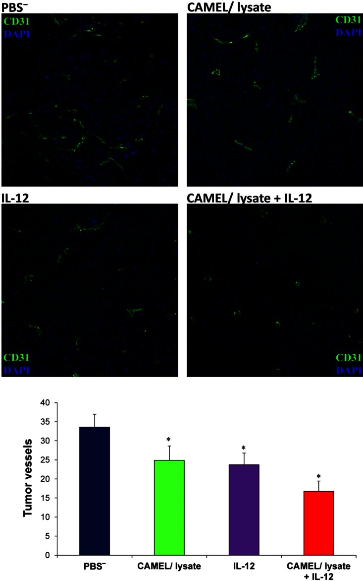
Tumor progression depends on tumor milieu, which influences neovasculature formation and immunosuppression. Combining immunotherapy with antiangiogenic/antivascular therapy might be an effective therapeutic approach. The aim of our study was to elaborate an anticancer therapeutic strategy based on the induction of immune response which leads to polarization of tumor milieu. To achieve this, we developed a tumor cell-based vaccine. CAMEL peptide was used as a B16-F10 cell death-inducing agent. The lysates were used as a vaccine to immunize mice bearing B16-F10 melanoma tumors. To further improve the therapeutic effect of the vaccine, we combined it with interleukin (IL)-12 gene therapy. IL-12, a cytokine with antiangiogenic properties, activates nonspecific and specific immune responses. We observed that combined therapy is significantly more effective (as compared with monotherapies) in inhibiting tumor growth. Furthermore, the tested combination polarizes the tumor microenvironment, which results in a switch from a proangiogenic/immunosuppressive to an antiangiogenic/immunostimulatory one. The switch manifests itself as a decreased number of tumor blood vessels, increased levels of tumor-infiltrating CD4(+), CD8(+) and NK cells, as well as lower level of suppressor lymphocytes (Treg). Our results suggest that polarizing tumor milieu by such combined therapy does inhibit tumor growth and seems to be a promising therapeutic strategy.
Keywords: CAMEL; Combined anti-tumor therapy; IL-12; Polarization of tumor microenvironment; Tumor cell-based vaccine.
Figures


References
-
- Budryk M, Wilczyńska U, Szary J, et al. Direct transfer of IL-12 gene into growing Renca tumors. Acta Biochim Pol. 2000;47:385–391. - PubMed
Publication types
MeSH terms
Substances
LinkOut - more resources
Full Text Sources
Other Literature Sources
Medical
Research Materials
Miscellaneous
