Review
. 2015 May;21(5):319-29.
doi: 10.1016/j.molmed.2015.02.004. Epub 2015 Mar 20.

The perfect storm: HLA antibodies, complement, FcγRs, and endothelium in transplant rejection

Affiliations
Review

The perfect storm: HLA antibodies, complement, FcγRs, and endothelium in transplant rejection

Kimberly A Thomas et al. Trends Mol Med. 2015 May.

Abstract

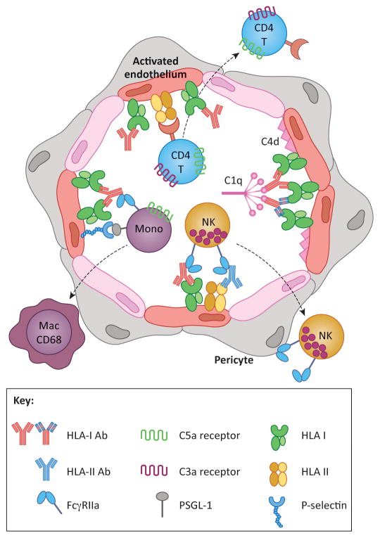
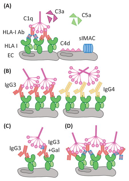
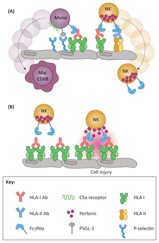
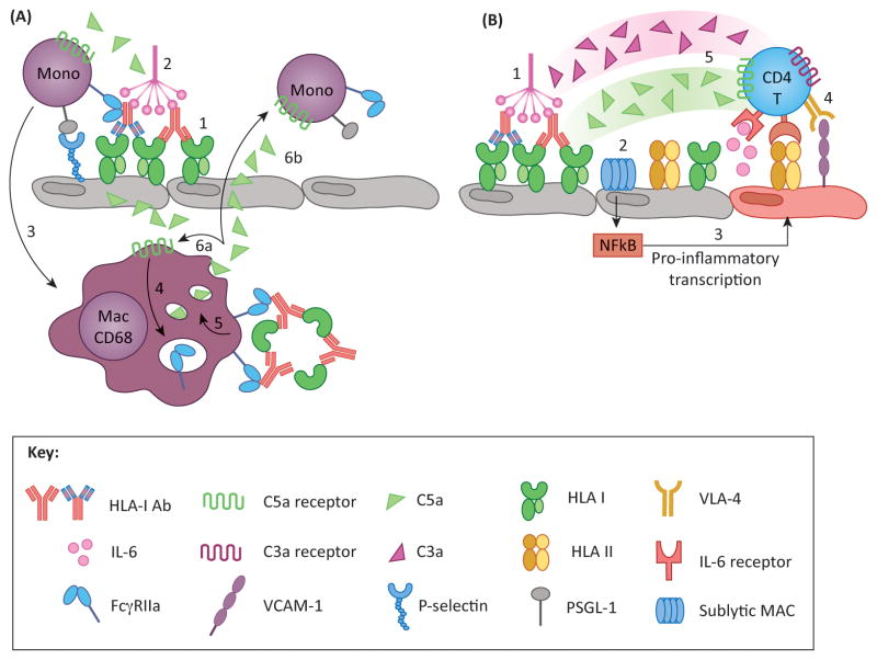
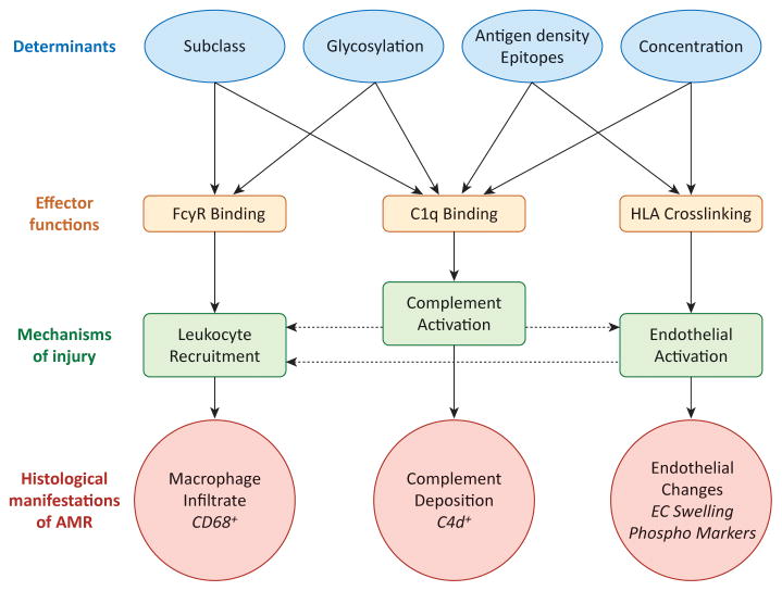
The pathophysiology of antibody-mediated rejection (AMR) in solid organ transplants is multifaceted and predominantly caused by antibodies directed against polymorphic donor human leukocyte antigens (HLAs). Despite the clearly detrimental impact of HLA antibodies (HLA-Abs) on graft function and survival, the prevention, diagnosis, and treatment of AMR remain a challenge. The histological manifestations of AMR reflect the signatures of HLA-Ab-triggered injury, specifically endothelial changes, recipient leukocytic infiltrate, and complement deposition. We review the interconnected mechanisms of HLA-Ab-mediated injury that might synergize in a 'perfect storm' of inflammation. Characterization of antibody features that are critical for effector functions may help to identify HLA-Abs that are more likely to cause rejection. We also highlight recent advances that may pave the way for new, more effective therapies.

Keywords: HLA antibodies; antibody-mediated rejection; classical complement pathway; organ transplantation.


Figures

Figure 1
Figure 1. HLA antibodies cause graft injury by inducing phenotypic changes in the donor vasculature
HLA crosslinking by antibodies of any subclass causes intracellular signaling leading to endothelial cell (EC) activation. Activated ECs express P-selectin, which promotes recruitment of leukocytes via interactions with PSGL-1. Recruited monocytes differentiate into CD68+ macrophages, which can be detected histologically in the capillaries and subendothelial space. Crosslinking of HLA molecules also enhances EC immunogenicity to recipient CD4 T cells, which proliferate and differentiate in response to alloantigen HLA class II. Complement activating antibodies trigger the classical pathway through binding of C1q, resulting in production of anaphylatoxins C3a and C5a, which have the potential to directly augment leukocyte recruitment and T cell alloresponses. Complement activation can be detected by immunohistochemical staining for C4d. Monocytes, neutrophils and NK cells also express FcγRs, which can interact with the heavy chain of HLA antibodies bound to donor ECs. FcγR functions augment leukocyte recruitment, and mediate phagocytosis and antibody-dependent cellular cytotoxicity. Taken together, the pleiotropic functions of HLA antibodies on the allograft ECs cause microvascular inflammation characteristic of antibody-mediated rejection. Antibodies in the figure with the same coloration of the Fc region are of the same subclass, whereas the varied colors within the F(ab′)2 denote unique antigenic specificities.
Figure 2
Figure 2. Complement activation by antibody/antigen determinants
(A) Activation of the classical complement pathway by HLA-Ab is mediated by C1q recognition of the Fc region of IgG. Through a series of subsequent enzymatic cleavages, the complement pathway yields the soluble anaphylatoxins, C3a and C5a, which are potent chemoattractants and stimulators of immune responses. C4d is covalently linked to the cell surface, and is a defining marker of AMR in renal and cardiac transplants. Additionally, sublytic MAC (slMAC), the terminal complex bound to cells but unable to induce lysis, is proving to be an important mediator of endothelial cell (EC) activation. Differences in antibody clonality, as demonstrated by the antibodies of varying specificity (red or blue F(ab′)2 region), allow for increased ratios of IgG:HLA, allowing for more C1q binding. (B) Antibody subclass determines the propensity of C1q binding as IgG3, a prominent complement fixer, is recognized by C1q, whereas the structure of IgG4 makes it a poor C1q binding partner. (C) Differential patterning of the N297 glycan (blue square) of IgG also modulates the level of C1q interaction. Terminal galactose residues confer maximal C1q binding to antibodies. (D) The density of HLA antigen on the surface of the cell, as well as the number of epitopes, heavily dictates the level of complement activation. The proximity of antibody Fc regions is increased when multiple antibodies can bind the same molecule of HLA. Patients with high titer polyclonal DSA may be predisposed to exacerbated complement activation during times of heightened inflammation, such as infection, when HLA expression is increased on the surface of endothelium.
Figure 3
Figure 3. Fc-mediated functions which contribute to graft injury
(A) Increased leukocytic infiltrate is a hallmark feature of AMR, and this occurrence is mediated by the Fc region of donor specific antibodies (DSA). Upon DSA binding to HLA, DSA-Fc are recognized by FcγR expressed on myeloid and NK cells. Additionally, monocytes are also able to interact with the endothelium through HLA-induced P-selectin to enhance tethering and extravasation. (B) An important feature of FcγR is their role in antibody-dependent cell-mediated cytotoxicity (ADCC). DSA bind to HLA on the surface of the endothelium, facilitating Fc interaction with FcγR expressed by myeloid and NK cells. This can lead to perforin-mediated lysis of target cells, in this case, endothelium, resulting in damage to the allograft.
Figure 4
Figure 4. Proposed models of DSA-induced inflammatory loops
(A) Macrophages perpetuate activation and recruitment via complement and FcγR pathways. DSA crosslinking of HLA on endothelium results in P-selectin mobilization to the cell surface, and provides a binding platform for C1q (1). Classical complement activation produces C5a (2), which has two functions: (i) C5a recruits monocytes to activated EC, which tether to P-selectin via PSGL-1, promoting graft infiltration and differentiation into macrophages (3); and (ii) C5a may act on intragraft CD68+ macrophages and induce FcγR expression (4). These cells can recognize immune complexes (IC) via FcγR, which can upregulate C5a production (5). Newly synthesized C5a may signal in either an autocrine (6a) or paracrine (6b) fashion, mediating further activation of intragraft macrophages and recruitment and activation of monocytes from the periphery, respectively. (B) Recent studies have identified a novel role for endothelial cells and complement in antigen presentation and stimulation of allogeneic T cells. Under inflammatory conditions (such as IFNγ activation) endothelial cells express HLA class II as well as ICAM-1, VCAM-1 and IL-6, molecules that are critical for promoting allo CD4 T cell proliferation (4) and differentiation into Th17 and Treg subsets. Preliminary work has shown that HLA antibodies modulate endothelial alloimmunogenicity through activation of the classical complement pathway (1) resulting in deposition of sublytic MAC (2). MAC triggers non-canonical NFκB signaling leading to inflammatory gene expression (3) and stimulation of allogeneic CD4 T cells (4). T cells also express receptors for complement split products C3a and C5a, which provide costimulatory signals and augment T cell proliferation. Therefore, it is likely that the presence of these anaphylatoxins at the endothelial-T cell interface might enhance T cell alloimmunity (5).
Figure 5
Figure 5. Mechanisms of DSA in graft pathogenesis
Features of antibody-antigen and antibody-effector system interactions that influence pathogenic functions and mechanisms of injury are shown. Variable factors regulating antibody-antigen interactions (blue ovals) directly influence the capacity of an antibody to trigger effector functions (green boxes), and mechanisms causing graft injury (purple boxes), which ultimately manifest in the graft as common histological features (red bursts). Linear effects are indicated by solid arrows. The functional endpoints of antibody-mediated injury are interrelated (with potential inflammatory loops indicated by dashed arrows), and likely synergize to cause maximal inflammation during AMR. For example, direct endothelial cell activation by HLA antibodies triggers adhesion of leukocytes, which can be enhanced when those leukocytes bind antibody through FcγRs. Activation of complement at the endothelial cell surface may cause production of anaphylatoxins C3a and C5a, which can act on leukocytes as chemoattractants, or enhance endothelial activation.

References

    1. Gupta G, et al. Late antibody-mediated rejection in renal allografts: outcome after conventional and novel therapies. Transplantation. 2014;97:1240–1246. doi: 10.1097/01.TP.0000442503.85766.91. - DOI - PubMed
    1. Sellares J, et al. Molecular diagnosis of antibody-mediated rejection in human kidney transplants. American journal of transplantation : official journal of the American Society of Transplantation and the American Society of Transplant Surgeons. 2013;13:971–983. doi: 10.1111/ajt.12150. - DOI - PubMed
    1. Sis B, et al. Endothelial gene expression in kidney transplants with alloantibody indicates antibody-mediated damage despite lack of C4d staining. American journal of transplantation : official journal of the American Society of Transplantation and the American Society of Transplant Surgeons. 2009;9:2312–2323. doi: 10.1111/j.1600-6143.2009.02761.x. - DOI - PubMed
    1. Wu GW, et al. Asymptomatic antibody-mediated rejection after heart transplantation predicts poor outcomes. The Journal of heart and lung transplantation : the official publication of the International Society for Heart Transplantation. 2009;28:417–422. doi: 10.1016/j.healun.2009.01.015. - DOI - PMC - PubMed
    1. Kfoury AG, et al. A longitudinal study of the course of asymptomatic antibody-mediated rejection in heart transplantation. The Journal of heart and lung transplantation : the official publication of the International Society for Heart Transplantation. 2012;31:46–51. doi: 10.1016/j.healun.2011.10.009. - DOI - PubMed

Publication types