Review
. 2015 Jul;93(7):1079-92.
doi: 10.1002/jnr.23583. Epub 2015 Mar 20.

Glucose, lactate, and shuttling of metabolites in vertebrate retinas

Affiliations
Review

Glucose, lactate, and shuttling of metabolites in vertebrate retinas

James B Hurley et al. J Neurosci Res. 2015 Jul.

Abstract

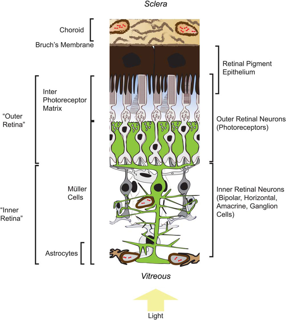
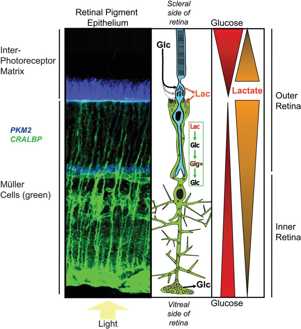
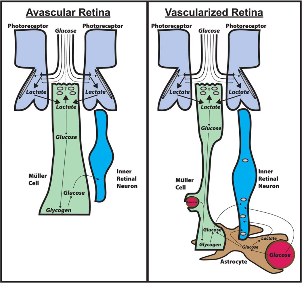
The vertebrate retina has specific functions and structures that give it a unique set of constraints on the way in which it can produce and use metabolic energy. The retina's response to illumination influences its energy requirements, and the retina's laminated structure influences the extent to which neurons and glia can access metabolic fuels. There are fundamental differences between energy metabolism in retina and that in brain. The retina relies on aerobic glycolysis much more than the brain does, and morphological differences between retina and brain limit the types of metabolic relationships that are possible between neurons and glia. This Mini-Review summarizes the unique metabolic features of the retina with a focus on the role of lactate shuttling.

Keywords: Müller cell; astrocyte neuronal lactate shuttle; glia; glucose; lactate shuttle; neuron; photoreceptor; retina.


Figures

Fig. 1
Fig. 1
Key structural features that influence production and consumption of metabolic energy in a vascularized retina. See text for descriptions of the metabolic roles of these features. [Color figure can be viewed in the online issue, which is available at wileyonlinelibrary.com.]
Fig. 2
Fig. 2
Phosphocreatine shuttle in photoreceptors in nonvascularized retinas. When mitochondria produce ATP, mtCK converts it to PCr, which shuttles the high-energy phosphate past ion pumps in the cell body of the photoreceptor. After it has reached the synaptic terminal, the PCr is used to make ATP. [Color figure can be viewed in the online issue, which is available at wileyonlinelibrary.com.]
Fig. 3
Fig. 3
Differences in glutamate distribution in conventional synapses compared with photoreceptor synapses. In brain and in inner retinal neurons (left), glutamate (black dots) released by a presynaptic neuron can be taken up by glia and used to synthesize glutamine. At the photoreceptor synapse (right), glutamate is rapidly sequestered back into the photoreceptor before it can escape from the synapse. [Color figure can be viewed in the online issue, which is available at wileyonlinelibrary.com.]
Fig. 4
Fig. 4
Model for distribution of metabolic fuels in an avascular retina. A z-stack of confocal images of a fixed mouse retina stained with antibodies to CRALBP (green) and PKM2 (blue) is shown for reference next to a schematic diagram showing a rod photoreceptor (blue) and a Müller glial cell (green). Evidence has been reported (MacGregor et al., 1986; Adler and Southwick, 1992) that there are gradients of glucose and lactate in the IPM, as shown at right. With this model we are hypothesizing that there also is a gradient of lactate in the retina, highest at the photoreceptor side and decreasing toward the ganglion cell side of the retina (consistent with Fig. 3 of Du et al., 2013). Evidence cited in the text shows that Müller cells can convert lactate to glucose and glycogen. The model includes this as a source of glucose in the inner retina. This model would generate a glucose gradient similar to that detected in nonvascularized rabbit retinas by MacGregor et al. (1986). [Color figure can be viewed in the online issue, which is available at wileyonlinelibrary.com.]
Fig. 5
Fig. 5
Shuttling mechanisms that distribute glucose and lactate in retinas. Left: Shuttling in avascular retinas. Most of the glucose from the IPM is taken up by photoreceptors and converted to lactate. The lactate is released from photoreceptors, taken up by Müller glia, and converted to glucose by gluconeogenesis. Müller cells store the glucose as glycogen primarily at their endfeet, where it can be broken down to glucose when required to fuel inner retinal neurons. Right: In vascularized retinas the retinas are thicker and the Müller cells are thinner. Astrocytes and Müller cells wrap around blood vessels in the inner retina. In addition to the Müller cells synthesizing glucose and storing glycogen, blood vessels supply glucose to astrocytes and neurons. The astrocytes can redistribute glucose and lactate to other retinal glia and neurons. [Color figure can be viewed in the online issue, which is available at wileyonlinelibrary.com.]

References

    1. Adler AJ, Southwick RE. Distribution of glucose and lactate in the interphotoreceptor matrix. Ophthalmic Res. 1992;24:243–252. - PubMed
    1. Alvarez Y, Cederlund ML, Cottell DC, Bill BR, Ekker SC, Torres-Vazquez J, Weinstein BM, Hyde DR, Vihtelic TS, Kennedy BN. Genetic determinants of hyaloid and retinal vasculature in zebrafish. BMC Dev Biol. 2007;7:114. - PMC - PubMed
    1. Ames A, 3rd, Li YY, Heher EC, Kimble CR. Energy metabolism of rabbit retina as related to function: high cost of Na+ transport. J Neurosci. 1992;12:840–853. - PMC - PubMed
    1. Arshavsky VY, Lamb TD, Pugh EN., Jr G proteins and phototrans-duction. Annu Rev Physiol. 2002;64:153–187. - PubMed
    1. Barnett NL, Pow DV, Robinson SR. Inhibition of Muller cell glutamine synthetase rapidly impairs the retinal response to light. Glia. 2000;30:64–73. - PubMed

LinkOut - more resources