. 2015 May 1;194(9):4328-38.
doi: 10.4049/jimmunol.1500018. Epub 2015 Mar 23.

The STAT5-GATA2 pathway is critical in basophil and mast cell differentiation and maintenance

Affiliations

The STAT5-GATA2 pathway is critical in basophil and mast cell differentiation and maintenance

Yapeng Li et al. J Immunol. .

Abstract

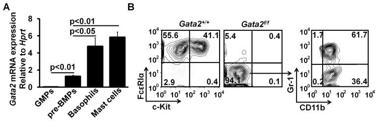
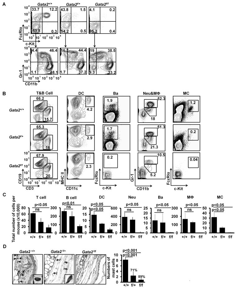
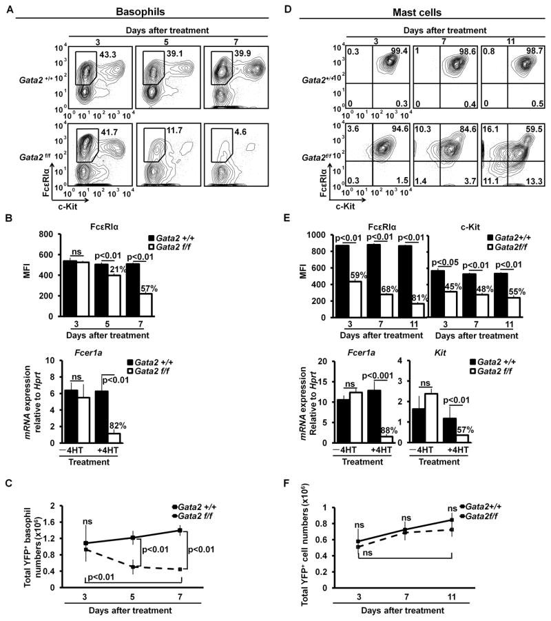
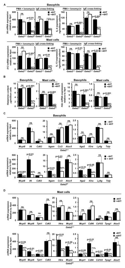
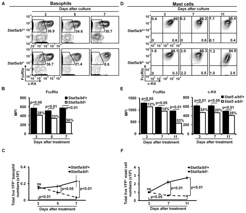
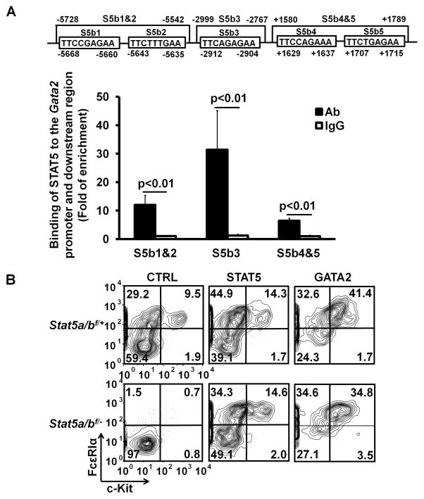
Transcription factor GATA binding protein 2 (GATA2) plays critical roles in hematopoietic stem cell survival and proliferation, granulocyte-monocyte progenitor differentiation, and basophil and mast cell differentiation. However, precise roles of GATA2 in basophil and mast cell differentiation and maintenance have not been delineated. We have identified GATA2 as an essential transcription factor in differentiation of newly identified common basophil and mast cell progenitors into basophils and mast cells. We observed Gata2 haploinsufficiency for mast cell differentiation, but not for basophil differentiation. We examined the precise role of GATA2 in maintaining the expression of a wide range of genes that are important for performing basophil or mast cell functions. The effects of GATA2 on gene expression were broadly based. We demonstrated that GATA2 was required for maintaining Fcer1a mRNA and FcεRIα protein expression on both basophils and mast cells, as well as for maintaining Kit mRNA and c-Kit protein expression on mast cells. GATA2 was required for histamine synthesis and was also critical for Il4 mRNA expression in basophils and Il13 mRNA expression in mast cells. We demonstrate a STAT5-GATA2 connection, showing that the STAT5 transcription factor directly bound to the promoter and an intronic region of the Gata2 gene. Overexpression of the Gata2 gene was sufficient to direct basophil and mast cell differentiation in the absence of the Stat5 gene. Our study reveals that the STAT5-GATA2 pathway is critical for basophil and mast cell differentiation and maintenance.


Conflict of interest statement

Disclosures

The authors have no financial conflicts of interest.

Figures

FIGURE 1
FIGURE 1
GATA2 is essential for the differentiation of pre-BMPs into both basophils and mast cells. (A) Regular GMPs (FcεRIα GMPs) and pre-BMPs were FACS-sorted from bone marrow cells of B6 mice. Basophils were purified from bone marrow cells of IL-3C-injected B6 mice. Mast cells were cultured from bone marrow cells of B6 mice in the presence of IL-3 for four weeks. Gata2 mRNA expression in GMPs, pre-BMPs, basophils, and mast cells was measured by qPCR (mean ± SD, triplicates). Data represent two independent experiments with similar results. (B) The FACS-sorted pre-BMPs from Gata2f/fRosaYfp/YfpTgCreErt2hemi (Gata2f/f) mice and Gata2+/+RosaYfp/YfpTgCreErt2hemi (Gata2+/+) mice were cultured in 1% methylcellulose containing medium in the presence of IL-3 with 25nM 4HT for 9 days. Then, the treated cells were collected and analyzed by FACS. YFP+ cells are shown. Data represent two independent experiments with similar results.
FIGURE 2
FIGURE 2
GATA2 haploid insufficiency is observed for mast cell differentiation but not for basophil differentiation. (A) Bone marrow cells (not FACS-sorted) of Gata2+/+, Gata2f/+ mice or Gata2f/f mice were cultured in methylcellulose containing medium in the presence of IL-3 and 25 nM 4HT for 9 days. YFP+ cells are shown in the FACS plots. (B) FACS analysis of cells from the tamoxifen-treated mice. YFP+ cells are shown. T cells, B cells, and dendritic cells (DC) were prepared from spleen; basophils (Ba), neutrophils (Neu), and macrophages (MΦ) from bone marrow cells; and mast cells (MC) from peritoneal cavity of the treated mice. Data represent two independent experiments with similar results. (C) Total numbers of YFP+ cells (mean ± SD, n=6). (D) Toluidine blue staining of ear sections (40×; insert, 100×). Mast cells are indicated by arrows. The right panel shows the average number of mast cells in ten different fields (40×) randomly selected from the sections of ears (mean ± SD, n=3). The percentages indicate the percentages of reduction in mast cell numbers in the ear sections.
FIGURE 3
FIGURE 3
GATA2 is critical for maintaining FcεRIα expression on basophils and FcεRIα and c-Kit expression on mast cells. (A) Bone marrow cells from IL-3C-injected Gata2+/+or Gata2f/f mice were cultured with IL-3 for three days. Then, the cells were treated with 25nM 4HT. Three, 5, and 7 days after the initial 4HT treatment, the cells were analyzed by FACS. (B) MFIs of FcεRIα expression on YFP+ Gata2+/+or Gata2−/− basophils (mean ± SD, n=6) (upper panel). The lower panel shows qPCR analysis of Fcer1a mRNA expression in the FACS-sorted YFP+ Gata2+/+or Gata2−/− basophils at 5 days after the initial 4HT treatment (mean ± SD, triplicates). Data represent three independent experiments with similar results. (C) Total numbers of YFP+ Gata2+/+ or Gata2−/− basophils (mean ± SD, n=6). (D) FACS analysis of 4-week BMMCs treated with 25nM 4HT. (E) MFIs of FcεRIα and c-Kit expression on YFP+ Gata2+/+ mast cells (FcεRIα+ c-Kit+) or “Gata2−/− mast cells” (including FcεRIα+/− and c-Kit+/− cells) (mean ± SD, n=4, upper panel). The lower panel shows qPCR analysis of Fcer1a and c-Kit mRNA expression in the FACS-sorted YFP+ Gata2+/+ mast cells or “Gata2−/− mast cells” at 11 days after the initial 4HT treatment (mean ± SD, triplicates). Data represent three independent experiments with similar results. (F) Total numbers of YFP+ Gata2+/+ mast cells or “Gata2−/− mast cells” (mean ± SD, n=4). YFP+ cells are shown (A and D). The percentages of reduction are indicated (B and E).
FIGURE 4
FIGURE 4
GATA2 is crucial for maintaining the expression of genes that are important in carrying out basophil or mast cell functions and for histamine synthesis. (A) qPCR and ELISA analyses of Il4 mRNA and IL-4 protein in basophils or Il13 mRNA and IL-13 protein in mast cells not treated or treated with 4HT. YFP+ Gata2+/+ or Gata2−/− basophils at day 5 after the initial 4HT treatment and YFP+ Gata2+/+ mast cells or “Gata2−/− mast cells” at day 11 after the initial 4HT treatment were FACS-sorted and stimulated with PMA and ionomycin for 6 hours or stimulated with IgE cross-linking overnight. (B) ELISA measurement of histamine content and qPCR analysis of Hdc mRNA expression in the FACS-sorted YFP+ Gata2−/− basophils and “Gata2−/− mast cells”. (C) qPCR analysis of mRNA expression of basophil genes in the FACS-sorted YFP+ Gata2+/+ or Gata2−/− basophils at day 5 after the initial 4HT treatment. Different scales were used to present data generated in the same experiment. (D) qPCR analysis of mRNA expression of mast cell genes in the FACS-sorted YFP+ Gata2+/+ mast cells or “Gata2−/− mast cells” at day 11 after the initial 4HT treatment. The percentages of reduction are indicated (A through D). Data represent mean ± SD (triplicates) and two independent experiments with similar results (A through D).
FIGURE 5
FIGURE 5
STAT5 is required for maintaining FcεRIα expression on basophils and FcεRIα and c-Kit expression on mast cells. (A) Bone marrow cells prepared from the IL-3C-injected Stat5a/bf/+RosaCreErt2/Yfp (Stat5a/bf/+) or Stat5a/bf/−RosaCreErt2/Yfp (Stat5a/bf/−) mice were cultured in the presence of IL-3 for 3 days. The resulting cells were treated with 25 nM 4HT for an additional 3 days. Three, 5, or 7 days after the initial 4HT treatment, the cells were analyzed by FACS. (B) MFIs of FcεRIα expression on YFP+ Stat5a/b+/− or Stat5a/b−/− basophils (mean ± SD, n=6). (C) Total numbers of YFP+ Stat5a/b+/− or Stat5a/b−/− basophils (mean ± SD, n=6). (D) FACS analysis of BMMCs treated with 4HT. (E) MFIs of FcεRIα and c-Kit expression on YFP+ Stat5a/b+/− or Stat5a/b−/− mast cells (mean ± SD, n=6). (F) Total numbers of YFP+ Stat5a/b+/− or Stat5a/b−/− mast cells (mean ± SD, n=6). YFP+ cells are shown (A and D). The percentages of reduction are indicated (B and E).
FIGURE 6
FIGURE 6
STAT5 directly regulates the Gata2 gene and overexpression of the Gata2 gene is sufficient to direct basophil and mast cell differentiation in the absence of the Stat5 gene. (A) STAT5-binding sites (S5bs1-5, upper panel) and ChIP analysis of STAT5 binding to the Gata2 promoter and downstream region (lower panel). BMMCs without stimulation were used for the ChIP analysis. (B) Bone marrow cells of Stat5a/bf/+ and Stat5a/bf/− mice were infected with retrovirus containing Stat5a, Gata2, or Thy1a (CTRL) gene. Twenty-four hours post infection, the infected cells were treated with 25nM 4HT for three days. Ten days after the initial 4HTtreatment, the cells were analyzed by FACS. YFP+ Thy1.1+ cell populations are shown. Data represent two independent experiments with similar results.

Similar articles

Cited by

References

    1. Stone KD, Prussin C, Metcalfe DD. IgE, mast cells, basophils, and eosinophils. J Allergy Clin Immunol. 2010;125:S73–80. - PMC - PubMed
    1. Galli SJ, Kalesnikoff J, Grimbaldeston MA, Piliponsky AM, Williams CM, Tsai M. Mast cells as “tunable” effector and immunoregulatory cells: recent advances. Annu Rev Immunol. 2005;23:749–786. - PubMed
    1. Siracusa MC, Kim BS, Spergel JM, Artis D. Basophils and allergic inflammation. The Journal of allergy and clinical immunology. 2013;132:789–801. quiz 788. - PMC - PubMed
    1. Karasuyama H, Mukai K, Obata K, Tsujimura Y, Wada T. Nonredundant roles of basophils in immunity. Annu Rev Immunol. 2011;29:45–69. - PubMed
    1. Kim HY, DeKruyff RH, Umetsu DT. The many paths to asthma: phenotype shaped by innate and adaptive immunity. Nat Immunol. 2010;11:577–584. - PMC - PubMed

Publication types

MeSH terms