. 2015 Mar 24:5:9449.
doi: 10.1038/srep09449.

Structure and function of a Clostridium difficile sortase enzyme

Affiliations

Structure and function of a Clostridium difficile sortase enzyme

Christopher J Chambers et al. Sci Rep. .

Abstract

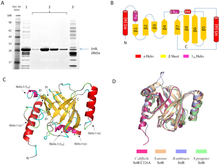
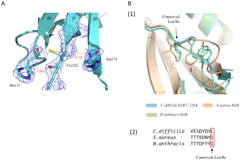
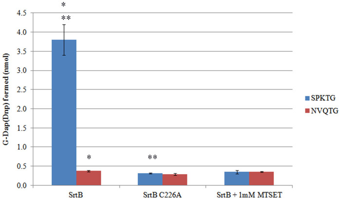
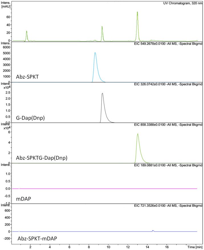
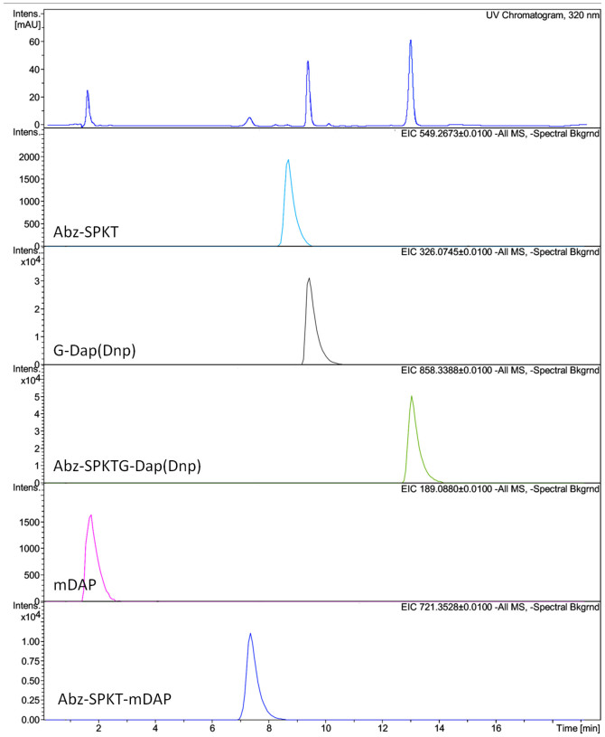
Sortase enzymes are responsible for covalent anchoring of specific proteins to the peptidoglycan of the cell wall of gram-positive bacteria. In some gram-positive bacteria (e.g. Staphylococcus aureus), sortases have been found to be essential for pathogenesis and their inhibitors are under development as potential novel therapeutics. Here we provide the first report on the structural characterisation of the C. difficile sortase. An active site mutant was crystallised and its structure determined to 2.55 Å by X-ray diffraction to provide structural insight into its catalytic mechanism. In order to elucidate the role of the sortase in the cell wall biogenesis, a C. difficile sortase knockout strain was constructed by intron mutagenesis. Characterisation of this mutant led to the discovery that the putative adhesin CD0386 is anchored to the peptidoglycan of C. difficile by the sortase SrtB and that an SPKTG peptide motif is involved in the transpeptidation reaction with the C. difficile peptidoglycan. In an animal model for C. difficile infection, the SrtB mutant caused disease at a similar rate of onset as the wild type strain. In conclusion, our detailed study shows that the SrtB enzyme from C. difficile does not play an essential role in pathogenesis.


Figures

Figure 1
Figure 1
(A) Final purification of recombinant C. difficile SrtB C226A mutant. Nickel affinity purified SrtB (1) was applied to a 5 ml Butyl sepharose column in a buffer containing 1 M (NH4)2SO4. SrtB flowed through the column at >95% purity (2) while the majority of contaminants bound to the column and were eluted on application of a low ionic strength buffer (3). (B) Schematic of C. difficile SrtB C226A coloured by secondary structure. A schematic of secondary structure elements clearly shows the non-contiguous nature of the barrel from β4 onwards. (C) C. difficile SrtB C226A Coloured by secondary structure. The structure of C. difficile SrtB incorporates three α-helices, two 310-helices and eight β-strands. (D) Superposition of C. difficile SrtB with several class B sortase structures. The C. difficile SrtB C226A structure superposed with the class B sortases of S. aureus (PDB 1NG5), B. anthracis (PDB 1RZ2) and Streptococcus pyogenes (PDB 3PSQ).
Figure 2
Figure 2
(A) The modelled active site of C. difficile SrtB. Portion of the electron density map (blue mesh) is shown contoured to 1.0 Sigma. Clear and continuous electron density is visible for His133 and Arg234. Electron density is clearly absent for the modelled Cys266. (B) The putative CWSS peptide binding loop of three superposed SrtB enzymes. (1) The β6-H5 loop of C. difficile SrtB, S. aureus SrtB and B. anthracis SrtB all contain a conserved Leu/Ile residue and a structurally semi-conserved tyrosine residue. (2) The conserved polar nature of the amino acids preceding the conserved Leu/Ile is clear in the primary sequence of the region.
Figure 3
Figure 3. Cleavage of SPKTG and NVQTG Peptides by SrtB.
Mean values (n = 3) are plotted with error bars representing +/− 1SD. Asterisks (*) and (**) indicate a p value of <0.01 as determined by Student's T-test.
Figure 4
Figure 4. Extracted ion chromatographs for Abz-SPKTG-Dap(Dnp) cleavage by SrtB in absence of mDAP.
The UV chromatograph is shown (top) followed by EICs for each of the analytes. The mass of each analyte is shown at the top right of the EIC and its putative assignment at the bottom left. Note that intensity scales are not equivalent between EICs.
Figure 5
Figure 5. Extracted ion chromatographs for cleavage and transpeptidation of Abz-SPKTG-Dap(Dnp) by SrtB in the presence of mDAP.
The UV chromatograph is shown (top) followed by EICs for each of the analytes. The mass of each analyte is shown at the top right of the EIC and its putative assignment at the bottom left. Note that intensity scales are not equivalent between EICs.
Figure 6
Figure 6
(A) Interruption of srtB by intron mutagenesis. (1) DNA fragments generated by PCR amplification utilising primers flanking the srtB gene, demonstrating an insertion of ~1.9 kb in strain 630 srtB::erm corresponding to the size of the Ll.LtrB intron. (2) Western blotting of cell fractions from 630 and 630 srtB::erm with antibodies raised against recombinant SrtB. (B) Cell fractionation and localisation of CD0386. Membrane and Wall fractions from C. difficile 630 and 630 srtB::erm after 7, 12 or 24 hrs of growth were analysed by SDS-PAGE, transferred to a nitrocellulose membrane and blotted with anti-CD0386 serum. Normalisation was effected by determining the protein concentration of each sample and loading an equal mass of protein from wild type and mutant fractions. The major bands in the figure correspond to CD0386. Note- A loss of CD0386 from the membrane fraction is observed. (C) Kaplan Meier plot of hamster challenge with C. difficile 630 and C. difficile 630 srtB::erm. Plotted points indicate the day on which hamsters were euthanized due to level 3 symptomatic scoring. No significant difference was observed, indicating that the sortase enzyme is not required for disease in the hamster model.

Similar articles

Cited by

References

    1. Pepin J. et al. Clostridium difficile- associated diarrhea in a region of Quebec from 1991 to 2003: a changing pattern of disease severity. CMAJ. 171, 466–472 (2004). - PMC - PubMed
    1. Starr J. M., Martin H., McCoubrey J., Gibson G. & Poxton I. R. Risk factors for Clostridium difficile colonisation and toxin production. Age Ageing 32, 657–660 (2003). - PubMed
    1. Stabler R. A. et al. Comparative genome and phenotypic analysis of Clostridium difficile 027 strains provides insight into the evolution of a hypervirulent bacterium. Genome Biol 10, R102 (2009). - PMC - PubMed
    1. Peterfreund G. L. et al. Succession in the gut microbiome following antibiotic and antibody therapies for Clostridium difficile. PLoS One 7, e46966 (2012). - PMC - PubMed
    1. Just I. et al. The enterotoxin from Clostridium difficile (ToxA) monoglucosylates the Rho proteins. J Biol Chem 270, 13932–13936 (1995). - PubMed

Publication types

MeSH terms

Associated data

LinkOut - more resources